. 2019 Sep 1;75(Pt 9):831-840.
doi: 10.1107/S2059798319010866. Epub 2019 Aug 22.

Structures of the substrate-binding protein YfeA in apo and zinc-reconstituted holo forms

Affiliations

Structures of the substrate-binding protein YfeA in apo and zinc-reconstituted holo forms

Christopher D Radka et al. Acta Crystallogr D Struct Biol. .

Abstract

In the structural biology of bacterial substrate-binding proteins (SBPs), a growing number of comparisons between substrate-bound and substrate-free forms of metal atom-binding (cluster A-I) SBPs have revealed minimal structural differences between forms. These observations contrast with SBPs that bind substrates such as amino acids or nucleic acids and may undergo >60° rigid-body rotations. Substrate transfer in these SBPs is described by a Venus flytrap model, although this model may not apply to all SBPs. In this report, structures are presented of substrate-free (apo) and reconstituted substrate-bound (holo) YfeA, a polyspecific cluster A-I SBP from Yersinia pestis. It is demonstrated that an apo cluster A-I SBP can be purified by fractionation when co-expressed with its cognate transporter, adding an alternative strategy to the mutagenesis or biochemical treatment used to generate other apo cluster A-I SBPs. The apo YfeA structure contains 111 disordered protein atoms in a mobile helix located in the flexible carboxy-terminal lobe. Metal binding triggers a 15-fold reduction in the solvent-accessible surface area of the metal-binding site and reordering of the 111 protein atoms in the mobile helix. The flexible lobe undergoes a 13.6° rigid-body rotation that is driven by a spring-hammer metal-binding mechanism. This asymmetric rigid-body rotation may be unique to metal atom-binding SBPs (i.e. clusters A-I, A-II and D-IV).

Keywords: X-ray crystallography; Yersinia pestis; YfeA; cluster A-I; substrate transfer; substrate-binding protein.


Figures

Figure 1
Figure 1
Analysis of YfeA from the periplasm of E. coli cells expressing the Yfe transporter. (a) EDS spectra of YfeA crystals grown with EDTA in the crystallization buffer (EDTA) and after 50°C partial denaturation in the presence of EDTA with EDTA in the crystallization buffer (50°C + EDTA), and of purified YfeA from the periplasm of E. coli cells expressing the Yfe transporter in minimal medium (Apo). Data are represented as the mean of three data sets, with bars indicating the standard error of the mean. EDTA: YfeA from LB co-incubated with 2 mM EDTA during crystallization. 50°C + EDTA: 30 s incubation of YfeA with 2 mM EDTA at 50°C. Apo: optimal fractionated YfeA overexpressed by pYFE3 autoinduction in cells grown in M9 minimal medium. (b, c) Analysis of the periplasmic fraction from E. coli cells expressing the Yfe transporter. (b) SDS–PAGE showing 9 h time-course fractionation after inoculating cells into M9 minimal medium. A representative gel is shown; the experiment was performed three times. An asterisk denotes the electrophoretic mobility position to probe for YfeA. (c) Densitometry calculation for the relative abundance of YfeA in the periplasm. Relative abundance is expressed as the percentage of YfeA signal relative to the total periplasm signal in each lane.
Figure 2
Figure 2
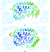
YfeA model fit. (a) Apo YfeA. The model, colored by B factor, is overlaid with anomalous difference electron density contoured at 5σ (magenta mesh) and electron density calculated from a 2F oF c map contoured at 1σ (blue mesh). Missing or broken electron density is apparent for helix 7. (b) Reconstituted holo YfeA. The model, colored by B factor, is overlaid with anomalous difference electron density contoured at 5σ (magenta mesh) and electron density calculated from a 2F oF c map contoured at 1σ (blue mesh). Contiguous electron density is apparent for helix 7.
Figure 3
Figure 3
The spring-hammer mechanism in YfeA. The holo YfeA reconstitution experiment reveals zinc binding at site 1 triggers the spring hammer. (a, b) Enlarged images show the model overlaid with anomalous difference electron density contoured at 5σ (magenta mesh) and electron density calculated from a 2F oF c map contoured at 1σ (blue mesh) at site 1. The Zn atom is omitted to emphasize the increase in anomalous difference electron density and peak height (magenta text) after reconstitution of the holo form. A placeholder water molecule (red sphere) is visible coordinating site 1 and the nearby Glu256 in the apo structure (a) (green) as site 1 residue Glu207 is disengaged from the other zinc-coordinating residues. Glu207 replaces the placeholder water molecule and engages site 1 in the reconstituted holo structure (b) (yellow).
Figure 4
Figure 4
Atomic ordering of the YfeA flexible lobe. The holo YfeA reconstitution experiment reveals atomic ordering of flexible lobe helix 7. (a, b) Enlarged images showing the model overlaid with anomalous difference electron density contoured at 5σ (magenta mesh) and electron density calculated from a 2F oF c map contoured at 1σ (blue mesh). The Zn atom is omitted to emphasize the increase in anomalous difference electron density across soaks, and the site 1 placeholder water molecule (red sphere) and residues (gray sticks) are shown. In the apo structure (a) (green), 2042 out of 2153 protein atoms are modeled and there is a visible gap between two flexible lobe residues where Pro226–Val238 should be. In the reconstituted holo structure (b) (yellow), with the spring-hammer having engaged the Zn atom, all amino acids are resolved as 2153 out of 2153 protein atoms are modeled.
Figure 5
Figure 5
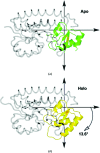
Rigid-body rotation of the YfeA flexible lobe. The DynDom Protein Domain Motion Analysis server (Taylor et al., 2014 ▸) calculates a mechanical hinge in the carboxy-terminal domain flexible lobe. The relative position of the hinge axis is at the origin in each coordinate plane along the z axis that would project out of the page. Relative to its position in the apo structure (a) (green), the flexible lobe undergoes a 13.6° rigid-body rotation in the reconstituted holo structure (b) (yellow).
Figure 6
Figure 6
Structural alignment of apo and holo cluster A-I SBPs colored by r.m.s.d. (a) SitA, PDB entries 4oxr (apo)/4oxq (holo). (b) ZnuA, PDB entries 2ps3 (apo)/2ps0 (holo). (c) TroA, PDB entries 1k0f (apo)/1toa (holo). (d) PsaA, PDB entries 3zk7 (apo)/3ztt (holo). (e) MntC, PDB entries 4nnp (apo)/5hdq (holo). (f) YfeA, apo quasi-native YfeA/reconstituted holo quasi-native YfeA. The distances between aligned Cα atoms are colored by a spectrum from minimum pairwise r.m.s.d. (blue) to maximum pairwise r.m.s.d. (red). Unaligned Cα atoms are colored gray.

References

    1. Abate, F., Malito, E., Cozzi, R., Lo Surdo, P., Maione, D. & Bottomley, M. J. (2014). Biosci. Rep. 34, e00154. - PMC - PubMed
    1. Adams, P. D., Afonine, P. V., Bunkóczi, G., Chen, V. B., Davis, I. W., Echols, N., Headd, J. J., Hung, L.-W., Kapral, G. J., Grosse-Kunstleve, R. W., McCoy, A. J., Moriarty, N. W., Oeffner, R., Read, R. J., Richardson, D. C., Richardson, J. S., Terwilliger, T. C. & Zwart, P. H. (2010). Acta Cryst. D66, 213–221. - PMC - PubMed
    1. Ahuja, S., Rougé, L., Swem, D. L., Sudhamsu, J., Wu, P., Russell, S. J., Alexander, M. K., Tam, C., Nishiyama, M., Starovasnik, M. A. & Koth, C. M. (2015). Structure, 23, 713–723. - PubMed
    1. Bearden, S. W. & Perry, R. D. (1999). Mol. Microbiol. 32, 403–414. - PubMed
    1. Bearden, S. W., Staggs, T. M. & Perry, R. D. (1998). J. Bacteriol. 180, 1135–1147. - PMC - PubMed