. 2019 Dec 24;13(12):13853-13865.
doi: 10.1021/acsnano.9b04397. Epub 2019 Sep 10.

Tumor-Derived Extracellular Vesicles Breach the Intact Blood-Brain Barrier via Transcytosis

Affiliations

Tumor-Derived Extracellular Vesicles Breach the Intact Blood-Brain Barrier via Transcytosis

Golnaz Morad et al. ACS Nano. .

Abstract

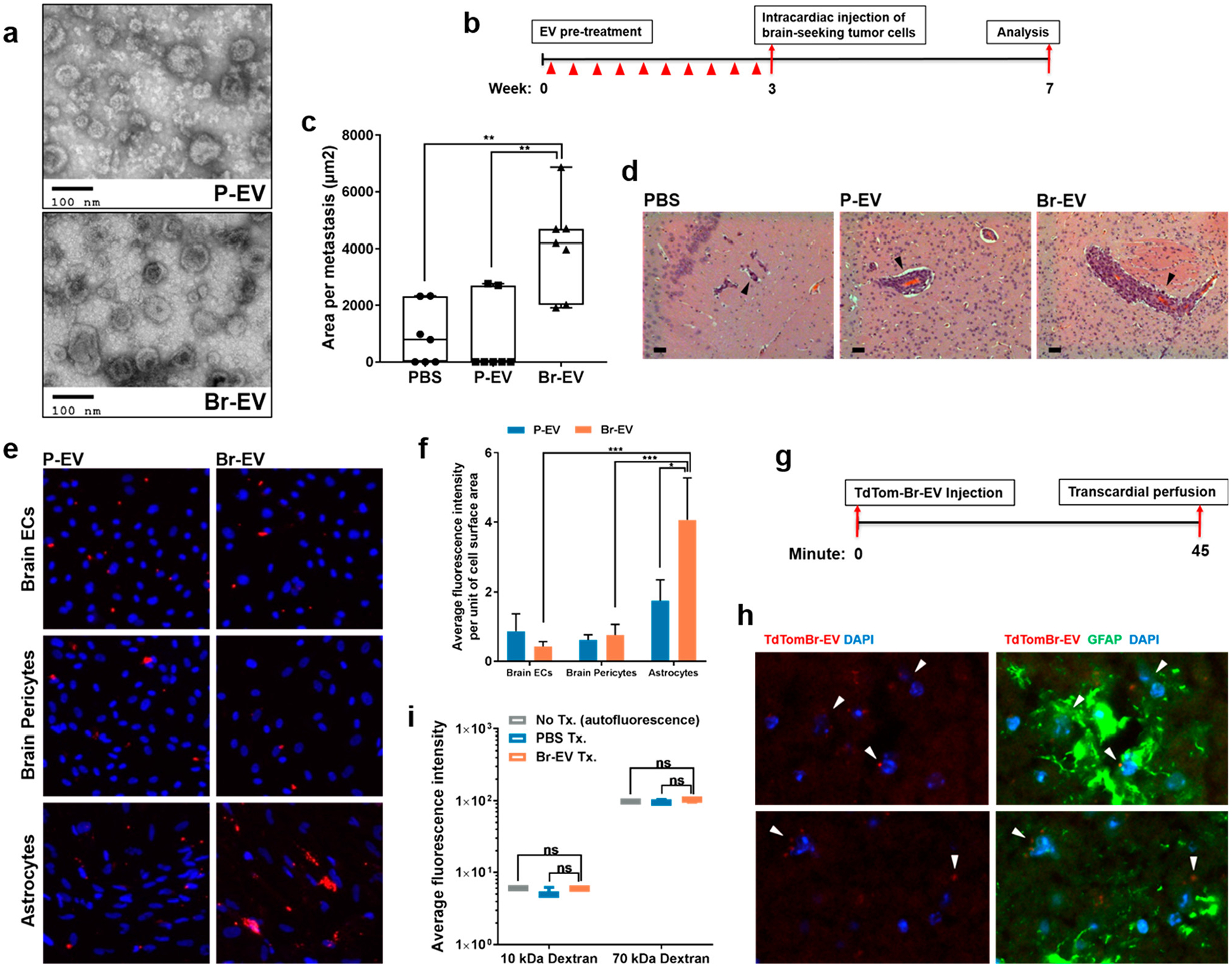
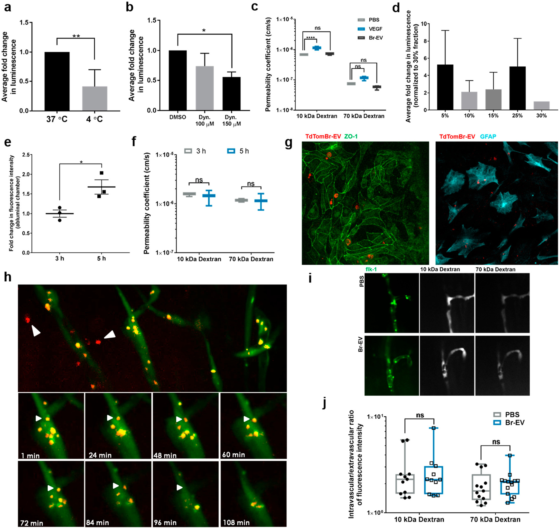
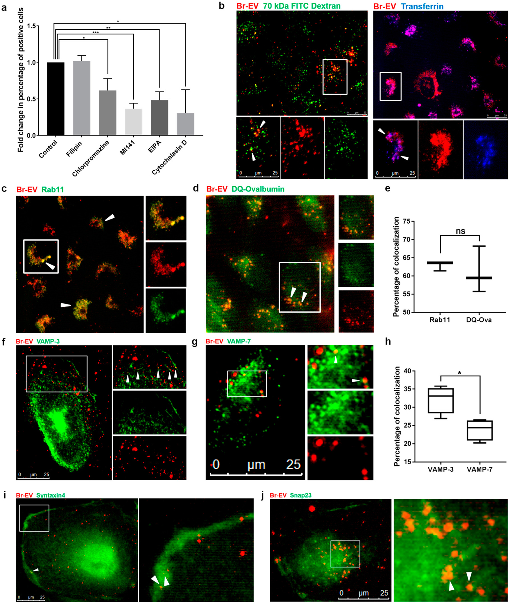
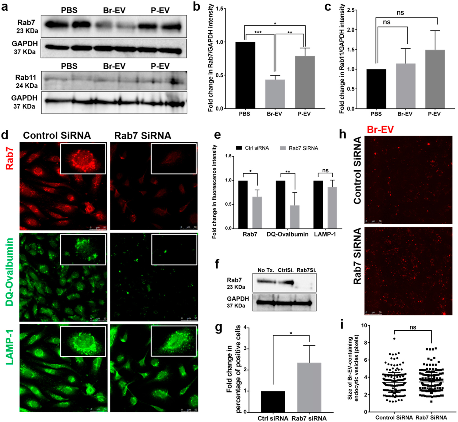
The restrictive nature of the blood-brain barrier (BBB) creates a major challenge for brain drug delivery with current nanomedicines lacking the ability to cross the BBB. Extracellular vesicles (EVs) have been shown to contribute to the progression of a variety of brain diseases including metastatic brain cancer and have been suggested as promising therapeutics and drug delivery vehicles. However, the ability of native tumor-derived EVs to breach the BBB and the mechanism(s) involved in this process remain unknown. Here, we demonstrate that tumor-derived EVs can breach the intact BBB in vivo, and by using state-of-the-art in vitro and in vivo models of the BBB, we have identified transcytosis as the mechanism underlying this process. Moreover, high spatiotemporal resolution microscopy demonstrated that the endothelial recycling endocytic pathway is involved in this transcellular transport. We further identify and characterize the mechanism by which tumor-derived EVs circumvent the low physiologic rate of transcytosis in the BBB by decreasing the brain endothelial expression of rab7 and increasing the efficiency of their transport. These findings identify previously unknown mechanisms by which tumor-derived EVs breach an intact BBB during the course of brain metastasis and can be leveraged to guide and inform the development of drug delivery approaches to deliver therapeutic cargoes across the BBB for treatment of a variety of brain diseases including, but not limited to, brain malignancies.

Keywords: blood−brain barrier; brain metastasis; breast cancer; drug delivery; exosomes; extracellular vesicles; transcytosis.


Conflict of interest statement

The authors declare no competing financial interest.

Figures

Figure 1.
Figure 1.
Brain metastasis-promoting breast cancer EVs breach the BBB. (a) Electron microscopy images of EVs isolated from parental and brain-seeking MDA-MB-231 breast cancer cells (P-EV and Br-EV, respectively). (b) Schematic depicting the in vivo brain metastasis study design. (c) Average surface area per brain metastasis (mean ± SD; n = 7 mice per group). Statistical analysis was performed using Mann- Whitney test. (d) Representative H&E images of brain metastases. All metastases demonstrated a vessel co-option pattern of growth (black arrows). Scale bar, 50 μm. (e) Representative fluorescent microscopy images (×200) and (f) quantification of the in vitro uptake of TdTom-EVs by the components of the BBB (mean ± SD; 3 independent experiments). Statistical analysis was performed using two-way ANOVA with Sidak’s multiple comparison tests. (g) Schematic showing the EV distribution study design. (h) Two representative fluorescence images of anti-GFAP immunostaining (green) of brain sections of mice that received retro-orbital injection of TdTom-Br-EVs (red). Arrows demonstrate Br-EVs taken up by astrocytes (×400, n = 3 mice). (i) Average fluorescence intensity in perfused brain tissue homogenates collected 45 min following injection of a combination of PBS or Br-EV with 10 kDa Alexa647 dextran and 70 kDa FITC dextran (mean ± SD; n = 3 mice per group). Statistical analysis was performed using Mann-Whitney test. In all panels, ns = not significant; *P ≤ 0.05; **P ≤ 0.01; ***P ≤ 0.001.
Figure 2.
Figure 2.
Br-EVs cross the brain endothelium via transcytosis. (a) Fold change in luminescent signal in the media from abluminal chamber of a Transwell BBB model under the effect of temperature and (b) endocytosis inhibition (mean ± SD; 3 independent experiments). Statistical analyses were performed using (a) unpaired two-tailed Student’s t test and (b) one-way ANOVA with Tukey’s multiple comparison test. (c) Effect of Br-EVs and VEGF (positive control) on the permeability coefficient of the endothelial monolayer to 10 and 70 kDa dextran (mean ± SD; 3 independent experiments). Statistical analysis was performed using two-way ANOVA with Sidak’s multiple comparison tests. (d) Fold change in luminescence intensity of the density gradient fractions of the media from the abluminal chamber. Luminescent signal was normalized to that of the 30% fraction, which does not contain EVs. The 15% and 25% fractions correspond to EV density of 1.105–1.184 g/mL (mean ± SD; 3 independent experiments). (e) Time-dependent increase in fluorescent signal in the abluminal channel of an in vitro BBB chip (mean ± SD; 3 independent experiments). Statistical analyses were performed using unpaired t test with Welch’s correction. (f) Effect of Br-EVs on the permeability of the BBB model to 10 and 70 kDa dextran (mean ± SD; 3 independent experiments). Statistical analysis was performed using two-way ANOVA with Sidak’s multiple comparison tests. (g) Fluorescent microscopy images of TdTom-Br-EVs taken up by endothelial cells (left panel) and astrocytes (right panel) in the BBB-on-a-chip model. (h, upper panels) Representative fluorescent images of the zebrafish brain (area selected by black square), 1 h after EV injection. White arrows demonstrate EVs in brain parenchyma. (h, lower panels) Time-lapse images of the interaction of Br-EV-containing endocytic vesicles (white arrows) with the endothelial abluminal plasma membrane (3 independent experiments). (i) Representative fluorescent images of dextran distribution in zebrafish brain vasculature. (j) Intravascular to extravascular ratio of fluorescence intensity in zebrafish brain following injection of dextran (mean ± SD; 10 kDa dextran, 11 fish per group; 70 kDa dextran, 14 fish per group; 3 independent experiments combined). Statistical analysis was performed using two-way ANOVA with Sidak’s multiple comparison tests. In all panels, ns = not significant; *P ≤ 0.05; **P ≤ 0.01; ***P ≤ 0.001; ****P ≤ 0.0001.
Figure 3.
Figure 3.
Br-EV transcytosis involves caveolin-independent endocytosis, recycling endosomes, and basolateral SNAREs. (a) Flow cytometry quantification of TdTom-Br-EV uptake by brain endothelial cells in the presence of chemical inhibitors of different pathways of endocytosis (mean ± SD; 3 independent experiments). Statistical analysis was performed using unpaired two-tailed Student’s t test. (b) Representative fluorescence microscopy images of the colocalization of TdTom-Br-EVs with 70 kDa FITC Dextran (marker of macropinocytosis, left panel) and Alexa647 transferrin (marker of clathrin-dependent endocytosis, right panel) from 3 independent experiments. The bottom panels show magnification of the area selected by the white square. White arrows indicate colocalization. Scale bar, 25 μm. Representative fluorescence microscopy images of the colocalization of TdTom-Br-EVs with (c) rab 11, (d) DQ-Ovalbumin, (f) VAMP-3, and (g) VAMP-7. The right panels show magnification of the area selected by white square. White arrows indicate colocalization. Scale bar, 25 μm. Quantification of the percentage of colocalized Br-EV-containing vesicles with rab11, DQ-Ovalbumin (e) and VAMP-3 and VAMP-7 (h) (mean ± SD; 3 independent experiments). Statistical analyses were performed using unpaired two-tailed Student’s t test. (i,j) Representative fluorescence microscopy images of the colocalization of TdTom-Br-EVs with syntaxin 4 (i) and Snap23 (j) from 3 independent experiments. The right panels show magnification of the area selected by white square. White arrows indicate colocalization. Scale bar, 25 μm. In all panels, ns = not significant; *P ≤ 0.05; **P ≤ 0.01; ***P ≤ 0.001.
Figure 4.
Figure 4.
Br-EVs downregulate the endothelial Rab7 to facilitate their transport. (a–c) Western blot images and quantification of rab7 and rab11 expression in brain endothelial cells following treatment with EVs in vitro (mean ± SD; duplicates in 3 independent experiments). Statistical analyses were performed using one-way ANOVA with Tukey’s multiple comparison test. (d,e) Representative fluorescent microscopy images and quantification of the effect of rab7 KD in brain endothelial cells (upper panel) on the transfer of DQ-ovalbumin to lysosomes for degradation (middle panel) and the expression of LAMP1 lysosomal marker (lower panel) (mean ± SD; 3 independent experiments). Scale bar, 25 μm. Statistical analyses were performed using unpaired two-tailed Student’s t test. (f) Western blot images of rab7 knockdown in brain endothelial cells. (g) Flow cytometry quantification of TdTom-Br-EV uptake by brain endothelial cells with or without rab7 KD (mean ± SD; 3 independent experiments). Statistical analyses were performed using unpaired two-tailed Student’s t test. (h) Representative fluorescent microscopy images of TdTom-Br-EV uptake by rab7 KD brain endothelial cells and (i) quantification of the size of Br-EV-containing endosomal vesicles (mean ± SD; 3 independent experiments). Scale bar, 25 μm. Statistical analyses were performed using unpaired two-tailed Student’s t test. In all panels, ns = not significant; *P ≤ 0.05; **P ≤ 0.01; ***P ≤ 0.001.

References

    1. Tang W; Fan W; Lau J; Deng L; Shen Z; Chen X Emerging Blood-Brain-Barrier-Crossing Nanotechnology for Brain Cancer Theranostics. Chem. Soc. Rev 2019, 48, 2967–3014. - PubMed
    1. Chow BW; Gu C The Molecular Constituents of the Blood- Brain Barrier. Trends Neurosci 2015, 38, 598–608. - PMC - PubMed
    1. Abbott NJ; Ronnback L; Hansson E Astrocyte-Endothelial Interactions at the Blood-Brain Barrier. Nat. Rev. Neurosci 2006, 7, 41–53. - PubMed
    1. Armulik A; Genove G; Mae M; Nisancioglu MH; Wallgard E; Niaudet C; He L; Norlin J; Lindblom P; Strittmatter K; Johansson BR; Betsholtz C Pericytes Regulate the Blood-Brain Barrier. Nature 2010, 468, 557–561. - PubMed
    1. Banks WA Characteristics of Compounds that Cross the Blood-Brain Barrier. BMC Neurol 2009, 9, S3–S8. - PMC - PubMed

Publication types