. 2019 Nov 1;129(11):4739-4744.
doi: 10.1172/JCI128205.

Peptide-conjugated oligonucleotides evoke long-lasting myotonic dystrophy correction in patient-derived cells and mice

Affiliations

Peptide-conjugated oligonucleotides evoke long-lasting myotonic dystrophy correction in patient-derived cells and mice

Arnaud F Klein et al. J Clin Invest. .

Abstract

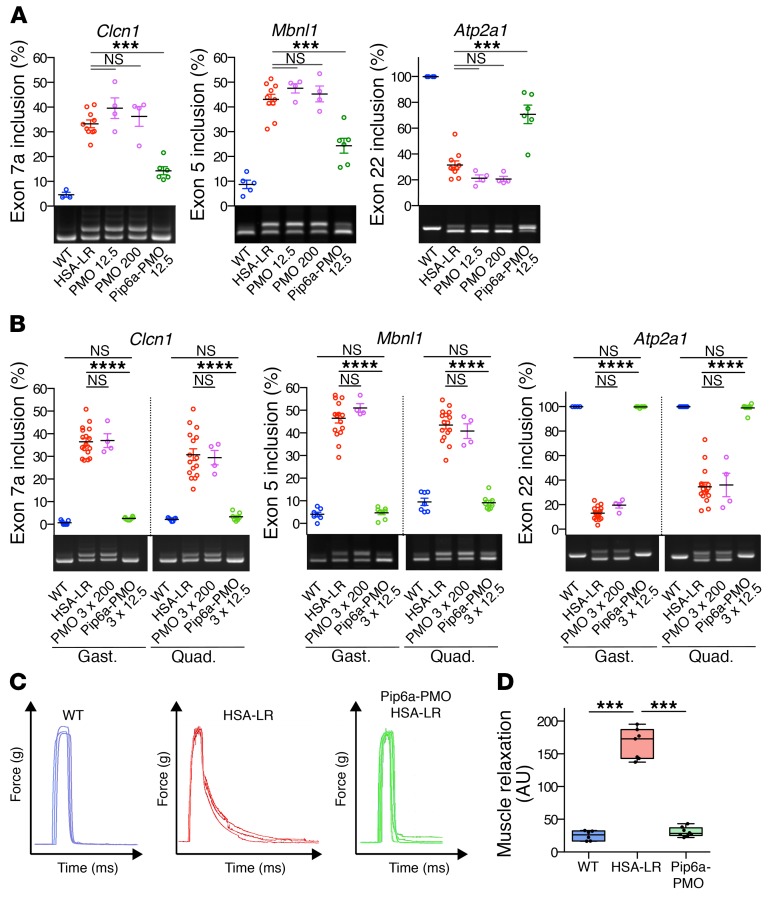
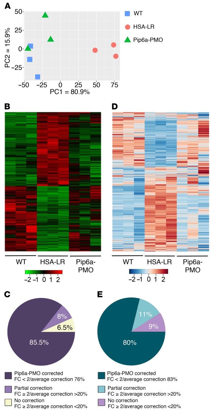
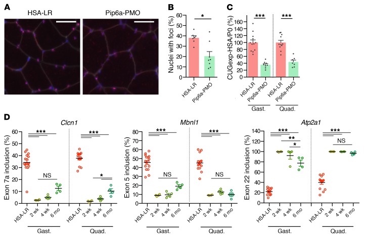
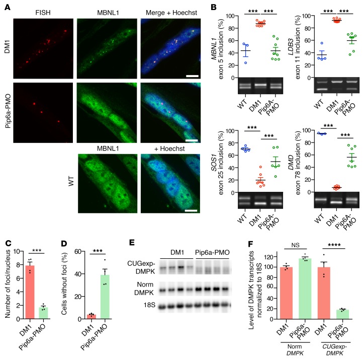
Antisense oligonucleotides (ASOs) targeting pathologic RNAs have shown promising therapeutic corrections for many genetic diseases including myotonic dystrophy (DM1). Thus, ASO strategies for DM1 can abolish the toxic RNA gain-of-function mechanism caused by nucleus-retained mutant DMPK (DM1 protein kinase) transcripts containing CUG expansions (CUGexps). However, systemic use of ASOs for this muscular disease remains challenging due to poor drug distribution to skeletal muscle. To overcome this limitation, we test an arginine-rich Pip6a cell-penetrating peptide and show that Pip6a-conjugated morpholino phosphorodiamidate oligomer (PMO) dramatically enhanced ASO delivery into striated muscles of DM1 mice following systemic administration in comparison with unconjugated PMO and other ASO strategies. Thus, low-dose treatment with Pip6a-PMO-CAG targeting pathologic expansions is sufficient to reverse both splicing defects and myotonia in DM1 mice and normalizes the overall disease transcriptome. Moreover, treated DM1 patient-derived muscle cells showed that Pip6a-PMO-CAG specifically targets mutant CUGexp-DMPK transcripts to abrogate the detrimental sequestration of MBNL1 splicing factor by nuclear RNA foci and consequently MBNL1 functional loss, responsible for splicing defects and muscle dysfunction. Our results demonstrate that Pip6a-PMO-CAG induces long-lasting correction with high efficacy of DM1-associated phenotypes at both molecular and functional levels, and strongly support the use of advanced peptide conjugates for systemic corrective therapy in DM1.

Keywords: Genetic diseases; Muscle; Therapeutics.


Conflict of interest statement

Conflict of interest: MJG and MJAW are cofounders of PepGen, a company committed to the enhanced peptide delivery of oligonucleotide therapeutics for treatment of neuromuscular and other diseases.

Figures

Figure 1
Figure 1. Pip6a-PMO corrects molecular and functional defects in HSA-LR mice.
HSA-LR mice were injected with single or multiple doses of naked PMO-CAG7 (PMO) or Pip6a-PMO-CAG7 (Pip6a-PMO) by intravenous injection and analyzed 2 weeks after treatment. (A) Quantification of splicing correction induced by a single 12.5 mg/kg dose of Pip6a-PMO, or 12.5 or 200 mg/kg dose of PMO in gastrocnemius of treated mice (WT n = 4; HSA-LR n = 10; PMO n = 4; Pip6a-PMO n = 6). (B) Representative RT-PCR results and quantification of alternative splicing profiles in gastrocnemius (gast.) and quadriceps (quad.) of HSA-LR mice treated with 3 injections of PMO at 200 mg/kg or Pip6a-PMO at 12.5 mg/kg (WT n = 7; HSA-LR n = 16; PMO n = 4; Pip6a-PMO n = 8). (C) Representative maximal force/time curves obtained by in situ force measurements of Pip6a-PMO–treated mice (3 times 12.5 mg/kg) compared with WT and HSA-LR mice. (D) Correction of myotonia (measured as the area under the force/time curve during relaxation after maximal muscle contraction) in Pip6a-PMO–injected HSA-LR mice (WT n = 6; HSA-LR n = 7; Pip6a-PMO n = 8). Data are expressed as mean ± SEM, except for D (boxes, 25th–75th percentile; whiskers, min to max; black line, the mean). ***P < 0.001; ****P < 0.0001 by 1-way ANOVA with Newman-Keuls post hoc test. NS, not significant.
Figure 2
Figure 2. Treatment with Pip6a-PMO normalizes global transcriptome at both expression and splicing levels.
Transcriptomic analysis by RNA sequencing was performed on total RNA isolated from gastrocnemius muscles of treated HSA-LR mice compared with HSA-LR and WT mice (n = 3). (A and B) Principal component analysis (A) and heatmap graphic (B) of all significantly expressed transcripts (adj. P < 0.1) reveal a global correction of the gene expression profile with Pip6a-PMO treatment. (C) Global correction of gene expression by Pip6a-PMO treatment (n = 376; FC ≥ 2, adj. P < 0.1): 85.5% of transcripts return to FC < 2 with an average correction index of 76%; 8% of transcripts remain at FC ≥ 2 but with correction index > 20%; 6.5% of transcripts are not corrected. (D) Heatmap graphic of all significant deregulated exon_bin (normalized counts) reveals a global correction of missplicing events with Pip6a-PMO treatment. (E) Overall correction of alternative splicing profiles by Pip6a-PMO treatment (n = 339 splicing events; FC ≥ 2, adj. P < 0.1): 80% of events return to FC < 2 with an average correction index of 83%; 11% remain at FC ≥ 2 but with correction index > 20%; 8% are not corrected.
Figure 3
Figure 3. Treatment with Pip6a-PMO normalizes DM1-specific phenotype.
HSA-LR mice were injected in the tail vein with three 12.5 mg/kg doses of Pip6a-PMO and analyzed 2 weeks after treatment. (A) Representative images of gastrocnemius muscle section stained for CUGexp foci (FISH, red), fiber membranes (WGA, gray), and nuclei (Hoechst, blue). Scale bars: 50 μm. (B) Quantification of the number of nuclei with foci in gastrocnemius muscle sections (HSA-LR n = 7; Pip6a-PMO n = 8). (C) Quantification of HSA transcripts levels normalized to murine Rlp0 (P0) by qRT-PCR (HSA-LR n = 12; Pip6a-PMO n = 8). (D) Quantification of splicing correction in gastrocnemius (gast.) and quadriceps (quad.) at 2 weeks, 4 weeks, and 6 months after treatment (HSA-LR n = 16; n = 4 for each time point of Pip6a-PMO). Data are expressed as mean ± SEM. *P < 0.05; **P < 0.01; ***P < 0.001 by Mann-Whitney test (B) or 1-way ANOVA with Newman-Keuls post hoc test (C and D). NS, not significant.
Figure 4
Figure 4. Pip6a-PMO corrects DM1-specific molecular symptoms in DM1 muscle cells.
Four-day-differentiated immortalized DM1 myoblasts (2600 CTG repeats) were treated with 1 μM Pip6a-PMO and analyzed after 24 hours. (A) Combined FISH (Cy3-CAG, red) and immunofluorescence (MBNL1, green) on DM1 or WT differentiated myoblasts. Scale bars: 10 μm. (B) Quantification of splicing corrections by RT-PCR (WT n = 4; DM1 and Pip6a-PMO n = 7). (C) Quantification of mean number of foci per nucleus in treated DM1 differentiated myoblasts (n = 4; >500 nuclei per n). (D) Quantification of the number of nuclei without foci in treated DM1 differentiated myoblasts (n = 4; >500 nuclei per n). (E and F) Levels of mutant DMPK and normal DMPK transcripts analyzed by Northern blot using a DMPK probe (n = 4). Data are expressed as mean ± SEM. ***P < 0.001; ****P < 0.0001 by 1-way ANOVA with Newman-Keuls post hoc test (B and F) or Mann-Whitney test (C and D). NS, not significant.

Comment in

References

    1. Harper PS. Myotonic Dystrophy. London, UK: Oxford University Press; 2001.
    1. Mahadevan M, et al. Myotonic dystrophy mutation: an unstable CTG repeat in the 3’ untranslated region of the gene. Science. 1992;255(5049):1253–1255. doi: 10.1126/science.1546325. - DOI - PubMed
    1. Brook JD, et al. Molecular basis of myotonic dystrophy: expansion of a trinucleotide (CTG) repeat at the 3’ end of a transcript encoding a protein kinase family member. Cell. 1992;68(4):799–808. doi: 10.1016/0092-8674(92)90154-5. - DOI - PubMed
    1. Fu YH, et al. An unstable triplet repeat in a gene related to myotonic muscular dystrophy. Science. 1992;255(5049):1256–1258. doi: 10.1126/science.1546326. - DOI - PubMed
    1. Taneja KL, McCurrach M, Schalling M, Housman D, Singer RH. Foci of trinucleotide repeat transcripts in nuclei of myotonic dystrophy cells and tissues. J Cell Biol. 1995;128(6):995–1002. doi: 10.1083/jcb.128.6.995. - DOI - PMC - PubMed

Publication types

MeSH terms