. 2019 Sep 3;15(9):e1008378.
doi: 10.1371/journal.pgen.1008378. eCollection 2019 Sep.

NME5 frameshift variant in Alaskan Malamutes with primary ciliary dyskinesia

Affiliations

NME5 frameshift variant in Alaskan Malamutes with primary ciliary dyskinesia

Linda Anderegg et al. PLoS Genet. .

Abstract

Primary ciliary dyskinesia (PCD) is a hereditary defect of motile cilia in humans and several domestic animal species. Typical clinical findings are chronic recurrent infections of the respiratory tract and fertility problems. We analyzed an Alaskan Malamute family, in which two out of six puppies were affected by PCD. The parents were unaffected suggesting autosomal recessive inheritance. Linkage and homozygosity mapping defined critical intervals comprising ~118 Mb. Whole genome sequencing of one case and comparison to 601 control genomes identified a disease associated frameshift variant, c.43delA, in the NME5 gene encoding a sparsely characterized protein associated with ciliary function. Nme5-/- knockout mice exhibit doming of the skull, hydrocephalus and sperm flagellar defects. The genotypes at NME5:c.43delA showed the expected co-segregation with the phenotype in the Alaskan Malamute family. An additional unrelated Alaskan Malamute with PCD and hydrocephalus that became available later in the study was also homozygous mutant at the NME5:c.43delA variant. The mutant allele was not present in more than 1000 control dogs from different breeds. Immunohistochemistry demonstrated absence of the NME5 protein from nasal epithelia of an affected dog. We therefore propose NME5:c.43delA as the most likely candidate causative variant for PCD in Alaskan Malamutes. These findings enable genetic testing to avoid the unintentional breeding of affected dogs in the future. Furthermore, the results of this study identify NME5 as a novel candidate gene for unsolved human PCD and/or hydrocephalus cases.


Conflict of interest statement

The authors have declared that no competing interests exist.

Figures

Fig 1
Fig 1. PCD phenotype in Alaskan Malamutes.
(A) Left lateral view of the thorax of one of the affected dogs at the age of 15 months with bronchopneumonia in the right middle lung lobe (encircled). (B) Endoscopic image of the trachea. Mucosal hyperemia with cobblestone appearance covered with a large amount of mucopurulent secretion. (C) Endoscopic image of the right caudal lung. Hyperemic bronchi with large amount of mucopurulent secretions is evident. (D, E) Endoscopic images of the nasal cavity. Severe turbinate lysis, hyperemic mucosa and mucopurulent mucus is evident. The bleeding on the left is from the endoscope and represents an iatrogenic lesion.
Fig 2
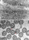
Fig 2. Transmission electron micrographs of bronchial mucosa from a PCD affected Alaskan Malamute.
(A) Overview of a ciliated airway demonstrating an overall reduced ciliation (upper part) with prominent basal bodies (white arrows). (B) A cross section of cilia is shown at higher magnification. In normal cilia, there is a 9 + 2 arrangement of microtubules with two single microtubules in the center and nine pairs of peripheral microtubules. In the affected dog, extra peripheral microtubule singlets appeared occasionally (red arrows). Furthermore, some of the outer dynein arms and most of the inner dynein arms were shortened or entirely absent.
Fig 3
Fig 3. Pedigree of the Alaskan Malamute family with two PCD cases.
Filled symbols represent dogs affected by PCD. The solitary symbol in the square represent the additional PCD affected dog (AM021) previously reported in the USA of which no pedigree data was available. For genotyping, DNA of this dog was extracted from FFPE tissues [10]. Other lab numbers indicate dogs, of which blood samples were available. Genotypes of the NME5:c.43delA variant for these dogs are shown. Two Inbreeding loops are visible in this pedigree.
Fig 4
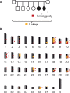
Fig 4. Combined linkage analysis with homozygosity mapping.
(A) Parametric linkage analysis was performed with eight family members of one Alaskan Malamute family. Homozygosity mapping was made with two affected dogs from this family. (B) Linked regions >1 Mb are marked in yellow and all shared homozygous regions are marked in red. Twenty regions on different chromosomes showed overlapping linked and homozygous regions (arrows) which are designated as critical intervals.
Fig 5
Fig 5. Sanger sequencing of the NME5:c.43delA variant.
Electropherograms from dogs with the three different genotypes confirm the presence of the variant.
Fig 6
Fig 6. NME5 protein expression.
(A) NME5 immunohistochemistry shows a strong signal at the ciliated epithelium from nasal mucosa of a control dog. (B) In a PCD affected Alaskan Malamute, the same polyclonal anti-NME5 antibody does not give any detectable reaction. (C-H) NME5 immunogold transmission electron microscopy of nasal mucosa, ciliary cross sections. (C-E) Nasal mucosa of a control dog with positive binding of gold particles to (C,D) outer microtubules at the inner dynein arm location and to (E) the central microtubules. (F-H) Nasal mucosa of a PCD affected Alaskan Malamute with single positive gold particles binding to different locations within the cilia, possibly due to non-specific binding of the antibody.

References

    1. Lucas JS, Burgess A, Mitchison HM, Moya E, Williamson M, Hogg C. Diagnosis and management of primary ciliary dyskinesia. Archives of disease in childhood. 2014;99(9):850–6. Epub 2014/04/29. 10.1136/archdischild-2013-304831 - DOI - PMC - PubMed
    1. Horani A, Ferkol TW, Dutcher SK, Brody SL. Genetics and biology of primary ciliary dyskinesia. Paediatric respiratory reviews. 2016;18:18–24. Epub 2015/10/20. 10.1016/j.prrv.2015.09.001 - DOI - PMC - PubMed
    1. Horani A, Ferkol TW. Advances in the Genetics of Primary Ciliary Dyskinesia: Clinical Implications. Chest. 2018;154(3):645–52. Epub 2018/05/26. 10.1016/j.chest.2018.05.007 - DOI - PMC - PubMed
    1. Kartagener M. Zur Pathogenese der Bronchiektasien: Bronchiektasien bei Situs viscerum inversus. Beiträge zur Klinik der Tuberkulose. 1933;83(4):498–501.
    1. Heuser T, Raytchev M, Krell J, Porter ME, Nicastro D. The dynein regulatory complex is the nexin link and a major regulatory node in cilia and flagella. The Journal of cell biology. 2009;187(6):921–33. Epub 2009/12/17. 10.1083/jcb.200908067 - DOI - PMC - PubMed

Publication types

Substances