

Brain iron levels in attention-deficit/hyperactivity disorder normalize as a function of psychostimulant treatment duration
- PMID: 31479897
- PMCID: PMC6726915
- DOI: 10.1016/j.nicl.2019.101993
Brain iron levels in attention-deficit/hyperactivity disorder normalize as a function of psychostimulant treatment duration
Abstract
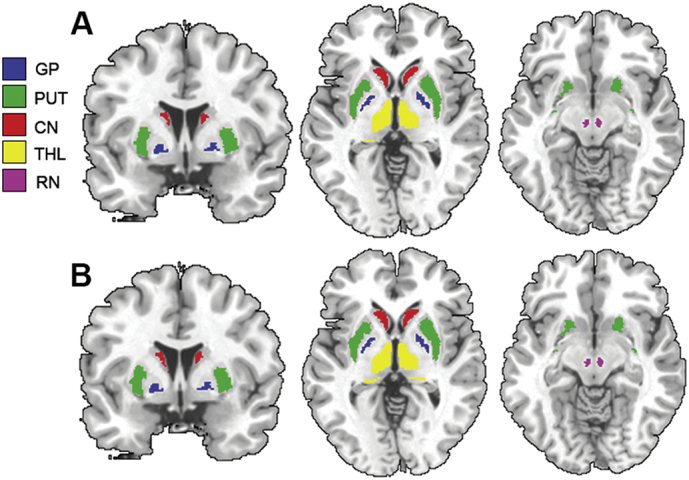
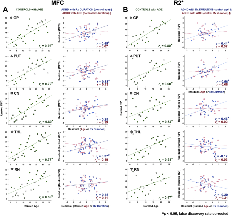
Brain iron homeostasis is a dopamine-related mechanism that may be modified with long-term psychostimulant treatment in attention-deficit/hyperactivity disorder (ADHD). We previously reported that while medication-naïve youth with ADHD have reduced brain iron compared to controls and psychostimulant-medicated patients, no differences were detected between the latter groups. In this follow-up study, we examined whether the duration of psychostimulant treatment correlates with the degree of iron normalization. Brain iron was indexed with MRI using an advanced method called magnetic field correlation (MFC) imaging and the conventional R2* proton transverse relaxation rate method. MFC was acquired in 30 psychostimulant-medicated youth with comorbid-free ADHD and 29 age-matched controls (all males). R2* was acquired in a subset of these individuals. Region-of-interest analyses for MFC and R2* group differences and within-group correlations with age and years of psychostimulant treatment were conducted in the globus pallidus (GP), putamen (PUT), caudate nucleus (CN), thalamus (THL) and red nucleus. No significant MFC and R2* group differences were detected. However, while all regional MFC and R2* significantly increased with age in the control group, MFC and R2* increased in the GP, PUT, CN and THL with psychostimulant treatment duration in the ADHD group to a greater degree than with age. Our findings suggest that while youth with ADHD may have less prominent age-related brain iron increases than that seen in typical development, long-term use of psychostimulant medications may compensate through a normalizing effect on basal ganglia iron. Longitudinal studies following ADHD patients before and after long-term psychostimulant treatment are needed to confirm these findings.
Keywords: ADHD; Brain iron; MRI; Magnetic field correlation; Psychostimulants; R2*.
Published by Elsevier Inc.
Figures
References
-
- Adams J.G. Psychostimulants: concerns over long-term adverse side effects. J. Miss. State Med. Assoc. 2015;56:346–347. - PubMed
-
- Adisetiyo V., Jensen J.H., Tabesh A., Deardorff R.L., Fieremans E., Di Martino A., Gray K.M., Castellanos F.X., Helpern J.A. Multimodal MR imaging of brain Iron in attention deficit hyperactivity disorder: a noninvasive biomarker that responds to psychostimulant treatment? Radiology. 2014;272:524–532. - PMC - PubMed
-
- Adisetiyo V., McGill C.E., DeVries W.H., Jensen J.H., Hanlon C.A., Helpern J.A. Elevated brain Iron in cocaine use disorder as indexed by magnetic field correlation imaging. Biol. Psychiatry Cogn. Neurosci. Neuroimaging. 2018 - PubMed
-
- Adler L.D., Nierenberg A.A. Review of medication adherence in children and adults with ADHD. Postgrad. Med. 2010;122:184–191. - PubMed
Publication types
MeSH terms
Substances
LinkOut - more resources
Full Text Sources
Medical
