. 2020 Apr;33(4):547-555.
doi: 10.5713/ajas.19.0173. Epub 2019 Jul 1.

MiR-26a promotes apoptosis of porcine granulosa cells by targeting the 3β-hydroxysteroid-Δ24-reductase gene

Affiliations

MiR-26a promotes apoptosis of porcine granulosa cells by targeting the 3β-hydroxysteroid-Δ24-reductase gene

Xiaodong Zhang et al. Asian-Australas J Anim Sci. 2020 Apr.

Abstract

Objective: Apoptosis of ovarian granulosa cells (GCs) affects mammalian follicular development and fecundity. This study aimed to explore the regulatory relationship between microRNA-26a (miR-26a) and the 3β-hydroxysteroid-Δ24-reductase gene (DHCR24) gene in porcine follicular granular cells (pGCs), and to provide empirical data for the development of methods to improve the reproductive capacity of pigs.

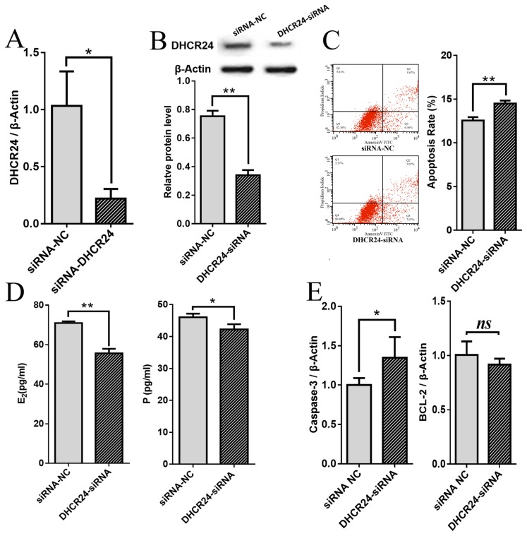
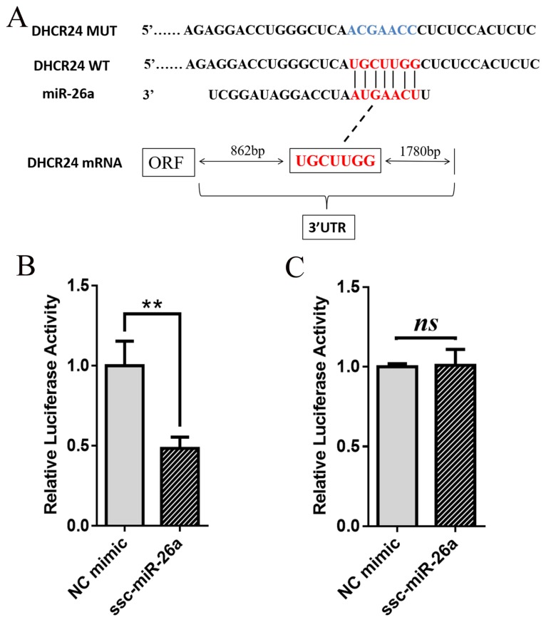
Methods: The pGCs were transfected with miR-26a mimic, miR-26a inhibitor and DHCR24-siRNA in vitro. The cell apoptosis rate of pGCs was detected by the flow cytometry. The secretion levels of estradiol (E2) and progesterone (P) in pGCs were detected by enzymelinked immunosorbent assay. Double luciferase validation system was used to detect the binding sites between miR-26a and DHCR24 3'-UTR region. Qualitative real-time polymerase chain reaction and Western blotting were used to verify the DHCR24 mRNA and protein expression in pGCs, respectively, after transfecting with miR-26a mimic and miR-26a inhibitor.

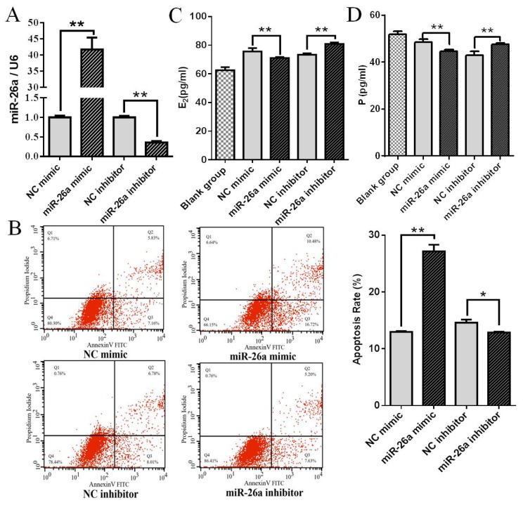
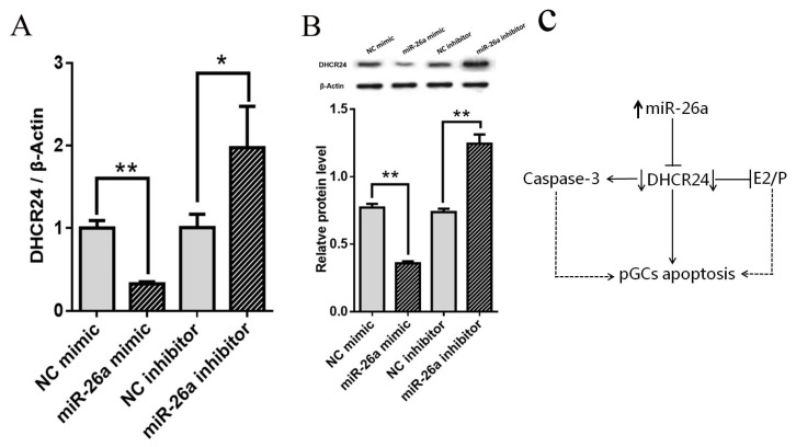
Results: Results showed that enhancement of miR-26a promoted apoptosis, and inhibited E2 and P secretion in pGCs. Meanwhile, inhibition of DHCR24 also upregulated the Caspase-3 expression, reduced the BCL-2 expression, promoted pGCs apoptosis, and inhibited E2 and P secretion in pGCs. There were the binding sites of miR-26a located within DHCR24 3'-UTR. Up-regulation of miR-26a inhibited DHCR24 mRNA and protein expression in pGCs.

Conclusion: This study demonstrates that miR-26a can promote cell apoptosis and inhibit E2 and P secretion by inhibiting the expression of DHCR24 in pGCs.

Keywords: 3β-hydroxysteroid-Δ24-reductase (DHCR24); Apoptosis; Granulosa Cells; Pig; miR-26a.


Conflict of interest statement

CONFLICT OF INTEREST

We certify that there is no conflict of interest with any financial organization regarding the material discussed in the manuscript.

Figures

Figure 1
Figure 1
miR-26a regulates apoptosis rate and E2 and P release in pGCs. (A) qPCR validation of miR-26a overexpression and inhibition using mimic and inhibitor. (B) The pGCs transfected with miR-26a mimic or inhibitor were subjected to Annexin V-FITC/PI double staining and flow cytometric analysis. (C, D) The levels of E2 and P were detected by ELISA. The blank group was the E2 or P content in the culture media supplemented with fetal bovine serum. Average results from three independent experiments are shown. E2, estradiol; P, progesterone; pGCs, porcine ovarian granulosa cells; qPCR, qualitative real-time polymerase chain reaction; ELISA, enzyme-linked immunosorbent assay; NC, negative control. * p<0.05, ** p<0.01.
Figure 2
Figure 2
Effects of DHCR24 on the apoptosis rate, E2 and P release and Caspase-3 and BCL-2 mRNA levels in pGCs. qPCR (A) and Western blot (B) analyses of DHCR24 mRNA and protein expression, respectively, in pGCs with NC-siRNA or DHCR24-siRNA. (C) pGCs transfected with DHCR24-siRNA or NC-siRNA were subjected to Annexin V-FITC/PI double staining and flow cytometric analysis. Knockdown of DHCR24 accelerates the apoptosis rate of pGCs. (D) pGCs transfected with DHCR24-siRNA or siRNA-NC. The levels of E2 and P were detected by ELISA. (E) qPCR analyses showed that knockdown of DHCR24 increased the mRNA level of Caspase-3, but did not affect the mRNA level of BCL-2 in pGCs. β-Actin was used as an internal control. Average results from three independent experiments are shown. DHCR24, 3β-hydroxysteroid-Δ24-reductase; E2, estradiol; P, progesterone; pGCs, porcine ovarian granulosa cells; qPCR, qualitative real-time polymerase chain reaction; NC, negative control; ELISA, enzyme-linked immunosorbent assay. * p<0.05, ** p<0.01.
Figure 3
Figure 3
DHCR24 is a direct target of miR-26a. (A) Schematic showing the interactions of miR-26a with wild-type DHCR24 3′-UTR (red) and the mutant version (blue). (B and C) Transfection of 293T cells with miR-26a mimic and a dual-luciferase reporter vector containing the wild-type DHCR24 3′-UTR (B) or the mutant version (C). Luciferase activity was measured and normalized to that in the NC mimic treated group. Average results from three independent experiments are shown. DHCR24, 3β-hydroxysteroid-Δ24-reductase; NC, negative control. * p<0.05, ** p<0.01.
Figure 4
Figure 4
Effects of miR-26a on the expression of DHCR24 in pGCs. (A) NC mimic, miR-26a mimic, NC inhibitor, or miR-26a inhibitor were transfected into pGCs. DHCR24 mRNA levels were detected by qPCR analysis. (B) NC mimic, miR-26a mimic, NC inhibitor, or miR-26a inhibitor were transfected into pGCs. DHCR24 protein expression was detected by western blotting. β-Actin was used as an internal control. NC means negative control. Average results from three independent experiments are shown. * p<0.05, ** p<0.01. (C) A model for miR-26a regulates pGCs apoptosis by targeting DHCR24. MiR-26a inhibits the expression of DHCR24 gene, which leads to an increase in expression of the proapoptotic Caspase-3 gene and inhibition in the secretion of E2/P and promotion of apoptosis level in pGCs. DHCR24, 3β-hydroxysteroid-Δ24-reductase; pGCs, porcine ovarian granulosa cells; qPCR, qualitative real-time polymerase chain reaction.

References

    1. McGee EA, Hsueh AJW. Initial and cyclic recruitment of ovarian follicles. Endocr Rev. 2000;21:200–14. doi: 10.1210/edrv.21.2.0394. - DOI - PubMed
    1. Pan Z, Zhang J, Lin F, Ma X, Wang X, Liu H. Expression profiles of key candidate genes involved in steroidogenesis during follicular atresia in the pig ovary. Mol Biol Rep. 2012;39:10823–32. doi: 10.1007/s11033-012-1976-2. - DOI - PubMed
    1. Peralta LE, Olarte MR, Arganaraz M, Ciocca D, Miceli DC. Progesterone receptors: Their localization, binding activity and expression in the pig oviduct during follicular and luteal phases. Domest Anim Endocrinol. 2005;28:74–84. doi: 10.1016/j.domaniend.2004.05.008. - DOI - PubMed
    1. Nie M, Yu S, Peng S, Fang Y, Wang H, Yang X. miR-23a and miR-27a promote human granulosa cell apoptosis by targeting SMAD5. Biol Reprod. 2015;93:98. doi: 10.1095/biolreprod.115.130690. - DOI - PubMed
    1. Xiong F, Hu L, Zhang Y, Xiao X, Xiao J. miR-22 inhibits mouse ovarian granulosa cell apoptosis by targeting SIRT1. Biol Open. 2016;5:367–71. doi: 10.1242/bio.016907. - DOI - PMC - PubMed