Case Reports
. 2019 Sep 3;19(1):218.
doi: 10.1186/s12883-019-1446-8.

Focal limb dystonia caused by a complication of the cerebellar developmental venous anomaly: a case report

Affiliations
Case Reports

Focal limb dystonia caused by a complication of the cerebellar developmental venous anomaly: a case report

Su Jin Chung. BMC Neurol. .

Abstract

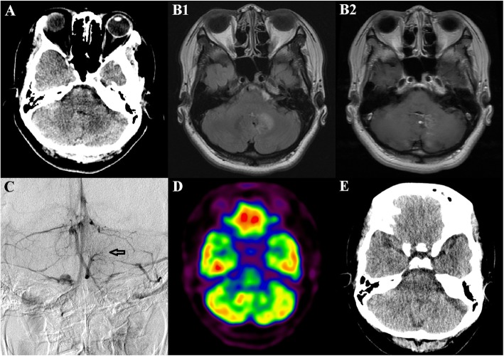
Background: There are no established theories regarding the role of the cerebellum in dystonia. We report a case of focal limb dystonia secondary to a vasogenic edema of the dentate nucleus caused by a symptomatic developmental venous anomaly.

Case presentation: A 44-year-old woman presented with sudden onset dystonia in her left arm for 1 week. Brain imaging revealed vasogenic edema in the deep white matter of the left cerebellar hemisphere, including the left dentate nucleus, secondary to a developmental venous anomaly. 18F-fluorodeoxyglucose positron emission tomography images showed hypometabolism in the corresponding cerebellar deep nuclei without the involvement of other brain regions. She was treated with a steroid. At the one-month follow-up, computed tomography scan demonstrated remission of the cerebellar edema, which was thought to be the cause of dystonia.

Conclusions: This case demonstrates that the cerebellum has an important role in the development of dystonia. Further studies are needed to elucidate the relationship between dystonia and cerebellar dysfunction.

Keywords: Cerebellum; Dentate nucleus; Developmental venous anomaly; Dystonia.


Conflict of interest statement

The authors declare that they have no competing interests.

Figures

Fig. 1
Fig. 1
Brain imaging results. a: A non-contrast computed tomography (CT) image shows a hypodense lesion in the left cerebellar deep nuclei. b: A fluid-attenuated inversion recovery image shows vasogenic edema within the deep white matter of the left cerebellum, especially affecting the left dentate nucleus (b1). These signal abnormalities are observed in multiple enhancing medullary veins with caput medusa appearance in a gadolinium-enhanced T1-weighted axial image (b2). c: Venous phase of the left vertebral artery digital subtraction angiography in anteroposterior view demonstrates umbrella-shaped collections of dilated medullary veins in the left cerebellum (arrow). d: Asymmetrically reduced glucose metabolism in the affected left cerebellum is revealed in 18F-fluorodeoxyglucose positron emission tomography. e: A follow-up brain CT scan 1 month later shows resolution of the cerebellar edema

Similar articles

Cited by

References

    1. Albanese A, Bhatia K, Bressman SB, Delong MR, Fahn S, Fung VS, et al. Phenomenology and classification of dystonia: a consensus update. Mov Disord. 2013;28(7):863–873. doi: 10.1002/mds.25475. - DOI - PMC - PubMed
    1. Shakkottai VG, Batla A, Bhatia K, Dauer WT, Dresel C, Niethammer M, et al. Current opinions and areas of consensus on the role of the cerebellum in dystonia. Cerebellum. 2017;16(2):577–594. doi: 10.1007/s12311-016-0825-6. - DOI - PMC - PubMed
    1. Alarcon F, Tolosa E, Munoz E. Focal limb dystonia in a patient with a cerebellar mass. Arch Neurol. 2001;58(7):1125–1127. doi: 10.1001/archneur.58.7.1125. - DOI - PubMed
    1. Baik JS. Neurological picture. Isolated cerebellar hemiatrophy related with focal dystonia. J Neurol Neurosurg Psychiatry. 2012;83(9):948. doi: 10.1136/jnnp-2012-303049. - DOI - PubMed
    1. Pereira VM, Geibprasert S, Krings T, Aurboonyawat T, Ozanne A, Toulgoat F, et al. Pathomechanisms of symptomatic developmental venous anomalies. Stroke. 2008;39(12):3201–3215. doi: 10.1161/STROKEAHA.108.521799. - DOI - PubMed

Publication types