. 2019 Oct 18;87(11):e00587-19.
doi: 10.1128/IAI.00587-19. Print 2019 Nov.

Yersiniabactin-Producing Adherent/Invasive Escherichia coli Promotes Inflammation-Associated Fibrosis in Gnotobiotic Il10-/- Mice

Affiliations

Yersiniabactin-Producing Adherent/Invasive Escherichia coli Promotes Inflammation-Associated Fibrosis in Gnotobiotic Il10-/- Mice

Melissa Ellermann et al. Infect Immun. .

Abstract

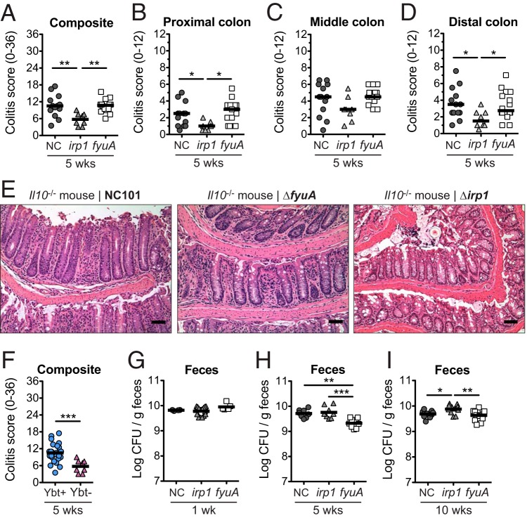
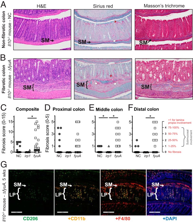
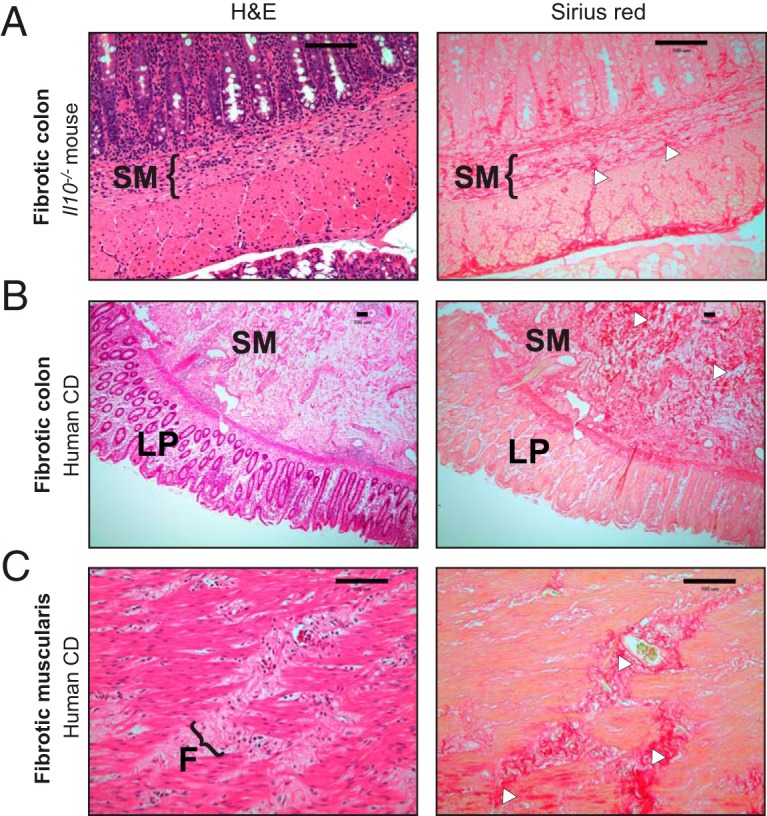
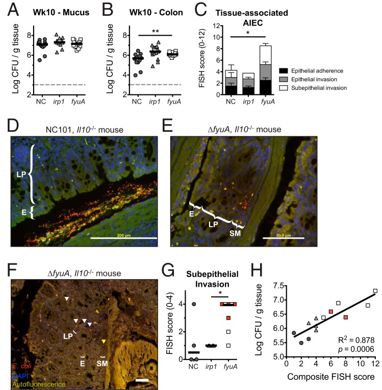
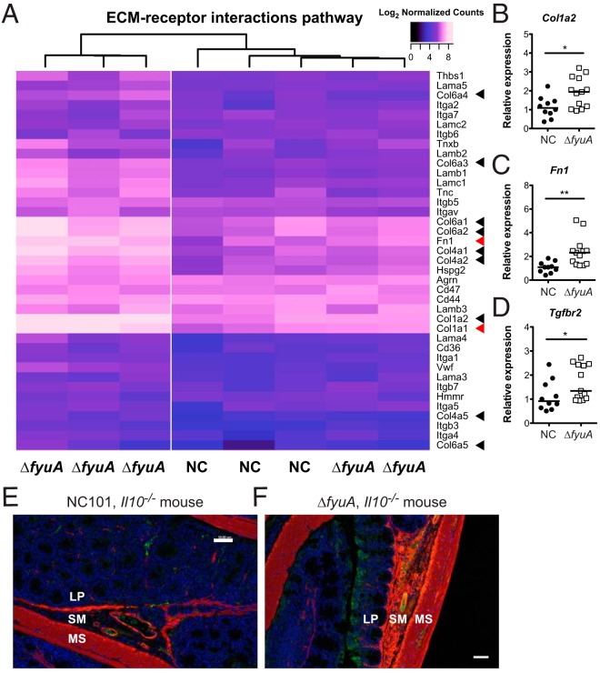
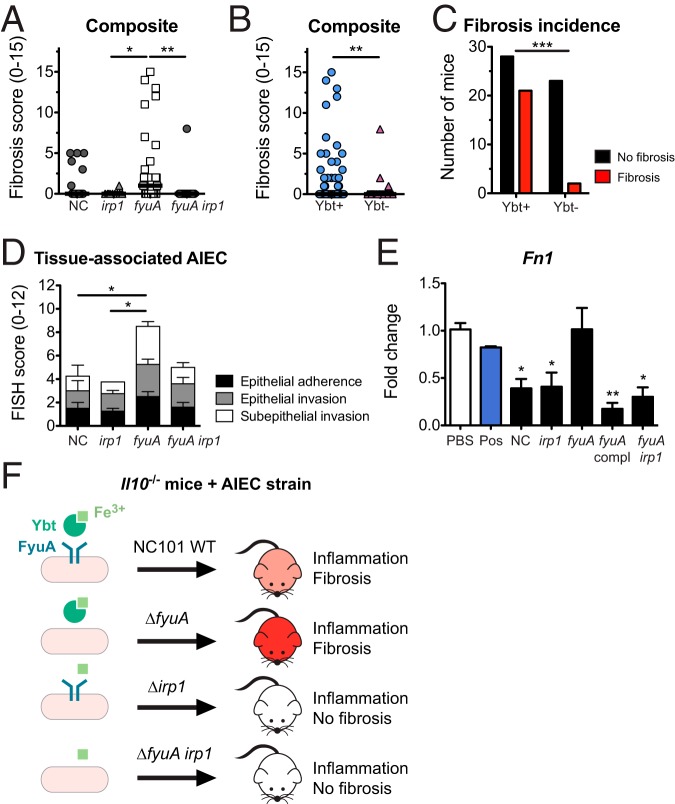
Fibrosis is a significant complication of intestinal disorders associated with microbial dysbiosis and pathobiont expansion, notably Crohn's disease (CD). Mechanisms that favor fibrosis are not well understood, and therapeutic strategies are limited. Here we demonstrate that colitis-susceptible Il10-deficient mice develop inflammation-associated fibrosis when monoassociated with adherent/invasive Escherichia coli (AIEC) that harbors the yersiniabactin (Ybt) pathogenicity island. Inactivation of Ybt siderophore production in AIEC nearly abrogated fibrosis development in inflamed mice. In contrast, inactivation of Ybt import through its cognate receptor FyuA enhanced fibrosis severity. This corresponded with increased colonic expression of profibrogenic genes prior to the development of histological disease, therefore suggesting causality. fyuA-deficient AIEC also exhibited greater localization within subepithelial tissues and fibrotic lesions that was dependent on Ybt biosynthesis and corresponded with increased fibroblast activation in vitro Together, these findings suggest that Ybt establishes a profibrotic environment in the host in the absence of binding to its cognate receptor and indicate a direct link between intestinal AIEC and the induction of inflammation-associated fibrosis.

Keywords: AIEC; Crohn’s disease; colitis; fibrosis; intestinal inflammation; microbiome; microbiota; yersiniabactin.


Figures

FIG 1
FIG 1
Yersiniabactin enhances the proinflammatory potential of AIEC in gnotobiotic Il10−/− mice. Germfree Il10−/− mice were monoassociated with the AIEC strain E. coli NC101 (NC) or the ΔfyuA or Δirp1 mutant for 5 weeks. (A to D) Composite (A) and regional (B to D) histopathology colitis scores. (E) Representative H&E histology of the colon. Scale bar, 50 μm. (F) Composite histopathology colitis scores of Il10−/− mice colonized with yersiniabactin (Ybt)-positive or Ybt-deficient NC101. Lines are at the medians. P values were determined by Kruskal-Wallis or Mann-Whitney test. (G to I) Quantitative bacterial culture from feces at 1 week (G), 5 weeks (H), or 10 weeks (I) postcolonization. Lines are at the means. P values were determined by one-way analysis of variance (ANOVA). Each symbol represents an individual mouse (n = 8 to 14). *, P < 0.05; **, P < 0.01; ***, P < 0.001.
FIG 2
FIG 2
Ybt+ AIEC promotes fibrosis development in colitic Il10−/− mice. Germfree Il10−/− mice were monoassociated with the Ybt+ AIEC strain NC and the ΔfyuA mutant or the Ybt Δirp1 strain for 10 weeks. (A and B) Representative colonic histology of gnotobiotic Il10−/− mice colonized with NC (A) or the ΔfyuA mutant (B). Colon sections were stained with H&E, Sirius red/fast green, or Masson’s trichrome. Regions of Sirius red binding are indicated by white arrowheads in the submucosa and red arrowheads in the lamina propria. (C to F) Composite (C) and regional (D to F) fibrosis histology scores. Each symbol represents an individual mouse (n = 11 to 29). Lines are at the medians. P values were determined by Kruskal-Wallis test. *, P < 0.05. (G) Representative colonic histology from gnotobiotic Il10−/− mice colonized with the ΔfyuA mutant for 5 weeks. Colonic sections were stained with antibodies against the established macrophage cell surface markers CD206, CD11b, and F4/80 and were counterstained with the DNA stain 4′,6-diamidino-2-phenylindole (DAPI). Scale bars, 200 μm. SM, submucosa; LP, lamina propria.
FIG 3
FIG 3
Fibrosis development in AIEC-colonized Il10−/− mice recapitulates histopathological features of fibrosis in human Crohn’s disease. (A) Representative colonic histology of ΔfyuA mutant-colonized fibrotic Il10−/− mice. (B and C) Representative histology of full-thickness colon cross sections from fibrotic Crohn’s disease patients, representative of 3 per group. (C) Magnification of the muscularis serosa. Colon sections were stained with H&E or Sirius red. Regions of Sirius red binding are indicated with white arrowheads. F, fibrotic lesion. Scale bars, 100 μm.
FIG 4
FIG 4
Inactivation of yersiniabactin transport enhances AIEC mucosal invasion. Germfree Il10−/− mice were monoassociated with NC or the ΔfyuA or Δirp1 mutant for 10 weeks. (A and B) Quantitative bacterial cultures of colonic mucus (A) and colonic (B) tissues. Each symbol represents an individual mouse (n = 11 to 15). Lines are at the medians. P values were determined by Kruskal-Wallis test. (C) FISH analysis of proximal colons (n = 4 to 8). P values were determined by Kruskal-Wallis test. (D to F) Representative FISH images of the proximal colon. Red, E. coli; blue, DAPI. Arrowheads in panel F indicate E. coli localized within the lamina propria (white) and submucosa (yellow). E, epithelium. Scale bars, 200 μm. (G) Subepithelial AIEC invasion scores as assessed by FISH in panel C. Red squares represent fibrotic mice as assessed by histopathology. Lines are at the medians. P values were determined by Kruskal-Wallis test. (H) Linear regression analysis of quantitative bacterial culture versus FISH score from colonic tissues. Red squares represent fibrotic mice as assessed by histopathology. *, P < 0.05; **, P < 0.01.
FIG 5
FIG 5
Deletion of fyuA in AIEC promotes transcriptome-wide changes in the colons of Il10−/− mice. Shown are the results of principal-component analysis of transcriptome-wide changes in the colons of NC- versus ΔfyuA mutant-colonized WT or Il10−/− mice after 10 weeks (A), NC- versus ΔfyuA mutant-colonized Il10−/− mice after 5 weeks (B), and NC- versus ΔfyuA mutant-colonized Il10−/− mice after 5 and 10 weeks (C).
FIG 6
FIG 6
Deletion of fyuA in AIEC promotes profibrotic host responses preceding fibrosis development. (A) Heat map of log2 normalized counts of genes in the ECM-receptor interaction KEGG pathway in NC- and ΔfyuA mutant-colonized Il10−/− mice at 5 weeks. (B to D) Relative colonic transcript levels of Col1a1 (B), Fn1 (C), and Tgfbr2 (D) in Il10−/− mice monoassociated with NC or the ΔfyuA mutant for 5 weeks. Each symbol represents an individual mouse (n = 10 to 13). Lines are at the medians. P values were determined by Mann-Whitney test. *, P < 0.05; **, P < 0.01. (E and F) Proximal colons from Il10−/− mice colonized with NC (E) or the ΔfyuA mutant (F) for 10 weeks were stained with α-SMA (red), CD31 (green), or DAPI. MS, muscularis serosa. Scale bars, 50 μm.
FIG 7
FIG 7
Yersiniabactin biosynthesis promotes fibrosis in AIEC-driven colitis. Germfree Il10−/− mice were monoassociated with the AIEC strain NC or the ΔfyuA, Δirp1, or ΔfyuA irp1 mutant for 10 weeks. (A) Composite fibrosis histology scores. Each symbol represents an individual mouse (n = 10 to 29). Lines are at the medians. P values were determined by Kruskal-Wallis test. (B) Composite histopathology colitis scores of Il10−/− mice colonized with Ybt-positive or Ybt-deficient AIEC. Lines are at the medians. P values were determined by Mann-Whitney test. (C) Fibrosis incidence rates of Il10−/− mice colonized with Ybt-positive or Ybt-deficient AIEC as assessed by H&E histology. P values were determined by Fisher’s exact test. (D) FISH analysis of proximal colons (n = 4 to 8). P values were determined by one-way ANOVA. (E) Swiss 3T3 fibroblasts were cocultured with NC or the Δirp1, ΔfyuA, ΔfyuA chromosomally complemented with fyuA under its native promoter, or ΔfyuA irp1 mutant. Fibroblasts stimulated with TGF-β served as a positive control (Pos). Data are represented as the means ± SEMs. P values were determined by Kruskal-Wallis test. (F) Working model. Data for NC-, ΔfyuA mutant-, and Δirp1 mutant-colonized mice are also presented in Fig. 2 and 4. *, P < 0.05; **, P < 0.01; ***, P < 0.001.

Similar articles

Cited by

References

    1. Sartor RB, Wu GD. 2017. Roles for intestinal bacteria, viruses, and fungi in pathogenesis of inflammatory bowel diseases and therapeutic approaches. Gastroenterology 152:327–339.e4. doi:10.1053/j.gastro.2016.10.012. - DOI - PMC - PubMed
    1. Peyrin-Biroulet L, Loftus EV, Colombel JF, Sandborn WJ. 2010. The natural history of adult Crohn’s disease in population-based cohorts. Am J Gastroenterol 105:289–297. doi:10.1038/ajg.2009.579. - DOI - PubMed
    1. Latella G, Rieder F. 2017. Intestinal fibrosis: ready to be reversed. Curr Opin Gastroenterol 33:239–245. doi:10.1097/MOG.0000000000000363. - DOI - PMC - PubMed
    1. Rieder F, Fiocchi C, Rogler G. 2017. Mechanisms, management, and treatment of fibrosis in patients with inflammatory bowel diseases. Gastroenterology 152:340–350.e6. doi:10.1053/j.gastro.2016.09.047. - DOI - PMC - PubMed
    1. van Tol EA, Holt L, Li FL, Kong FM, Rippe R, Yamauchi M, Pucilowska J, Lund PK, Sartor RB. 1999. Bacterial cell wall polymers promote intestinal fibrosis by direct stimulation of myofibroblasts. Am J Physiol 277:G245–G255. doi:10.1152/ajpgi.1999.277.1.G245. - DOI - PubMed

Publication types

LinkOut - more resources