. 2019 Oct 11;294(41):15176-15181.
doi: 10.1074/jbc.AC119.010236. Epub 2019 Sep 3.

Molecular interaction site on procoagulant myosin for factor Xa-dependent prothrombin activation

Affiliations

Molecular interaction site on procoagulant myosin for factor Xa-dependent prothrombin activation

Hiroshi Deguchi et al. J Biol Chem. .

Abstract

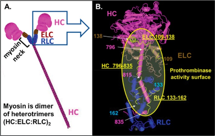
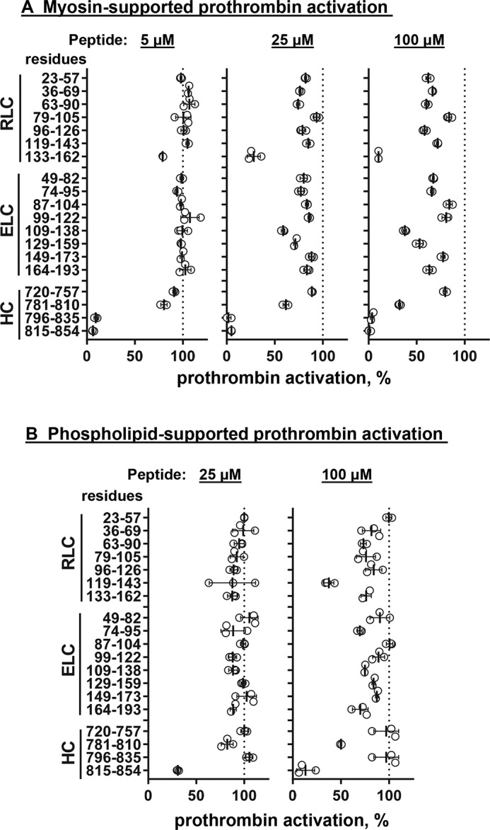
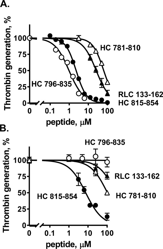
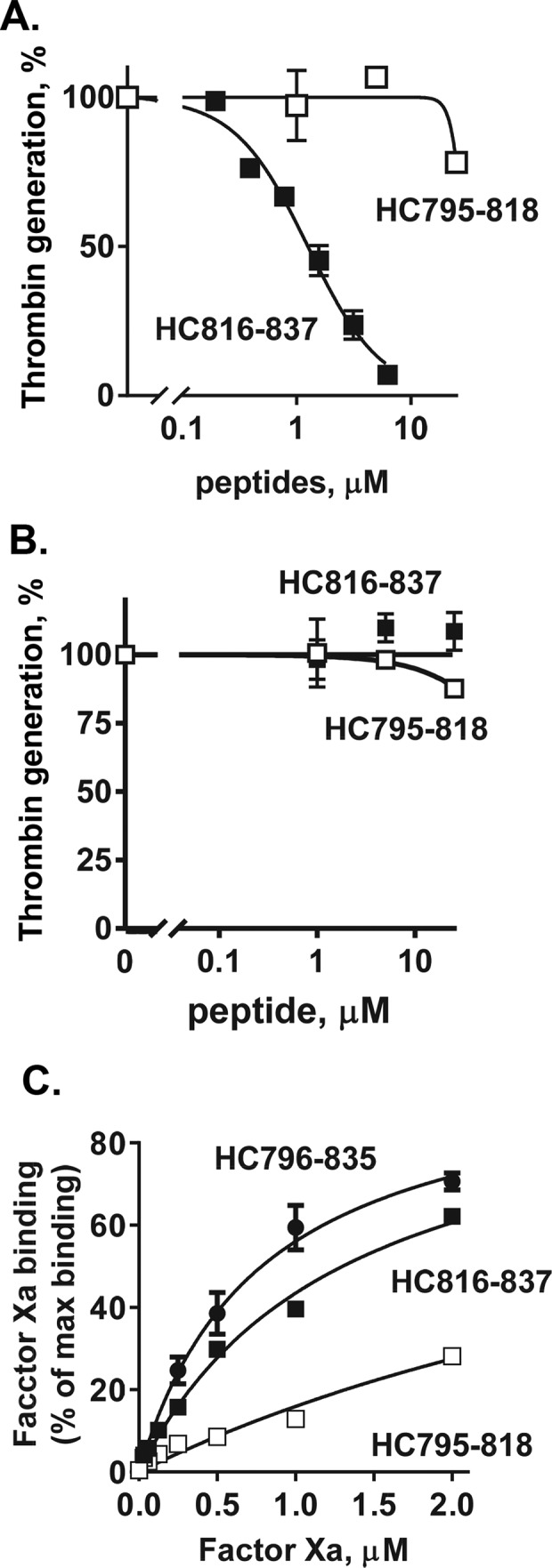
Skeletal muscle myosin has potent procoagulant activity that is based on its ability to enhance thrombin generation due to binding coagulation factors Xa and Va and accelerating prothrombin activation. A well-studied myosin inhibitor that binds to myosin's neck region inhibits myosin-dependent prothrombin activation. Hence, to identify a potential binding site(s) on skeletal muscle myosin for factor Xa, 19 peptides (25-40 residues) representing the neck region, which consists of a regulatory light chain, an essential light chain, and a heavy chain (HC), were screened for inhibition of myosin-supported prothrombin activation. Peptide HC796-835 comprising residues 796-835 of the heavy chain strongly inhibited myosin-enhanced prothrombin activation by factors Xa and Va (50% inhibition at 1.2 μm), but it did not inhibit phospholipid vesicle-enhanced prothrombin activation. Peptide inhibition studies also implicated several myosin light chain sequences located near HC796-835 as potential procoagulant sites. A peptide comprising HC796-835's C-terminal half, but not a peptide comprising its N-terminal half, inhibited myosin-enhanced prothrombin activation (50% inhibition at 1.2 μm). This inhibitory peptide (HC816-837) did not inhibit phospholipid-enhanced prothrombin activation, indicating its specificity for inhibition of myosin-dependent procoagulant mechanisms. Binding studies showed that purified factor Xa was bound to immobilized peptides HC796-835 and HC816-837 with apparent Kd values of 0.78 and 1.3 μm, respectively. In summary, these studies imply that HC residues 816-835 in the neck region of the skeletal muscle myosin directly bind factor Xa and, with contributions from light chain residues in this neck region, contribute to provision of myosin's procoagulant surface.

Keywords: coagulation factor; factor Xa; myosin; peptides; skeletal muscle; thrombin.


Conflict of interest statement

The Scripps Research Institute has intellectual property rights related to this study

Figures

Figure 1.
Figure 1.
Depiction of the three-dimensional structure of skeletal muscle myosin and location of its neck region where myosin's three polypeptide chains are clustered and where factor Xa is bound to promote prothrombin activation by factors Xa/Va. A, scheme for polypeptide structure of skeletal muscle myosin that is a dimer of heterotrimers, each trimer comprising an RLC, an ELC, and an HC. The neck region depicts where the three polypeptides are clustered and represents the potential region responsible for myosin-enhancement of prothrombin activation by the factor Xa/Va complex. B, expanded view of the neck region of skeletal muscle myosin containing coagulation factor-binding sites. Worm-style structures of rabbit skeletal muscle myosin (PDB code 5H53) are depicted using the NCBI Molecular Modeling Database and Cn3D software (17). Structures shown in pink, brown, and blue are the HC, ELC, and RLC, respectively. Pink and light blue numbers show residue numbers for HC and RLC, respectively. Peptides HC796–835, RLC133–162, and ELC109–138 are shown as yellow lines. Myosin's HC residues 815–835, RLC residues 133–162, and ELC residues 129–138 are suggested to be key for myosin's specific procoagulant activity. The yellow shaded area in the yellow circle shows the potential key region for myosin to exert procoagulant activity that includes a major binding site for factor Xa on HC residues 816–835.
Figure 2.
Figure 2.
Screening for the inhibitory effects of myosin neck region peptides on myosin-enhanced or phospholipid-enhanced prothrombin activation by factors Xa and Va. Skeletal muscle myosin (2 nm final) (A) or phospholipid vesicles (PC/PS, 80%/20%, w/w) (4 μm final) (B) were incubated with 19 myosin-derived peptides at three different peptide concentrations, 5, 25, and 100 μm, respectively, as indicated, with factor Va (5 nm final) and factor Xa (0.2 nm final) in TBSA plus 5 mm CaCl2 at room temperature. Thrombin generation was determined as described under “Experimental procedures.” 100% was the value for controls in the absence of added peptides. Each value represents the mean ± S.D. (error bars) of triplicate determinations.
Figure 3.
Figure 3.
Inhibition of myosin-enhanced prothrombin activation by myosin peptides and binding of peptide HC796–835 to factor Xa. Varying concentrations of peptides HC781–810, HC815–854, HC796–835, and RLC133–162 were incubated with factor Va (5 nm final) and factor Xa (0.2 nm final) in TBSA plus 5 mm CaCl2 at room temperature in the presence of skeletal muscle myosin (2 nm final) (A) or phospholipid vesicles (PC/PS, 80%/20%, w/w) (4 μm final) (B). Thrombin generation was initiated by the addition of prothrombin (0.75 μm) in TBSA containing 5 mm Ca2+. The reaction was quenched by adding EDTA (10 mm final). Thrombin generation was determined as described under “Experimental procedures.” 100% was the value for controls in the absence of added peptides. Each value represents the mean ± S.E. (error bars) of triplicate determinations.
Figure 4.
Figure 4.
Inhibition of myosin-enhanced prothrombin activation by myosin HC peptides and their binding of factor Xa. Varying concentrations of peptides HC795–818 and HC816–837 were incubated with factor Va (5 nm final) and factor Xa (0.2 nm final) in TBSA plus 5 mm CaCl2 at room temperature in the presence of skeletal muscle myosin (2 nm final) (A) or phospholipid vesicles (PC/PS, 80%/20%, w/w) (4 μm final) (B). Thrombin generation was then initiated by the addition of prothrombin (0.75 μm) in TBSA containing 5 mm Ca2+. The reaction was quenched by adding EDTA (10 mm final), and thrombin generation was determined as described under “Experimental procedures.” 100% was the value for controls in the absence of added peptides. C, peptides HC796–835, HC795–818, and HC816–837 were immobilized in microtiter plate wells, and the binding of factor Xa to HC796–835, HC795–818, and HC816–837 was determined as described under “Experimental procedures.” Each value represents the mean ± S.E. (error bars) of triplicate determinations.

References

    1. Deguchi H., Sinha R. K., Marchese P., Ruggeri Z. M., Zilberman-Rudenko J., McCarty O. J. T., Cohen M. J., and Griffin J. H. (2016) Prothrombotic skeletal muscle myosin directly enhances prothrombin activation by binding factors Xa and Va. Blood 128, 1870–1878 10.1182/blood-2016-03-707679 - DOI - PMC - PubMed
    1. Hayat M. D., Wyseure T., Mosnier L. O., and Griffin J. H. (2018) Skeletal muscle myosin has pro-hemostatic activity in vivo. Am. J. Hematol. 93, E25
    1. Baumann B. A., Taylor D. W., Huang Z., Tama F., Fagnant P. M., Trybus K. M., and Taylor K. A. (2012) Phosphorylated smooth muscle heavy meromyosin shows an open conformation linked to activation. J. Mol. Biol. 415, 274–287 10.1016/j.jmb.2011.10.047 - DOI - PMC - PubMed
    1. Huang W., Wilson G. J., Brown L. J., Lam H., and Hambly B. D. (1998) EPR and CD spectroscopy of fast myosin light chain conformation during binding of trifluoperazine. Eur. J. Biochem. 257, 457–465 10.1046/j.1432-1327.1998.2570457.x - DOI - PubMed
    1. Patel H., Margossian S. S., and Chantler P. D. (2000) Locking regulatory myosin in the off-state with trifluoperazine. J. Biol. Chem. 275, 4880–4888 10.1074/jbc.275.7.4880 - DOI - PubMed

Publication types