. 2019 Dec 1;79(23):5944-5957.
doi: 10.1158/0008-5472.CAN-19-1165. Epub 2019 Sep 3.

Syndecan-Mediated Ligation of ECM Proteins Triggers Proliferative Arrest of Disseminated Tumor Cells

Affiliations

Syndecan-Mediated Ligation of ECM Proteins Triggers Proliferative Arrest of Disseminated Tumor Cells

Tsukasa Shibue et al. Cancer Res. .

Abstract

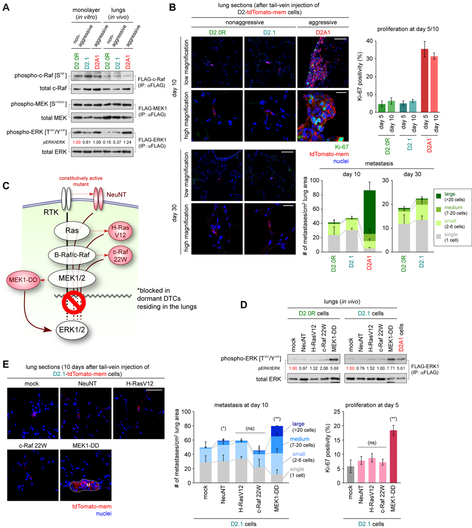
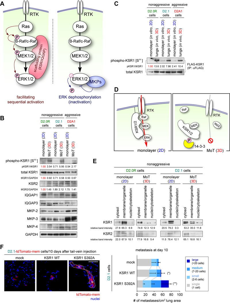
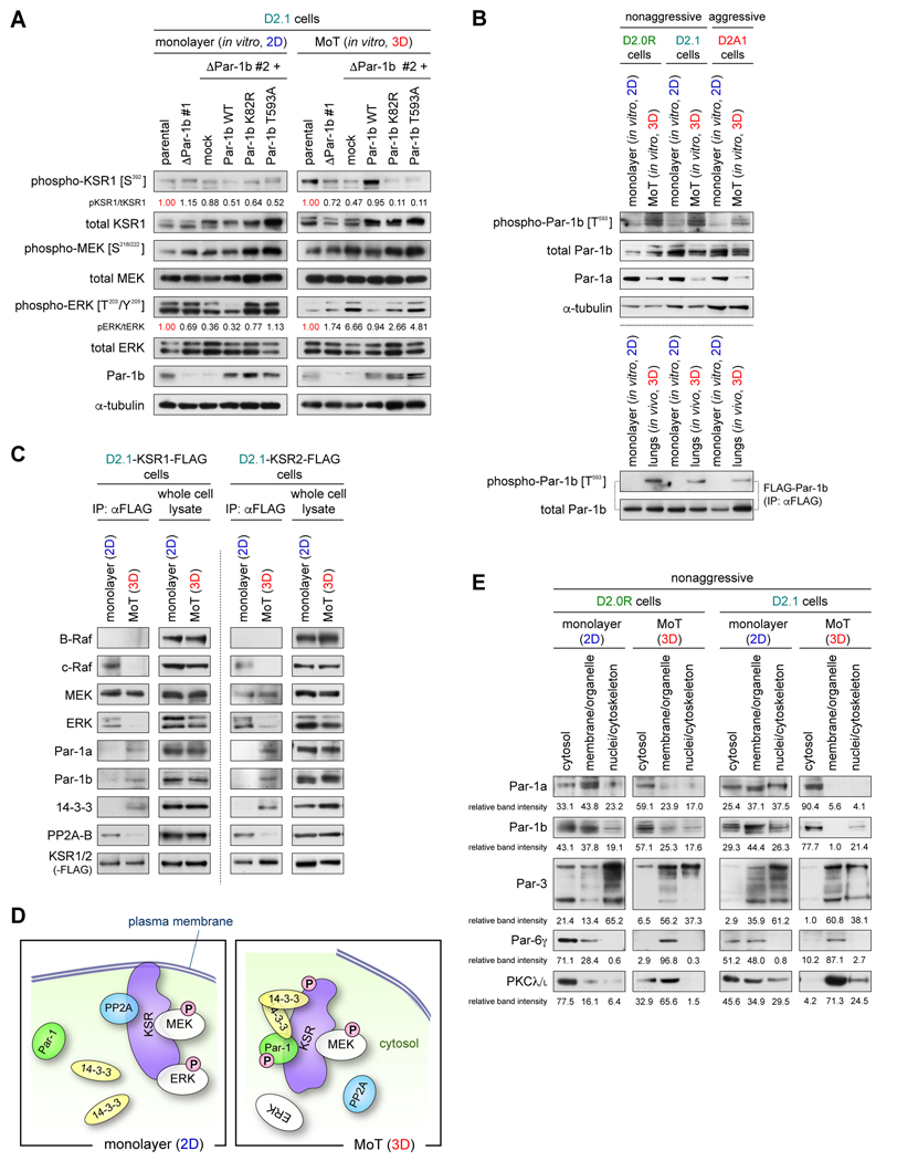
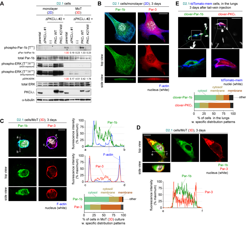
Systemic dissemination of tumor cells often begins long before the development of overt metastases, revealing the inefficient nature of the metastatic process. Thus, already at the time of initial clinical presentation, many patients with cancer harbor a myriad disseminated tumor cells (DTC) throughout the body, most of which are found as mitotically quiescent solitary cells. This indicates that the majority of DTCs fail, for still unknown reasons, to initiate rapid proliferation after entering foreign tissue, which likely contributes significantly to the inefficiency of metastasis formation. Here, we showed that extracellular matrix (ECM) components of the host parenchyma prevented proliferation of DTCs that had recently infiltrated foreign tissue by binding to syndecan receptors expressed on the surface of these cells. This led to the recruitment of the Par-3:Par-6:atypical PKC protein complex, a critical regulator of cell polarity, to the plasma membrane and release of Par-1 kinase into the cytosol. Cytosolic Par-1 bound, phosphorylated, and inactivated KSR scaffolding proteins ultimately inhibited Ras/ERK signaling and, in turn, cell proliferation. Inhibition of the syndecan-mediated signaling restored the proliferation of otherwise dormant DTCs, enabling these cells to efficiently colonize foreign tissues. Intriguingly, naturally aggressive cancer cells overcame the antiproliferative effect of syndecan-mediated signaling either by shutting down this signaling pathway or by activating a proproliferative signaling pathway that works independent of syndecan-mediated signaling. Collectively, these observations indicate that the proliferative arrest of DTCs is attributable, in part, to the syndecan-mediated ligation of ECM proteins. SIGNIFICANCE: This study identifies a novel signaling pathway that regulates the proliferative dormancy of individual disseminated tumor cells.Graphical Abstract: http://cancerres.aacrjournals.org/content/canres/79/23/5944/F1.large.jpg.


Conflict of interest statement

Disclosure of Potential Conflicts of Interest

No potential conflicts of interest were disclosed.

Figures

Figure 1.
Figure 1.. A defect of the Ras/ERK cascade observed in nonaggressive cancer cells
(A) Activity of the Ras/ERK cascade components. Cells were engineered to express a FLAG-c-Raf, FLAG-MEK1 or FLAG-ERK1 and then propagated under monolayer culture or tail-vein injected into Balb/c mice. Five days later, cells (or lungs) were harvested and analyzed by immunoprecipitation (IP)-immunoblotting (IB). (B) Metastatic behaviors of D2 cells. Cell were engineered to express tdTomato-mem plasma membrane marker and tail-vein injected into Balb/c mice. Subsequently, lung sections were prepared and stained for Ki-67 (green) and the nuclei (with DAPI; blue) (left). The sizes of metastases (bottom-right) and Ki-67-positivity were also scored (top-right). (C) Observed effects of Ras/ERK cascade regulation. After infiltrating the lung parenchyma, the nonaggressive D2.0R and D2.1 cells exhibited a defect in MEK-dependent ERK activation. MEK1-DD, but not the other constitutively active mutant oncoproteins tested, successfully circumvented this defect (see D and E). RTK, receptor tyrosine kinase. (D, E) Forced activation of the Ras/ERK cascade. D2.1 cells were engineered to express constitutively active mutants of Ras/ERK cascade components. In D, these cells were manipulated to express the FLAG-ERK1, tail-vein injected into Balb/c mice and then analyzed as in A. In E, these cells were manipulated to express tdTomato-mem, tail-vein injected into Balb/c mice, after which lungs were harvested and sectioned (left). The numbers and sizes of lung metastases (middle), as well as Ki-67-positivity of these cells (right), were scored. (*) p = 0.02, (**) p < 0.001, (ns) p > 0.05 (vs mock; for combined abundance of medium and large colonies [middle]). M, large colony. Values = means ± SD (n = 4: B [top-right], E [right]) or means + SD (n = 4: B [bottom-right], E [middle]). Bars = 100 μm (B, low magnification), 20 μm (B, high magnification), or 50 μm (E).
Figure 2.
Figure 2.. Functional inactivation of KSR scaffolding proteins under 3D conditions
(A) Regulation of Ras/ERK cascade by scaffolding proteins and phosphatases. The KSR and IQGAP scaffolding proteins (pink) positively regulate the Ras/ERK cascade by bringing the Raf, MAK, and ERK kinases in physical juxtaposition, while the MKP phosphatases (blue) negatively regulate this cascade by dephosphorylating ERK. (B) Expression levels of the regulators of Ras/ERK cascade progression in the D2 cells. (C) KSR1 phosphorylation in cancer cells residing in the lung parenchyma. Three different D2 cell types, all expressing FLAG-KSR1, were either propagated in monolayer culture or injected into Balb/c mice via the tail-vein. Five days later, cells (or lungs) were harvested, lysed and analyzed by IP-IB. (D) Effects of KSR phosphorylation. When the nonaggressive cells are growing in the 2D monolayer culture, less-phosphorylated KSR proteins successfully assemble the components of the Ras/ERK cascade at the plasma membrane, allowing efficient signal transduction. In contrast, when these cells are propagated in the 3D MoT culture, highly-phosphorylated KSR proteins get sequestered to the cytosol, preventing the assembly by them of the Ras/ERK cascade components. (E) Subcellular distribution of KSR proteins. After 5 days of culture, D2.0R and D2.1 cells were fractionated into indicated fractions and analyzed by IB. Also see Supplementary Fig. S5C. (F) Effects of KSR1 manipulation on lung metastasis formation. D2.1-tdTomato-mem cells, expressing either WT KSR1 or its S392A mutant, as well as control cells, were tail-vein injected into Balb/c mice. The numbers and sizes of lung metastases at day 10 were scored. Bar = 50 μm. Values = means + SD (n = 4). (*) p = 0.03, (**) p < 0.01 (vs mock; for combined abundance of medium and large colonies). m, medium colony; M, large colony.
Figure 3.
Figure 3.. The Par-1 kinases as mediators of KSR phosphorylation under 3D conditions
(A) Involvement of Par-1b in controlling KSR1 S392 phosphorylation. Parental and Par-1b-knockout (ΔPar-1b #1, 2) D2.1 cells, one of which (#2) was manipulated to express either WT, kinase-dead (K82R), or non-phosphorylatable (T593A) Par-1b or a mock vector, were propagated for 5 days and analyzed by IB. (B) Par-1b phosphorylation under different conditions. D2 cells were in vitro cultured for 5 days and analyzed by IB (top). These cells were also engineered to express FLAG-Par-1b and then either propagated under monolayer culture or tail-vein injected into Balb/c mice. Five days later, cells (or lungs) were harvested, lysed and analyzed by IP-IB (bottom). (C) Interactions between the KSR scaffolds and their binding partners. D2.1 cells, engineered to express either FLAG-KSR1 or FLAG-KSR2, were propagated for 5 days, lysed and analyzed by IP-IB. (D) Summary of the proposed interactions of KSR scaffolds with their binding partners. The association of KSR1/2 with protein phosphatase 2 (PP2A), which dephosphorylates KSR, is also illustrated. (E) Subcellular distribution of polarity-regulating proteins. After being propagated for 5 days, D2.0R and D2.1 cells were fractionated and analyzed. Also see Supplementary Fig. S5C.
Figure 4.
Figure 4.. Subcellular localization of the regulators of cell polarity in 2D vs 3D conditions
(A) PKCλ/ι as a mediator of Par-1b T593 phosphorylation. Parental and two clones of PKCλ/ι-knockout (ΔPKCλ/ι #1, 2) D2.1 cells, one of which (#2) was manipulated to express either WT or kinase-dead (K274W) PKCι, were propagated for 5 days and analyzed by IB. (B-E) Subcellular localization of Par-1b and Par-3. In B-D, D2.1 cells were propagated under either 2D monolayer (B) or 3D MoT (C,D) conditions and immunostained for Par-1b (green) and Par-3 (red). The actin cytoskeleton (F-actin; blue) and the nuclei (white) were also visualized. In C and D, fluorescence intensity along the indicated lines (a-b, c-d [C] and e-f [D]) was plotted (C, top-right; D, bottom). In addition, the localization patterns of Par-1b and Par-3 were classified into four different classes and plotted (C, bottom-right). n ≒ 300. In E, D2.1-tdTomato-mem cells were engineered to express either clover-Par-1b or clover-PKCι and then injected into Balb/c mice via the tail-vein. Subsequently, lungs were harvested, sectioned and stained for nuclei (white). The patterns of clover-Par-1b and clover-PKCι localization were also scored (bottom). n ≒ 200.Bars = 20 μm (B, E), 10 μm (C, D).
Figure 5.
Figure 5.. Par-3 as a critical controller of Ras/ERK signaling under 3D conditions
(A) Effect of Par-3 knockout on intracellular signaling. Parental and two clones of Par-3-knockout (ΔPar-3 #1, 2) D2.1 cells, one of which (#2) was manipulated to express either Par-3 or a mock vector, were propagated as indicated for 5 days and analyzed by IB. (B) Effect of Par-3 knockout on Par-1b localization. Parental and two clones of Par-3 knockout D2.1 cells were propagated under 3D MoT conditions and stained for Par-1b (green), F-actin (blue) and nuclei (white) (left). Fluorescence intensity along the indicated lines (a-b and c-d) was plotted (bottom-right). The patterns of Par-1b localization were also scored (top-right). n ≒ 300. (*) p < 3 × 10⁻¹⁶ (vs parental; by Fisher’s exact test). (C) Effect of Par-3 knockout on lung metastasis formation. Two clones of D2.1-ΔPar-3 cells and control cells, also expressing tdTomato-mem, were tail-vein injected into Balb/c mice. The numbers and sizes of lung metastases at day 10 were scored. (*) p < 0.003 (vs parental; for combined abundance of medium and large colonies). Values = means + SD (n = 4) (D) Tethering of Par-3 and Par-6γ to receptors of ECM proteins. D2.1 cells were engineered to express either FLAG-Par-3 or FLAG-Par-6γ and propagated for 5 days. Their lysates were subjected to IP-IB to identify interactions between Par-3/6γ and ECM receptors. Whole cell lysate represents 10% of input lysate. For syndecan-1 and-4, the bands were detected at approximately 70 kDa and 60 kDa, respectively, which are likely to represent syndecan homodimers. Bars = 10 μm (B) or 50 μm (C).
Figure 6.
Figure 6.. Cell polarity and signaling governed by syndecans under 3D conditions.
(A) Effects of syndecan knockout on subcellular localization of polarity-regulating proteins. Parental and a syndecan-1/3/4 triple knockout clone (ΔSdc #2) of D2.1 cells were propagated for 5 days, and then fractionated and analyzed. (B) Effect of syndecan knockout on Par-3 localization. Parental and two clones of triple syndecan knockout (ΔSdc #1, 2) D2.1 cells were propagated under 3D MoT conditions and stained for Par-3 (red), F-actin (blue) and nuclei (white) (left). Fluorescence intensity along the indicated lines (a-b and c-d) was plotted (center). The patterns of Par-3 localization were also scored (right). n = 300. Bar = 10 mm. (*) p < 3 × 10⁻¹⁶ (vs parental; by Fisher’s exact test). (C) Effects of syndecan knockout on signaling. Parental and ΔSdc #1, 2 versions of D2.1 cells, one of which (#2) was manipulated to ectopically express either a set of three syndecan isoforms (ΔSdc #2 + Sdc-1/3/4) or mock vectors, were propagated for 5 days and analyzed by IB.
Figure 7.
Figure 7.. Syndecans as critical negative regulators of metastatic outgrowth.
(A, B) Effect of syndecan knockout on lung metastasis formation. Parental and two clones of syndecan-1/3/4 triple knockout (ΔSdc #1, #2) D2.1 cells (A), as well as parental and syndecan-1/2/3/4 quadruple knockout (ΔSdc) versions of the EpH4-Ras cells and 4TO7 cells (B), all of which were also expressing tdTomato-mem, were tail-vein injected into Balb/c mice. The numbers and sizes of lung metastases at day 10 were scored. (*) p < 0.003, (**) p < 0.001 (vs parental; for combined abundance of medium and large metastases). (C-E) Effect of syndecan knockout on metastasis formation in multiple organs. In C and D, parental and ΔSdc versions of the 4TO7 cells, also expressing firefly luciferase, were injected into Balb/c mice via the left ventricle. Mice were then subjected to in vivo bioluminescence imaging (C). On day 20, target organs of metastasis were harvested and subjected to ex vivo bioluminescence imaging (D). In E, these versions of 4TO7 cells were implanted into the mammary fat pads of the NOD scid mice. 21 days later, target organs of metastasis were harvested and their metastatic burdens were scored (left). Primary tumors were also harvested and weighed (right). The red bar represents the mean value. (*) p = 0.02, (**) p < 0.007, (***) p = 0.04, (ns) p > 0.05. Bars = 50 μm. Values = means + SD (n = 4: A, B; n = 6: C).

Similar articles

Cited by

References

    1. Naumov GN, MacDonald IC, Weinmeister PM, Kerkvliet N, Nadkarni KV, Wilson SM, et al. Persistence of solitary mammary carcinoma cells in a secondary site: a possible contributor to dormancy. Cancer Res 2002;62(7):2162–8. - PubMed
    1. Pantel K, Alix-Panabieres C, Riethdorf S. Cancer micrometastases. Nat Rev Clin Oncol 2009;6(6):339–51. - PubMed
    1. Aguirre Ghiso JA, Kovalski K, Ossowski L. Tumor dormancy induced by downregulation of urokinase receptor in human carcinoma involves integrin and MAPK signaling. J Cell Biol 1999;147(1):89–104. - PMC - PubMed
    1. Barkan D, Kleinman H, Simmons JL, Asmussen H, Kamaraju AK, Hoenorhoff MJ, et al. Inhibition of metastatic outgrowth from single dormant tumor cells by targeting the cytoskeleton. Cancer Res 2008;68(15):6241–50. - PMC - PubMed
    1. Shibue T, Weinberg RA. Integrin beta1-focal adhesion kinase signaling directs the proliferation of metastatic cancer cells disseminated in the lungs. Proc Natl Acad Sci U S A 2009;106(25):10290–5. - PMC - PubMed

Publication types

Substances