. 2020 Jun;69(6):1027-1038.
doi: 10.1136/gutjnl-2019-318930. Epub 2019 Sep 3.

Single-cell transcriptional analyses of spasmolytic polypeptide-expressing metaplasia arising from acute drug injury and chronic inflammation in the stomach

Affiliations

Single-cell transcriptional analyses of spasmolytic polypeptide-expressing metaplasia arising from acute drug injury and chronic inflammation in the stomach

Kevin A Bockerstett et al. Gut. 2020 Jun.

Abstract

Objective: Spasmolytic polypeptide-expressing metaplasia (SPEM) is a regenerative lesion in the gastric mucosa and is a potential precursor to intestinal metaplasia/gastric adenocarcinoma in a chronic inflammatory setting. The goal of these studies was to define the transcriptional changes associated with SPEM at the individual cell level in response to acute drug injury and chronic inflammatory damage in the gastric mucosa.

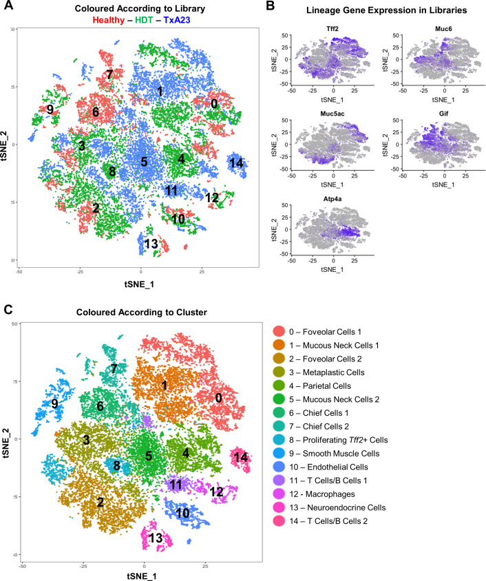
Design: Epithelial cells were isolated from the gastric corpus of healthy stomachs and stomachs with drug-induced and inflammation-induced SPEM lesions. Single cell RNA sequencing (scRNA-seq) was performed on tissue samples from each of these settings. The transcriptomes of individual epithelial cells from healthy, acutely damaged and chronically inflamed stomachs were analysed and compared.

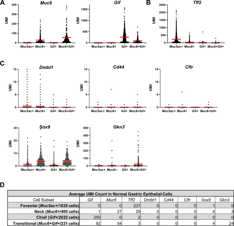
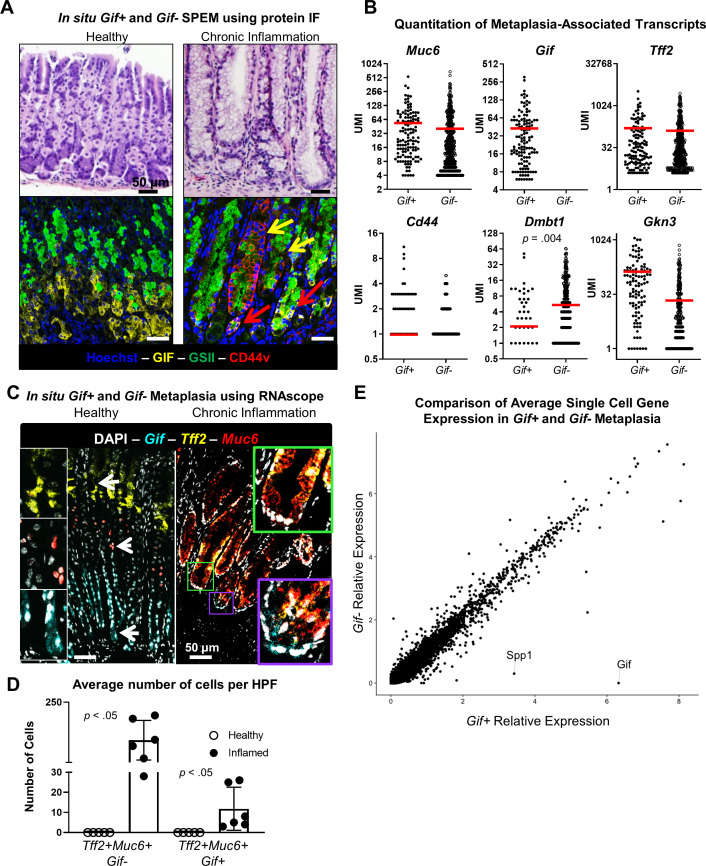
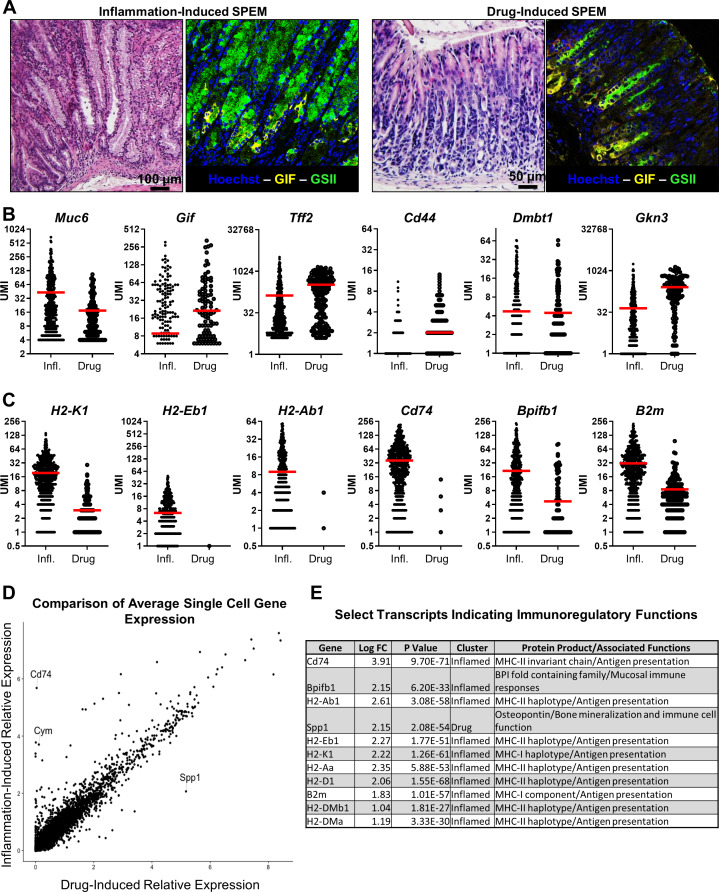
Results: scRNA-seq revealed a population Mucin 6 (Muc6)+gastric intrinsic factor (Gif)+ cells in healthy tissue, but these cells did not express transcripts associated with SPEM. Furthermore, analyses of SPEM cells from drug injured and chronically inflamed corpus yielded two major findings: (1) SPEM and neck cell hyperplasia/hypertrophy are nearly identical in the expression of SPEM-associated transcripts and (2) SPEM programmes induced by drug-mediated parietal cell ablation and chronic inflammation are nearly identical, although the induction of transcripts involved in immunomodulation was unique to SPEM cells in the chronic inflammatory setting.

Conclusions: These data necessitate an expansion of the definition of SPEM to include Tff2+Muc6+ cells that do not express mature chief cell transcripts such as Gif. Our data demonstrate that SPEM arises by a highly conserved cellular programme independent of aetiology and develops immunoregulatory capabilities in a setting of chronic inflammation.

Keywords: GASTRIC CANCER; GASTRIC METAPLASIA; GASTRITIS.


Conflict of interest statement

Competing interests: None declared.

Figures

Figure 1
Figure 1
Identification of gastric epithelial cell lineages using scRNA-seq and unbiased clustering. (A) t-Stochastic neighbour embedding (t-SNE) unbiased clustering of gastric epithelial cell suspensions isolated from BALB/c (red), TxA23 (blue) and HDT (green) mice. (B) Feature dot plots for Tff2, Muc6, Muc5ac, Gif and Atp4a. Cells with high relative expression for indicated target transcript denoted by intensity of purple colouring. (C) t-SNE plot of gastric epithelial cells from BALB/c, TxA23 and HDT mice in which algorithmically defined clusters are identified by colour. HDT, high dose tamoxifen.
Figure 2
Figure 2
Muc6+Gif+ cells present in normal gastric mucosa are not a metaplastic lineage. 1635 Muc5ac+ foveolar cells, 400 Muc6+Gif neck cells, 2032 Gif+Muc6− chief cells and 231 Muc6+Gif+ transitional cells were identified in single cell libraries generated from normal BALB/c gastric mucosa. (A) Per cell UMI counts of Muc6 and Gif transcripts in Muc5ac+, Muc6+, Gif+ and Muc6+Gif+ populations. (B) Per cell UMI counts of spasmolytic-polypeptide/Tff2 transcripts. (C) Per cell UMI counts of SPEM-associated transcripts Dmbt1, Cd44, Cftr, Sox9 and Gkn3. (D) Table of average UMI counts of indicated genes in the normal gastric epithelial cell populations. Red bar indicates average UMI count per cell in each cell subset. Significance determined unpaired Student’s t-test. Gif, gastric intrinsic factor; Muc6, Mucin 6; SPEM, spasmolytic polypeptide-expressing metaplasia; Tff2, trefoil factor 2; UMI, unique molecular identifier.
Figure 3
Figure 3
Gif+ SPEM and Gif− neck cell hyperplasia/hypertrophy have a similar metaplastic phenotype and transcriptional profile. (A) Representative bright field and immunofluorescent photomicrographs of gastric corpus of healthy BALB/c and chronically inflamed TxA23 mice at 4 months of age. Yellow arrows indicate GIF− neck cell hyperplasia/hypertrophy, red arrows indicate GIF+SPEM. n=3 mice per group. (B) Per cell UMI counts of metaplasia-associated transcripts Muc6, Gif, Tff2, Cd44, Dmbt1 and Gkn3. Red bar indicates mean UMI count per cell. Significance was determined using Wilcoxon ranked-sum test. (C) Representative fluorescent images of multicolor RNA in situ hybridisation of Tff2 transcripts (yellow), Gif transcripts (cyan) and Muc6 transcripts (red). White arrows indicate location of high magnification insets of Gif+, Muc6+ and Tff2+ cells. Green box indicates Gif-Muc6+Tff2+ cells, purple box indicates Tff2+Muc6+Gif+ canonical SPEM with corresponding high magnification inset images. (D) Quantitation of the average number of Muc6+Tff2+Gif- and Muc6+Tff2+Gif+ cells per HPF. Each dot represents the average number of cells counted from three HPFs in the gastric corpus of a single mouse. n=5–6 mice per group. Significance determined using an unpaired Student’s t test. (E) Dot plot of the relative expression of all detected transcripts calculated from the per-cell average relative expression from scRNA-seq data sets in Gif+ SPEM and Gif− neck cell hyperplasia/hypertrophy in chronic metaplasia. Gif, gastric intrinsic factor; HPF, high power field; Muc6, Mucin 6; SPEM, spasmolytic polypeptide-expressing metaplasia; Tff2, gastric intrinsic factor.
Figure 4
Figure 4
The metaplastic response is conserved and develops an immunoregulatory phenotype during chronic inflammation. Muc6+Tff2+ metaplastic cells were identified in libraries generated from the stomachs of mice with acute high dose tamoxifen-induced metaplasia and mice with chronic inflammatory metaplasia. (A) Representative bright field and immunofluorescent images of inflammation-induced and drug-induced SPEM. n=3 mice per group. (B) Per cell transcript counts of SPEM-associated transcripts Muc6, Gif, Tff2, Cd44, Dmbt1 and Gkn3. (C) Per cell transcript counts of genes significantly associated with inflammatory SPEM: H2-K1, H2-Eb1, Cd74, Reg3g and Chil4. Each dot represents one cell. Red bars indicate mean UMI count per cell. (D) Dot plot of the relative expression of all detected transcripts calculated from the per-cell average relative expression from scRNA-seq data sets in drug-induced and inflammation-induced SPEM cells. Gif, gastric intrinsic factor; Muc6, Mucin 6; SPEM, spasmolytic polypeptide-expressing metaplasia; Tff2, trefoil factor 2; UMI, unique molecular identifier.

References

    1. Nozaki K, Ogawa M, Williams JA, et al. . A molecular signature of gastric metaplasia arising in response to acute parietal cell loss. Gastroenterology 2008;134:511–22. 10.1053/j.gastro.2007.11.058 - DOI - PMC - PubMed
    1. Weis VG, Goldenring JR. Current understanding of SPEM and its standing in the preneoplastic process. Gastric Cancer 2009;12:189–97. 10.1007/s10120-009-0527-6 - DOI - PMC - PubMed
    1. Bray F, Ferlay J, Soerjomataram I, et al. . GLOBOCAN estimates of incidence and mortality worldwide for 36 cancers in 185 countries. CA Cancer J Clin 2018;2018:394–424. - PubMed
    1. Mills JC, Samuelson LC. Past questions and current understanding about gastric cancer. Gastroenterology 2018;155:939–44. 10.1053/j.gastro.2018.06.044 - DOI - PMC - PubMed
    1. Correa P, Shiao YH. Phenotypic and genotypic events in gastric carcinogenesis. Cancer Res 1994;54(7 Suppl):1941s–3. - PubMed

Publication types

MeSH terms