Review
. 2019 Oct;60(10):1350-1358.
doi: 10.2967/jnumed.119.228320. Epub 2019 Sep 3.

Multimodality Imaging of Prostate Cancer

Affiliations
Review

Multimodality Imaging of Prostate Cancer

Soleen Ghafoor et al. J Nucl Med. 2019 Oct.

Abstract

Prostate cancer is a very heterogeneous disease, and contemporary management is focused on identification and treatment of the prognostically adverse high-risk tumors while minimizing overtreatment of indolent, low-risk tumors. In recent years, imaging has gained increasing importance in the detection, staging, posttreatment assessment, and detection of recurrence of prostate cancer. Several imaging modalities including conventional and functional methods are used in different clinical scenarios with their very own advantages and limitations. This continuing medical education article provides an overview of available imaging modalities currently in use for prostate cancer followed by a more specific section on the value of these different imaging modalities in distinct clinical scenarios, ranging from initial diagnosis to advanced, metastatic castration-resistant prostate cancer. In addition to established imaging indications, we will highlight some potential future applications of contemporary imaging modalities in prostate cancer.

Keywords: PET/MRI; PSMA; fluciclovine; multiparametric MRI; theranostics; whole-body MRI.


Figures

FIGURE 1.
FIGURE 1.
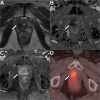
A 75-y-old patient with local recurrence of Gleason 3 + 4 PCa (arrows) treated with radical prostatectomy. (A–C) mpMRI shows masslike T2-hypointense thickening at right lateral vesicourethral anastomosis (A) with diffusion restriction on apparent diffusion coefficient image (B) and early arterial enhancement on dynamic contrast-enhanced image (C). (D) 18F-fluciclovine PET/CT shows asymmetric increased radiotracer uptake at that site (SUVmax, 5.5).
FIGURE 2.
FIGURE 2.
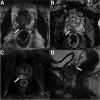
A 53-y-old patient with abnormal digital rectal findings, elevated PSA level, and lesion (arrows) found on imaging. (A–C) mpMRI shows circumscribed T2-hypointense lesion in right posterior base to mid gland peripheral zone (A) with marked diffusion restriction (B) and early arterial enhancement (C). Lesion measured 1.9 cm and involved the central zone and base of seminal vesicles. Final surgical pathology revealed 3 + 4 Gleason PCa with extracapsular extension and seminal vesicle invasion.
FIGURE 3.
FIGURE 3.
A 69-y-old patient with history of Gleason 7 PCa treated with high-intensity focus ultrasound therapy 1 year previously. (A, B, and D) Surveillance mpMRI does not reveal any findings concerning for recurrence. (C) On 68Ga-PSMA PET/MRI, area of focal intense uptake localized to right apex (arrow) is seen, corresponding to subsequently biopsy-proven recurrent Gleason 4 + 4 tumor.
FIGURE 4.
FIGURE 4.
A 68-y-old patient with BCR of Gleason 3 + 4 PCa after radical prostatectomy and pelvic LN dissection 11 years previously. PSA was 1.4 ng/mL at time of imaging. (A) CT portion of study does not reveal any abnormal LNs. (B) Fused 68Ga-PSMA PET/CT shows focal avid uptake in right external iliac region (arrow) corresponding to subcentimeter external iliac LN (arrow in A), suggestive of LN metastasis, which was confirmed on subsequent excisional biopsy.

References

    1. Yadav SS, Stockert JA, Hackert V, Yadav KK, Tewari AK. Intratumor heterogeneity in prostate cancer. Urol Oncol. 2018;36:349–360. - PubMed
    1. Pezaro C, Omlin A, Lorente D, et al. Visceral disease in castration-resistant prostate cancer. Eur Urol. 2014;65:270–273. - PMC - PubMed
    1. Turkbey B, Rosenkrantz AB, Haider MA, et al. Prostate imaging reporting and data system version 2.1: 2019 update of prostate imaging reporting and data system version 2. European Urology. - PubMed
    1. Turkbey B, Mani H, Shah V, et al. Multiparametric 3T prostate magnetic resonance imaging to detect cancer: histopathological correlation using prostatectomy specimens processed in customized magnetic resonance imaging based molds. J Urol. 2011;186:1818–1824. - PMC - PubMed
    1. Weinreb JC, Barentsz JO, Choyke PL, et al. PI-RADS prostate imaging: reporting and data system—2015, version 2. Eur Urol. 2016;69:16–40. - PMC - PubMed

Publication types

MeSH terms