

MCOLN1 Promotes Proliferation and Predicts Poor Survival of Patients with Pancreatic Ductal Adenocarcinoma
- PMID: 31481985
- PMCID: PMC6701426
- DOI: 10.1155/2019/9436047
MCOLN1 Promotes Proliferation and Predicts Poor Survival of Patients with Pancreatic Ductal Adenocarcinoma
Retraction in
-
RETRACTION: MCOLN1 Promotes Proliferation and Predicts Poor Survival of Patients with Pancreatic Ductal Adenocarcinoma.Dis Markers. 2025 May 6;2025:9850902. doi: 10.1155/dim/9850902. eCollection 2025. Dis Markers. 2025. PMID: 40435332 Free PMC article.
Abstract
Background: MCOLN1 (mucolipin subfamily, member 1) was first identified as an autophagic regulator, which was essential for efficient fusion of both autophagosomes and late endosomes with lysosomes. This study is aimed at investigating the role of MCOLN1 in the development of pancreatic ductal adenocarcinoma (PDAC).
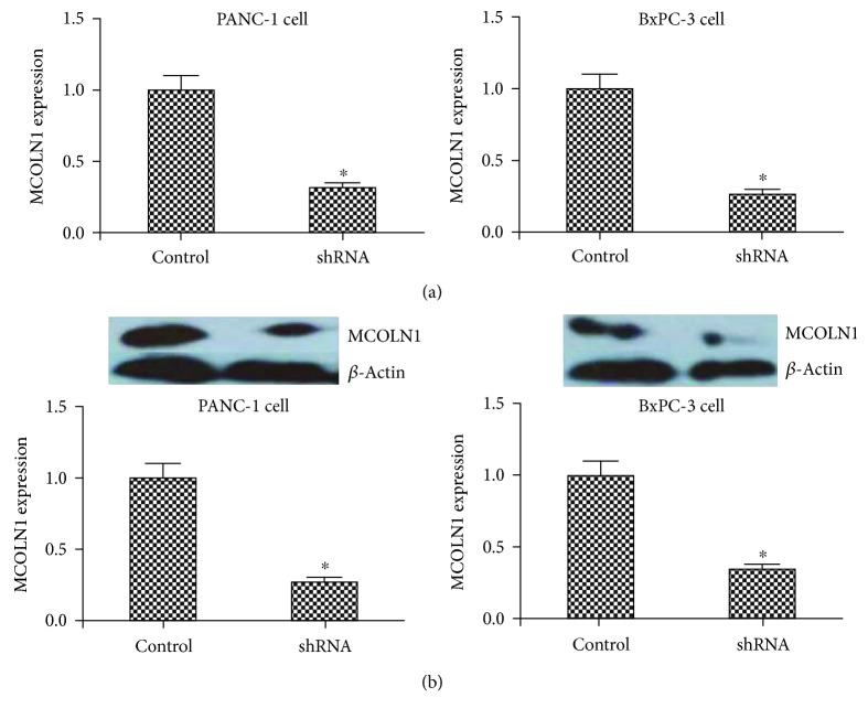
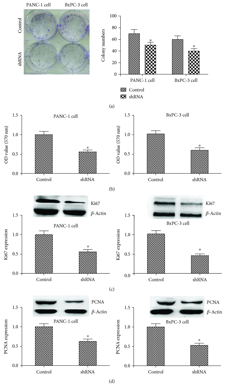
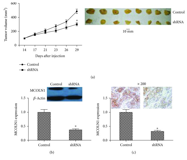
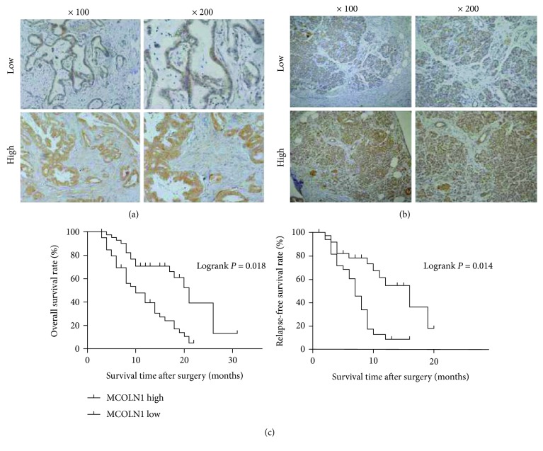
Methods: Immunohistochemistry (IHC) assay was conducted to evaluate the expression level of MCOLN1 in 82 human PDAC tumor tissues. Overall survival (OS) and recurrence-free survival (RFS) analysis was performed to assess the prognosis of patients. Colony formation and MTT assays [3-(4,5-dimethyl-2-thiazolyl)-2,5-diphenyl-2-H-tetrazolium bromide] were performed to measure the proliferation capacity of tumor cells. The expression level of related genes was measured by RT-PCR (reverse transcription polymerase chain reaction) and western blot assays. The animal model was used to examine the effects of indicated protein on tumorigenesis in vivo.
Results: The results of IHC showed that a high level of MCOLN1 expression was associated with the poor clinical characteristics of PDAC patients. OS and RFS were significantly worse in patients with high MCOLN1 expression. Silencing of MCOLN1 dramatically blocked the proliferation of PDAC cells. Mechanism studies confirmed that knockdown of MCOLN1 decreased the expression of Ki67 and PCNA (proliferating cell nuclear antigen), two markers of cell proliferation. In vivo, MCOILN1 depletion reduced the formation and growth of tumors in mice.
Conclusion: The high level of MCOLN1 expression was associated with poor clinical outcomes of PDAC patients. MCOLN1 ablation could inhibit PDAC proliferation of both in vitro and in vivo, which provide a new insight and novel therapeutic target for the treatment of PDAC.
Conflict of interest statement
The authors have declared that no competing financial interest exists.
Figures
References
-
- Rahib L., Smith B. D., Aizenberg R., Rosenzweig A. B., Fleshman J. M., Matrisian L. M. Projecting cancer incidence and deaths to 2030: the unexpected burden of thyroid, liver, and pancreas cancers in the United States. Cancer Research. 2014;74(11):2913–2921. doi: 10.1158/0008-5472.CAN-14-0155. - DOI - PubMed
Publication types
MeSH terms
Substances
LinkOut - more resources
Full Text Sources
Other Literature Sources
Medical
Miscellaneous
