

The human endosalpinx: anatomical three-dimensional study and reconstruction using confocal microtomography
- PMID: 31482002
- PMCID: PMC6717942
- DOI: 10.5114/pjr.2019.86824
The human endosalpinx: anatomical three-dimensional study and reconstruction using confocal microtomography
Abstract
Purpose: To evaluate in three dimensions (3D) the human endosalpinx and reconstruct its surface along its different anatomical segments, without the injection or insertion of luminal contrasts, using confocal microtomography (micro-CT).
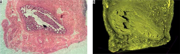
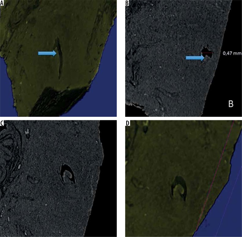
Material and methods: 15 fallopian tubes (FT) from 14 women in reproductive age from procedures for benign disease or sterilization were selected. The specimens were fixed in formalin and stained with Lugol solution. Micro-CT studies were conducted on the specimens using protocols adapted from biological studies, to acquire images to reconstruct in 3D the endosalpinx surface.
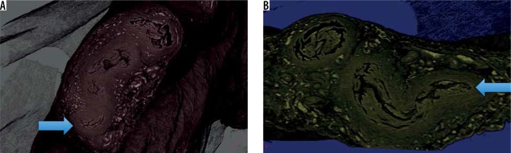
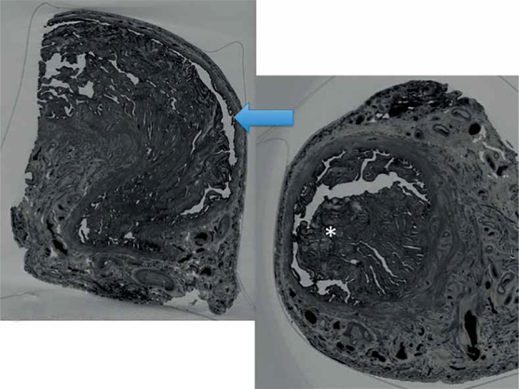
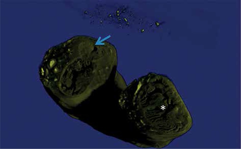
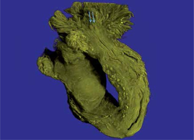
Results: From these specimens, 6 presented the intra-mural segment, 14 presented the isthmus and 15 presented the ampulla and fimbria segment of the FT. The specimen presented tissue definition, and contrast sufficient for FT endosalpinx morphological analysis and lumen definition. The intramural portion presented initially a mucosal projection toward the lumen, bending on its own axis, and increased numbers of projections towards the isthmic portion, where the projections become longer more numerous. The endosalpinx becomes more tortuous, the lumen diameter increases and the mucosal projections become more bulky in the ampullary portion, with the projections less present on the antimesenteric side. The infundibular portion is marked with the organized and predictable endosalpinx, the abdominal ostium is cleared demonstrated, with the reduction of the endosalpinx volume. The fimbria demonstrated a small relation between fringes and intratubal endosalpinx.
Conclusions: Microscopic anatomy of different segments of the human FT mucosa can be analyzed and reconstructed in 3D with histological correlation using micro-CT.
Keywords: anatomy; fallopian tubes; multislice computed tomography; three-dimensional imaging.
Conflict of interest statement
The authors report no conflict of interest.
Figures
References
-
- Li S, Winuthayanon W. Oviduct: roles in fertilization and early embryo development. J Endocrinol. 2017;232:R1–R26. - PubMed
-
- Hunter RHF. Their role in fertility and infertility. 1st ed. Berlin Heidelberg: Springer-Verlag; 1988. The Fallopian tubes.
-
- Yaniz JL, Lopez-Gatius F, Hunter RH. Scanning electron microscopic study of the functional anatomy of the porcine oviductal mucosa. Anat Histol Embryol. 2006;35:28–34. - PubMed
-
- Nayak RK, Ellington EF. Ultrastructural and ultracytochemical cyclic changes in the bovine uterine tube (oviduct) epithelium. Am J Vet Res. 1977;38:157–168. - PubMed
-
- Odor DL, Gaddum-Rosse P, Rumery RE, Blandau RJ. Cyclic variations in the oviuductal ciliated cells during the menstrual cycle and after estrogen treatment in the pig-tailed monkey, Macaca nemestrina. Anat Rec. 1980;198:35–57. - PubMed
LinkOut - more resources
Full Text Sources