. 2019 Aug 28;5(8):1387-1395.
doi: 10.1021/acscentsci.9b00369. Epub 2019 Aug 7.

Spontaneous Isomerization of Long-Lived Proteins Provides a Molecular Mechanism for the Lysosomal Failure Observed in Alzheimer's Disease

Affiliations

Spontaneous Isomerization of Long-Lived Proteins Provides a Molecular Mechanism for the Lysosomal Failure Observed in Alzheimer's Disease

Tyler R Lambeth et al. ACS Cent Sci. .

Abstract

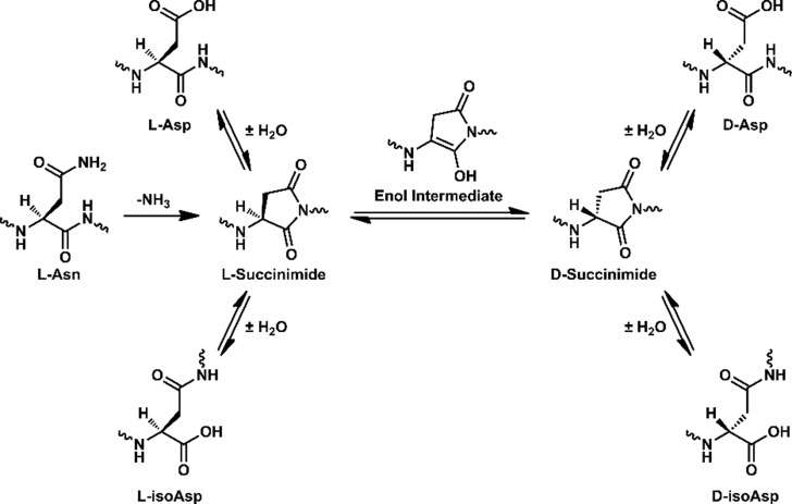
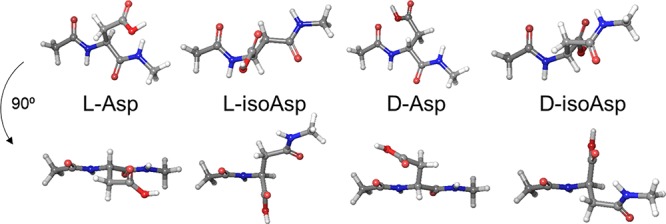
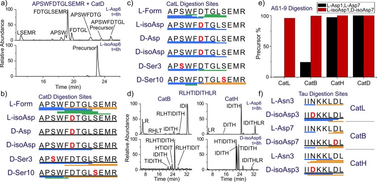
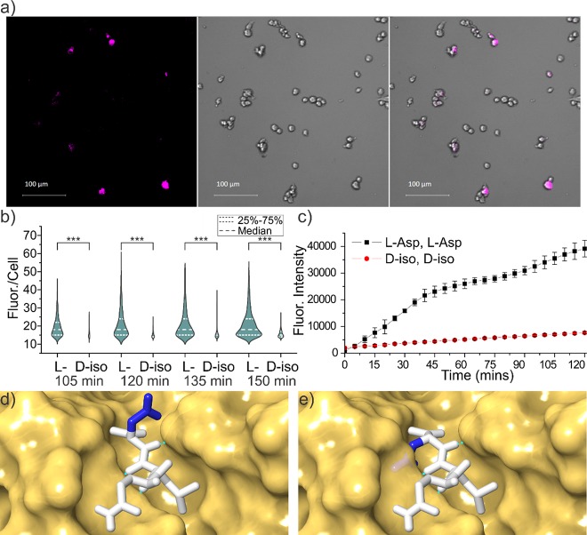
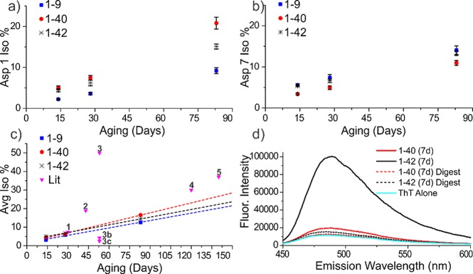
Proteinaceous aggregation is a well-known observable in Alzheimer's disease (AD), but failure and storage of lysosomal bodies within neurons is equally ubiquitous and actually precedes bulk accumulation of extracellular amyloid plaque. In fact, AD shares many similarities with certain lysosomal storage disorders though establishing a biochemical connection has proven difficult. Herein, we demonstrate that isomerization and epimerization, which are spontaneous chemical modifications that occur in long-lived proteins, prevent digestion by the proteases in the lysosome (namely, the cathepsins). For example, isomerization of aspartic acid into l-isoAsp prevents digestion of the N-terminal portion of Aβ by cathepsin L, one of the most aggressive lysosomal proteases. Similar results were obtained after examination of various target peptides with a full series of cathepsins, including endo-, amino-, and carboxy-peptidases. In all cases peptide fragments too long for transporter recognition or release from the lysosome persisted after treatment, providing a mechanism for eventual lysosomal storage and bridging the gap between AD and lysosomal storage disorders. Additional experiments with microglial cells confirmed that isomerization disrupts proteolysis in active lysosomes. These results are easily rationalized in terms of protease active sites, which are engineered to precisely orient the peptide backbone and cannot accommodate the backbone shift caused by isoaspartic acid or side chain dislocation resulting from epimerization. Although Aβ is known to be isomerized and epimerized in plaques present in AD brains, we further establish that the rates of modification for aspartic acid in positions 1 and 7 are fast and could accrue prior to plaque formation. Spontaneous chemistry can therefore provide modified substrates capable of inducing gradual lysosomal failure, which may play an important role in the cascade of events leading to the disrupted proteostasis, amyloid formation, and tauopathies associated with AD.


Conflict of interest statement

The authors declare no competing financial interest.

Figures

Scheme 1
Scheme 1. Pathways for Isomerization of Aspartic Acid and Deamidation of Asparagine
Figure 1
Figure 1
Model structures of the aspartic acid isomers, where the isostructure conformation closest to native backbone orientation is shown. Two views are illustrated for each isomer.
Figure 2
Figure 2
(a) LC chromatogram for digestion of APSWFDTGLSEMR by cathepsin D. Summary of digestion by (b) cathepsin D and (c) cathepsin L. Each bar represents a fragment detected in the LC-MS chromatogram, color-coded by N-terminal (blue), C-terminal (gold), and internal (green). Undigested precursor >50% relative intensity is represented by a black line. (d) LC chromatograms for digestion of RLHTIDITHLR by exopeptidases cathepsins B and H for the native isomer (upper traces) and d-isoAsp isomer (lower traces). (e) Summary of digestion of Aβ1–9 (l-Asp1, l-Asp7) vs (l-isoAsp1, d-isoAsp7) by major cathepsins. Only the canonical isomer is digested. (f) Summary of digestion of 594IINKKLDL601 from Tau using the same color scheme.
Figure 3
Figure 3
(a) Sample images of SIM-A9 mouse microglial cells after 150 min incubation with cleavable peptide target with all l-residues, fluorescence from 481 to 499 nm (left), bright-field (middle), and overlay (right). (b) Violin plot showing quantitative comparison of fluorescence intensity per cell from Aβ1–7 cleavage for canonical and the d-isoAsp1/d-isoAsp7 isomers as a function of incubation time. *** p < 0.001. (c) Fluorescence intensity as a function of time for incubation of same peptide with cathepsin L only. (d) Active site of cathepsin L with native peptide substrate bound and (e) mutated epimer with d-Asp side chain highlighting inherent steric clash if backbone orientation is maintained. Structures derived from PDB ID 3K24 with hydrogen bonds indicated by green dashed lines.
Figure 4
Figure 4
Isomerization % as a function of time for (a) Asp1 and (b) Asp7. (c) Average isomerization rate for Asp1 and Asp7 relative to rates from the literature. (d) ThT assay after 7 days confirming that any fibrils are largely digested during analysis. Data points: 1, 2, 3, 3b,c (estimated rate of the VYPDGA peptide from the literature point 3 modified to correspond to VYPDSA and VYPDAA based on known deamidation rates.), 4, and 5.

References

    1. Kaushik S.; Cuervo A. M. Proteostasis and Aging. Nat. Med. 2015, 21 (12), 1406–1415. 10.1038/nm.4001. - DOI - PubMed
    1. Mizushima N. Autophagy: Process and Function. Genes Dev. 2007, 21 (22), 2861–2873. 10.1101/gad.1599207. - DOI - PubMed
    1. Dikic I. Proteasomal and Autophagic Degradation Systems. Annu. Rev. Biochem. 2017, 86 (1), 193–224. 10.1146/annurev-biochem-061516-044908. - DOI - PubMed
    1. Luzio J. P.; Hackmann Y.; Dieckmann N. M. G.; Griffiths G. M. The Biogenesis of Lysosomes and Lysosome-Related Organelles. Cold Spring Harbor Perspect. Biol. 2014, 6 (9), a016840–a016840. 10.1101/cshperspect.a016840. - DOI - PMC - PubMed
    1. Bissa B.; Beedle A.; Govindarajan R. Lysosomal Solute Carrier Transporters Gain Momentum in Research. Clin. Pharmacol. Ther. 2016, 100 (5), 431–436. 10.1002/cpt.450. - DOI - PMC - PubMed