. 2019 Aug 29:2:323.
doi: 10.1038/s42003-019-0567-3. eCollection 2019.

Cold adaptation in the environmental bacterium Shewanella oneidensis is controlled by a J-domain co-chaperone protein network

Affiliations

Cold adaptation in the environmental bacterium Shewanella oneidensis is controlled by a J-domain co-chaperone protein network

Nathanael Jean Maillot et al. Commun Biol. .

Abstract

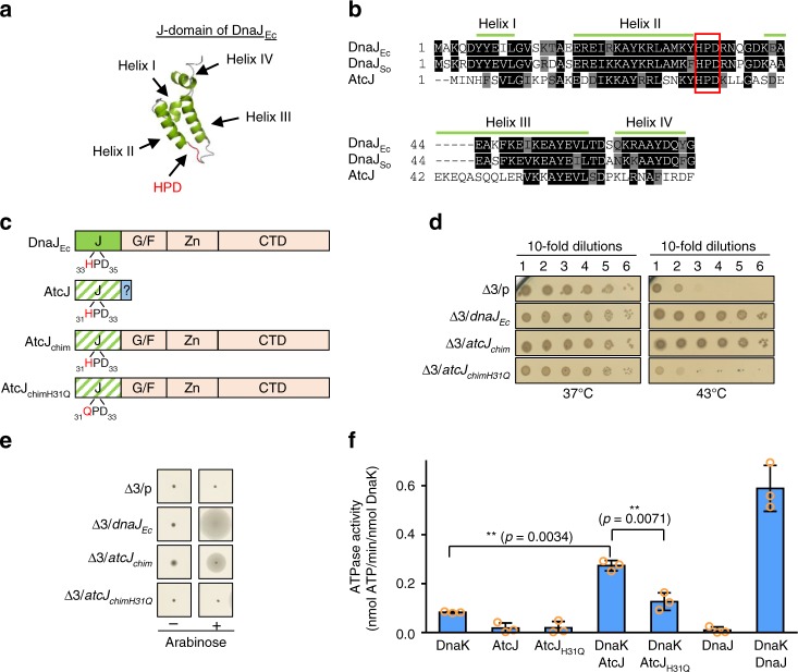
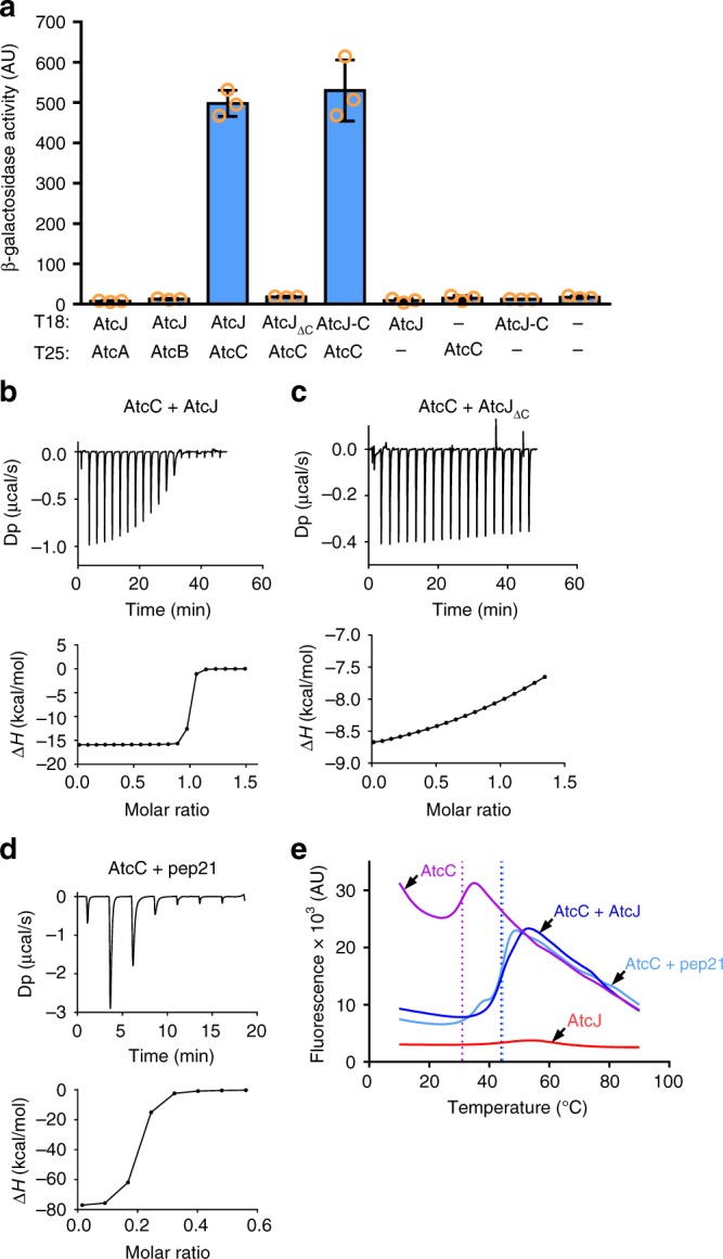
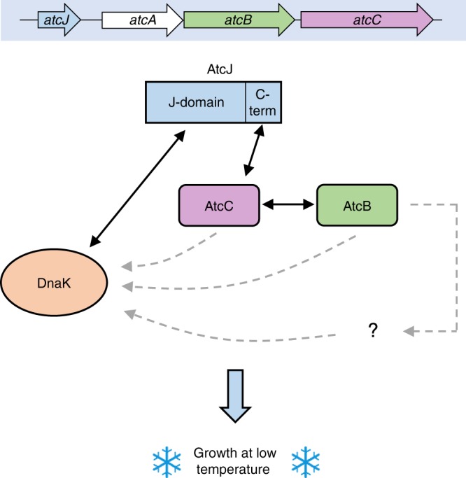
DnaK (Hsp70) is a major ATP-dependent chaperone that functions with two co-chaperones, a J-domain protein (JDP) and a nucleotide exchange factor to maintain proteostasis in most organisms. Here, we show that the environmental bacterium Shewanella oneidensis possesses a previously uncharacterized short JDP, AtcJ, dedicated to cold adaptation and composed of a functional J-domain and a C-terminal extension of 21 amino acids. We showed that atcJ is the first gene of an operon encoding also AtcA, AtcB and AtcC, three proteins of unknown functions. Interestingly, we found that the absence of AtcJ, AtcB or AtcC leads to a dramatically reduced growth at low temperature. In addition, we demonstrated that AtcJ interacts via its C-terminal extension with AtcC, and that AtcC binds to AtcB. Therefore, we identified a previously uncharacterized protein network that involves the DnaK system with a dedicated JDP to allow bacteria to survive to cold environment.

Keywords: Bacterial physiology; Chaperones.


Conflict of interest statement

Competing interestsThe authors declare no competing interests.

Figures

Fig. 1
Fig. 1
AtcJ is a functional J-domain protein. a Structure of the J-domain of E. coli DnaJ (PDB: 1XBL). Helices are shown in green, loops are in gray, and the conserved HPD tripeptide is in red. b Sequence alignment of the J-domains of E. coli DnaJ (DnaJEc), S. oneidensis DnaJ (DnaJSo), and AtcJ using the ClustalW/Omega method and Boxshade (ExPaSy). Black boxes indicate that the residues are strictly conserved and gray boxes that they have conserved substitutions. Green lines indicate the helices of DnaJEc shown in a. The HPD motif is framed in red. c Schematic of the chimera. The J-domain of DnaJEc is shown in green, and the glycine–phenylalanine-rich domain (G/F), the zinc-binding domain (Zn), and the C-terminal domain (CTD) are shown in light orange. The J-domain (J) of AtcJ is green hatched, and the C-terminal extremity of 21 residues is colored blue. The AtcJ chimera (AtcJchim) possesses the J-domain of AtcJ and the G/F, Zn and CTD domains of DnaJEc. The AtcJchimH31Q chimera has the H31Q point mutation in the J-domain of AtcJ. d Production of the wild-type chimera (AtcJchim) suppresses the growth phenotype of the E. coli Δ3 strain (dnaJ cbpA djlA) at high temperature. E. coli Δ3 strain containing the pBad33 vector (p) or plasmids as indicated were grown at 28 °C. Ten time serial dilutions were spotted on LB-agar plates containing 2% L-arabinose (w/v). Plates were incubated overnight at 37 °C and 43 °C. e Production of the wild-type chimera (AtcJchim) partially suppresses the motility phenotype of the E. coli Δ3 strain (dnaJ cbpA djlA). Same strains as in d were spotted on LB soft agar (0.3% (w/v)), and motility was observed by the circumference halo around the initial spot after 1 day at 28 °C. In d and e, plates are representative of at least three experiments. f Stimulation of the DnaK ATPase activity by AtcJ. ATPase activities were measured at 37 °C with 10 µM DnaK, 50 µM AtcJ or AtcJH31Q, or 2 µM DnaJ where indicated. The data from three replicates are shown as mean ± SD. t Test analysis shows that the difference measured is significant (**p < 0.01)
Fig. 2
Fig. 2
AtcJ allows cold adaptation. a Growth of S. oneidensis wild-type and mutant (ΔatcJ) strains at several temperatures on LB-agar plates. After initial growth at 28 °C, strains were diluted to OD600 = 1, and 10-time serial dilutions were spotted on LB-agar plates. Plates were incubated 10 days at 7 °C, 5 days at 15 °C, and 1 day at 28 °C and 35 °C. b Growth of S. oneidensis wild-type and mutant (ΔatcJ) strains at 7 °C and 28 °C in liquid media. After initial growth at 28 °C, strains as in A were diluted to OD600 = 0.05, and were incubated at 7 °C or 28 °C with shaking. Absorbance was measured with time. The data from three replicates are shown as mean ± SD. c Complementation of the growth phenotype. Wild-type or ΔatcJ strains containing as indicated the pBad33 vector (p) or the plasmids producing wild-type AtcJ, AtcJH31Q or AtcJΔC that lacks the last 21 residues were treated as in a, and dilutions were spotted on agar plates containing 0.2% arabinose. Plates were incubated at 7 °C (10 days) or at 28 °C (1 day). In a and c, plates are representative of at least three experiments. d The ΔatcJ strains containing the same plasmids as in c were grown overnight at 28 °C with 0.2% arabinose. Protein extracts were analyzed by western blot using anti-AtcJ antibody. The upper arrow indicates the bands corresponding to AtcJ-WT or H31Q mutant, and the lower arrow AtcJΔC. The HtpG protein (lower panel) was detected by a specific antibody to show that the same amount of the different extracts was loaded on the gel
Fig. 3
Fig. 3
atcJ is encoded in an operon crucial for cold resistance. a Schematic of the atc operon. Specific oligonucleotides indicated by little arrows were used to amplify DNA fragments from the cDNA matrix. The fragments correspond to atcJ (1), atcJ-atcA (2), atcA-atcB (3), and atcB-atcC (4). b cDNA amplification. The total RNA from wild-type S. oneidensis strain grown at 7 °C to exponential phase was extracted and retro-transcribed in cDNA. PCR were performed using cDNA as a matrix and the oligonucleotides indicated in a. No amplification (lane 5) was observed when the primers 1 (see panel a) were used in the PCR with RNA as a matrix, indicating that the RNA preparation was not contaminated by the chromosome. c Growth of the ΔatcA, ΔatcB, and ΔatcC mutant strains. After initial growth at 28 °C, S. oneidensis strains were diluted to OD600 = 1, and 10-time serial dilutions were spotted on LB-agar plates. Plates were incubated 10 days at 7 °C and 1 day at 28 °C. d Complementation of the growth phenotype. Wild-type, ΔatcB, or ΔatcC strains containing as indicated the pBad33 vector (p) or plasmids with the atcB or atcC gene were treated as in c, and dilutions were spotted on 0.2% arabinose LB-agar plates that were incubated at 7 °C for 10 days. In c and d, plates are representative of at least three experiments
Fig. 4
Fig. 4
AtcJ interacts with AtcC. a Bacterial two-hybrid assay showing that AtcJ and AtcC interact. E. coli Bth101 strain co-transformed as indicated with the T18 and T25 plasmids coding for protein fusion between T18 and AtcJ, AtcJΔC, or the last 21 amino acids of AtcJ (AtcJ-C), and protein fusion between T25 and AtcA, AtcB, or AtcC were grown overnight at 28 °C. β-galactosidase activity was measured as explained in the Methods section. The data from three replicates are shown as mean ± SD. bd ITC experiments. Binding assays were performed at 25 °C with 36 µM AtcC and 285 µM AtcJ (b), 36 µM AtcC and 285 µM AtcJΔC (c), or 36 µM AtcC and 285 µM of the pep21 peptide corresponding to the last 21 amino acids of AtcJ (d). Top panels show heat exchange upon ligand titration, and bottom panels show the integrated data with binding isotherms fitted to a single-site binding model. The data shown are representative of two independent experiments. e Thermal Shift Assay experiments were performed as indicated in Methods with 10 µM AtcC, 40 µM AtcJ, or 40 µM pep21. Proteins were mixed as indicated in the figure, SYPRO Orange was added, and the temperature was increased from 10 °C to 90 °C with a 0.5 °C step size. Fluorescence was measured at each temperature. Dot lines indicate the temperature melting point of AtcC alone (purple, 31 °C ± 0.5), with AtcJ (blue, 44 °C ± 0.5), or with pep21 (light blue, 44.5 °C ± 0.5). The data shown are representative of four independent experiments
Fig. 5
Fig. 5
AtcB interacts with AtcC. a Bacterial two-hybrid assay. E. coli Bth101 strain co-transformed as indicated with the T18 and T25 plasmids coding for protein fusion between T18 and AtcC or AtcB, and protein fusion between T25 and AtcA or AtcB were grown overnight at 28 °C. β-galactosidase activity was measured as explained in the Methods section. The data from three replicates are shown as mean ± SD. b Co-purification assay. AtcB with a CBP tag was produced with or without AtcC with a 6-His tag in the E. coli MG1655 strain. CBP-AtcB was purified using CBP affinity resin and bound proteins were analyzed by western blot with anti-CBP and anti-His antibodies as indicated. c In vitro co-purification assay with purified proteins. Purified AtcB (16 µg) or HtpG (16 µg) with a Strep-tag, and purified AtcC (100 µg) with a 6His-tag were mixed as indicated (final volume 200 µL). Proteins with a Strep-tag were pulled down on Strep-Tactin beads, washed several times, and proteins were separated on SDS-PAGE. The results shown in b and c are representative of three independent experiments
Fig. 6
Fig. 6
Model of the role of the Atc proteins in cold resistance. The AtcJ, AtcA, AtcB, and AtcC proteins are encoded from the atcJABC operon. AtcJ binds DnaK with its J-domain, and binds AtcC with its last 21 amino acids (C-term). In addition, AtcC interacts with AtcB. This network of interaction could allow the transfer of AtcC, AtcB, or another protein to DnaK, or could target the DnaK system to a specific location in the cell. Altogether, these proteins play a key role to support bacterial growth at low temperature. Black solid arrows indicate physical or functional interactions demonstrated in this study; gray dashed arrows indicate putative interactions

References

    1. Balchin D, Hayer-Hartl M, Hartl FU. In vivo aspects of protein folding and quality control. Science. 2016;353:aac4354. doi: 10.1126/science.aac4354. - DOI - PubMed
    1. Finka A, Mattoo RUH, Goloubinoff P. Experimental milestones in the discovery of molecular chaperones as polypeptide unfolding enzymes. Annu. Rev. Biochem. 2016;85:715–742. doi: 10.1146/annurev-biochem-060815-014124. - DOI - PubMed
    1. Finka A, Sharma SK, Goloubinoff P. Multi-layered molecular mechanisms of polypeptide holding, unfolding and disaggregation by HSP70/HSP110 chaperones. Front. Mol. Biosci. 2015;2:29. doi: 10.3389/fmolb.2015.00029. - DOI - PMC - PubMed
    1. Genevaux P, Georgopoulos C, Kelley WL. The Hsp70 chaperone machines of Escherichia coli: a paradigm for the repartition of chaperone functions. Mol. Microbiol. 2007;66:840–857. doi: 10.1111/j.1365-2958.2007.05961.x. - DOI - PubMed
    1. Mayer Matthias P., Gierasch Lila M. Recent advances in the structural and mechanistic aspects of Hsp70 molecular chaperones. Journal of Biological Chemistry. 2018;294(6):2085–2097. doi: 10.1074/jbc.REV118.002810. - DOI - PMC - PubMed

Publication types

Substances

Supplementary concepts