. 2019 Oct;145(1):23-34.
doi: 10.1007/s11060-019-03275-z. Epub 2019 Sep 3.

MicroRNA-29a inhibits glioblastoma stem cells and tumor growth by regulating the PDGF pathway

Affiliations

MicroRNA-29a inhibits glioblastoma stem cells and tumor growth by regulating the PDGF pathway

Yanzhi Yang et al. J Neurooncol. 2019 Oct.

Abstract

Background and purpose: microRNAs are small noncoding RNAs that play important roles in cancer regulation. In this study, we investigated the expression, functional effects and mechanisms of action of microRNA-29a (miR-29a) in glioblastoma (GBM).

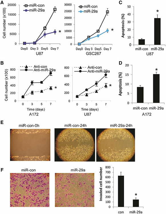
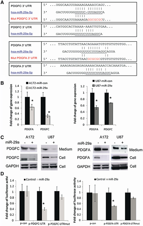
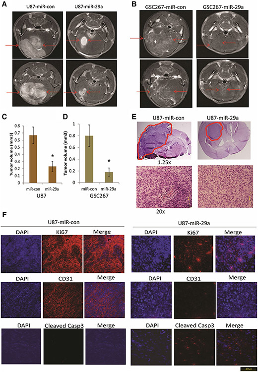
Methods: miR-29a expression levels in GBM cells, stem cells (GSCs) and human tumors as well as normal astrocytes and normal brain were measured by quantitative PCR. miR-29a targets were uncovered by target prediction algorithms, and verified by immunoblotting and 3' UTR reporter assays. The effects of miR-29a on cell proliferation, death, migration and invasion were assessed with cell counting, Annexin V-PE/7AAD flow cytometry, scratch assay and transwell assay, respectively. Orthotopic xenografts were used to determine the effects of miR-29a on tumor growth.

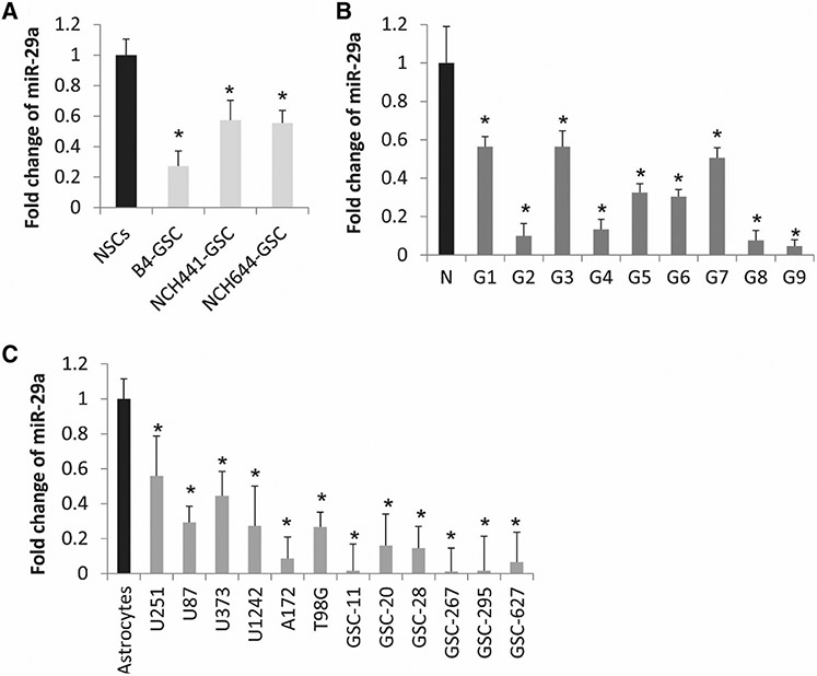
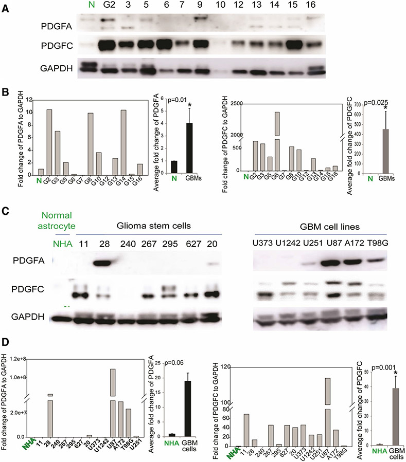
Results: Mir-29a was downregulated in human GBM specimens, GSCs and GBM cell lines. Exogenous expression of miR-29a inhibited GSC and GBM cell growth and induced apoptosis. miR-29a also inhibited GBM cell migration and invasion. PDGFC and PDGFA were uncovered and validated as direct targets of miR-29a in GBM. miR-29a downregulated PDGFC and PDGFA expressions at the transcriptional and translational levels. PDGFC and PDGFA expressions in GBM tumors, GSCs, and GBM established cell lines were higher than in normal brain and human astrocytes. Mir-29a expression inhibited orthotopic GBM xenograft growth.

Conclusions: miR-29a is a tumor suppressor miRNA in GBM, where it inhibits cancer stem cells and tumor growth by regulating the PDGF pathway.

Keywords: Glioblastoma (GBM); Glioblastoma stem cells (GSC); Platelet-derived growth factor (PDGF); microRNA (miRNA).


Figures

Fig. 1
Fig. 1
miR-29a expression is downregulated in GSCs, GBM cells, and human tumors. a Quantitative RT-PCR of miR-29a in GSCs and GBM cells showing lower expression levels in GSCs isolated from GBM tumors by CD133+ as compared to normal neural stem cells (NSCs) (CD133+ ) isolated from normal brain. b Quantitative RT-PCR of miR-29a in GBM surgical specimens (G) showing lower levels of miR-29a than in normal brain (N). c Quantitative RT-PCR of miR-29a in GBM cell lines (U251, U87, U373, U1242 and T98G) and GSCs (GSC-11, GSC-20, GSC-28, GSC-267, GSC-295 and GSC-627) showing lower levels of expression as compared to normal human astrocytes. *P < 0.05
Fig. 2
Fig. 2
miR-29a inhibits GBM cell and GSC proliferation, induces GBM cell apoptosis and inhibits GBM cell migration and invasion. a Proliferation assay showing the inhibition of GBM cell U87 and GSC-267 proliferation by pre-miR-29a. b miR-29a inhibitor (anti-miR-29a) transfection enhances GBM cell A172 and U87 proliferation. c, d AnnexinV-PE and 7-AAD flow-cytometric analysis of GBM cells U87 (c) and A172 (d) showing induction of apoptosis after pre-miR-29a transfection. e GBM cells A172 were transfected with either Pre-miR-29a or controls and assessed for migration with the wound healing assay. f Pre-miR-29a or control-transfected A172 cells were used in a transwell invasion assay. The left panel shows a representative invasion assay, the right panel shows the quantification of invading cells. The data show that miR-29a overexpression inhibits GBM cell migration and invasion. *P < 0.05
Fig. 3
Fig. 3
miR-29a regulates PDGFC and PDGFA expressions by directly binding to their mRNA 3′ UTRs. a Alignment of PDGFC, PDGFA 3′ UTRs and miR-29a sequences showing the predicted binding sites. The sites of targeted mutagenesis for the generation of mutant controls are shown in red. b Quantitative RT-PCR showing the inhibitory effects of miR-29a on the mRNA levels of PDGFC and PDGFA in GBM cells A172 and U87. c Immunoblots (left panel) showing the effects of miR-29a overexpression on the protein levels of secreted and intracellular PDGFC in GBM cells. The right panel shows that miR-29a downregulates the protein levels of PDGFA in growth medium and cell lystaes. d 3′ UTR luciferase assays for PDGFC (left panel) and PDGFA (right panel) showing the inhibition of luciferase activity by miR-29a in GBM cells. No significant alteration in luciferase activity was observed when 3′UTR binding sites were mutated. *P < 0.05
Fig. 4
Fig. 4
PDGFC and PDGFA are upregulated in GBM cells, GSCs and GBM tumors. a Immunoblots showing higher levels of PDGFA and PDGFC protein in a subset of GBM specimens (G) as compared to normal brain (N). b Quantification of PDGFA (left panel) and PDGFC level (middle panel) in GBM specimens as compared to normal brain (P = 0.01 and P = 0.025, respectively). c Immunoblots showing PDGFA and PDGFC levels in GSCs (left panel) and GBM cell lines (right panel) and normal human astrocytes (NHA). d Quantification of PDGFA (left panel) and PDGFC levels (right panel) in GBM cell lines, GSCs as compared to NHA (P = 0.06 and P < 0.001, respectively)
Fig. 5
Fig. 5
miR-29a inhibits GBM cell and GSC derived xenograft growth. a, b GBM cells U87 (a) and GSC-267 cells (b) were infected with lentivirus encoding pre-miR-29a or pre-miR-control for 24 h and implanted into the brains of immunodeficient mice (n = 6). After 3 weeks, the mice were subjected to MRI scan and tumor volumes were calculated (c, d). e H&E staining of U87 xenografts generated from Lenti-control or pre-miR-29a U87 cells (red lines indicate the tumors). f Immunohistochemistry staining of U87 xenograft sections with antibodies against Ki67, CD31 and Cleaved Caspase 3. The data show that miR-29a inhibits in vivo GBM xenograft growth, reduces Ki67 and CD31 levels and enhances Cleaved Caspase 3 levels. *P < 0.05

Similar articles

Cited by

References

    1. Bartel DP (2009) MicroRNAs: target recognition and regulatory functions. Cell 136(2):215–233 - PMC - PubMed
    1. Krichevsky AM, King KS, Donahue CP, Khrapko K, Kosik KS (2003) A microRNA array reveals extensive regulation of microRNAs during brain development. RNA 9(10):1274–1281 - PMC - PubMed
    1. Lee YS, Dutta A (2009) MicroRNAs in cancer. Annu Rev Pathol 4:199–227 - PMC - PubMed
    1. Caldas C, Brenton JD (2005) Sizing up miRNAs as cancer genes. Nat Med 11(7):712–714 - PubMed
    1. Calin GA, Croce CM (2006) MicroRNA signatures in human cancers. Nat Rev Cancer 6(11):857–866 - PubMed

MeSH terms