

Combination of ERK2 inhibitor VX-11e and voreloxin synergistically enhances anti-proliferative and pro-apoptotic effects in leukemia cells
- PMID: 31482470
- PMCID: PMC6823322
- DOI: 10.1007/s10495-019-01564-6
Combination of ERK2 inhibitor VX-11e and voreloxin synergistically enhances anti-proliferative and pro-apoptotic effects in leukemia cells
Abstract
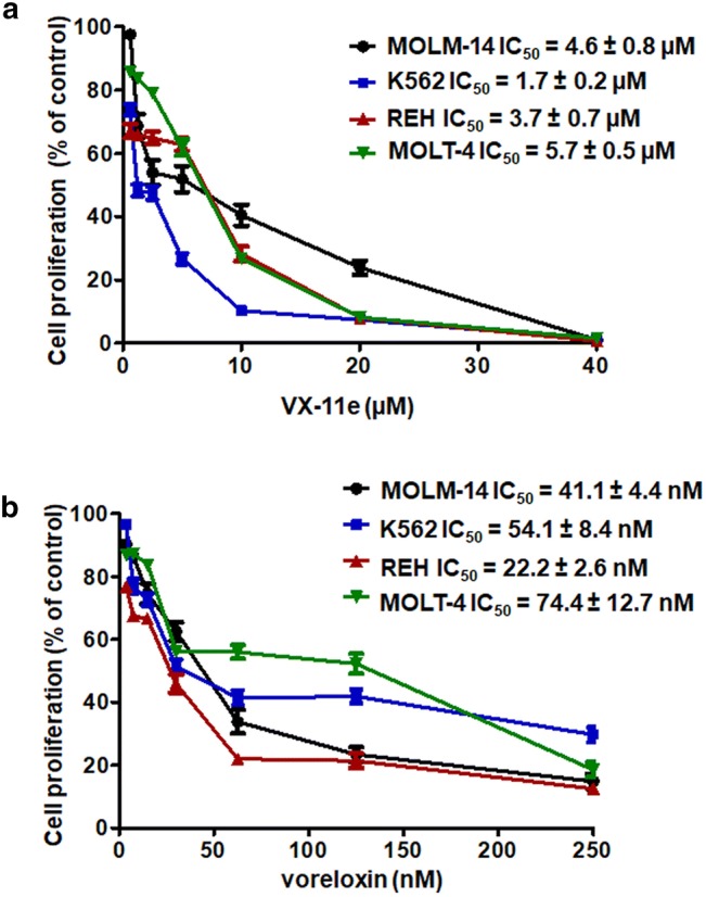
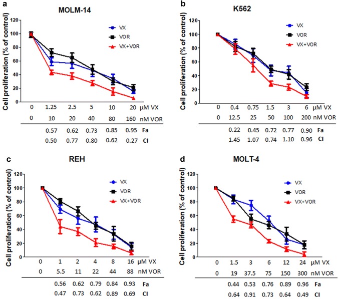
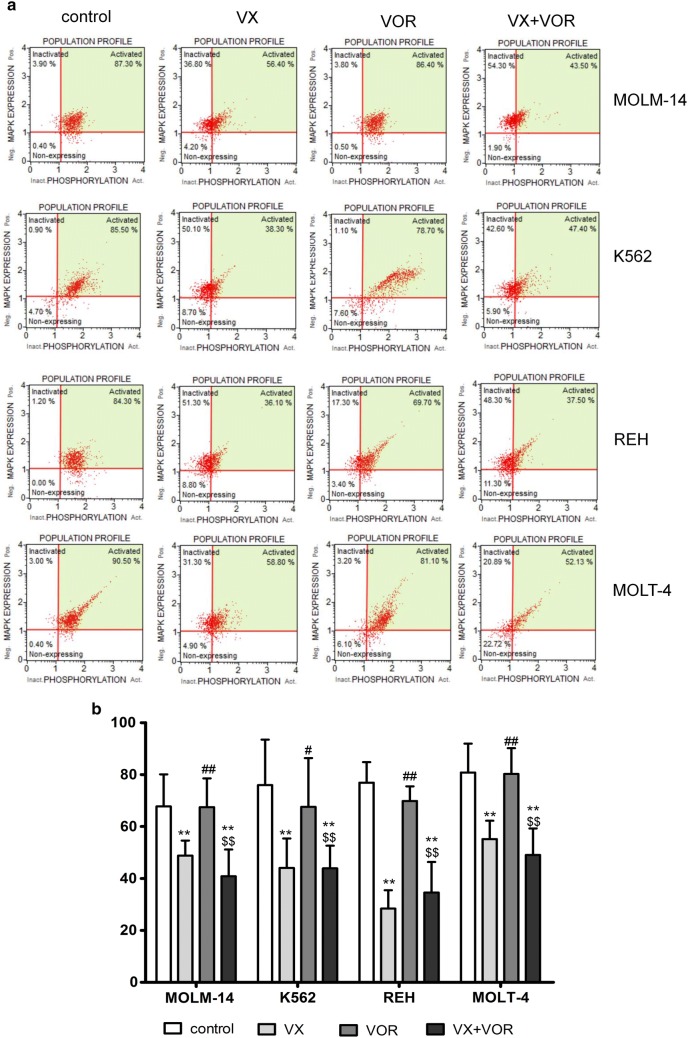
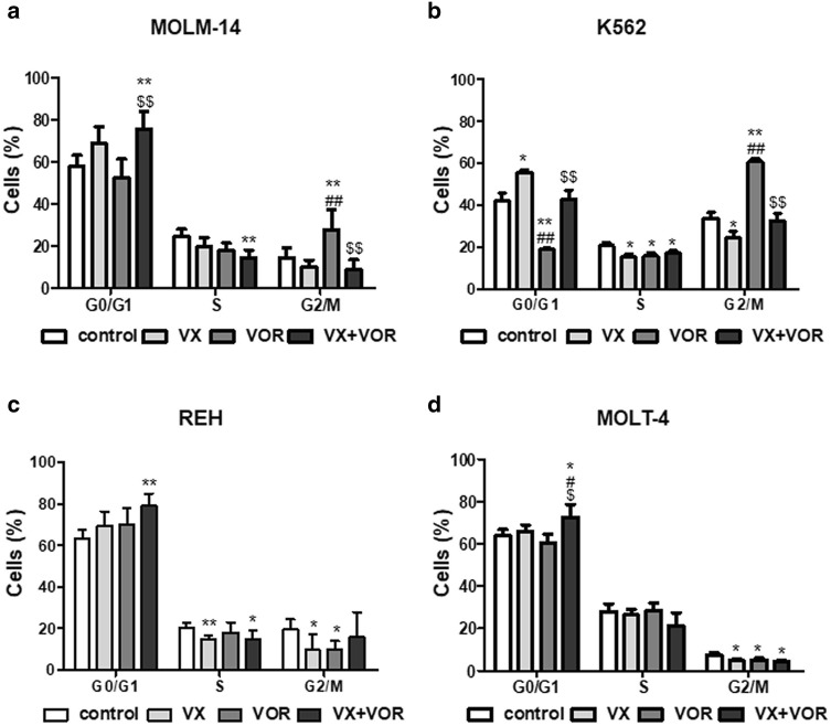
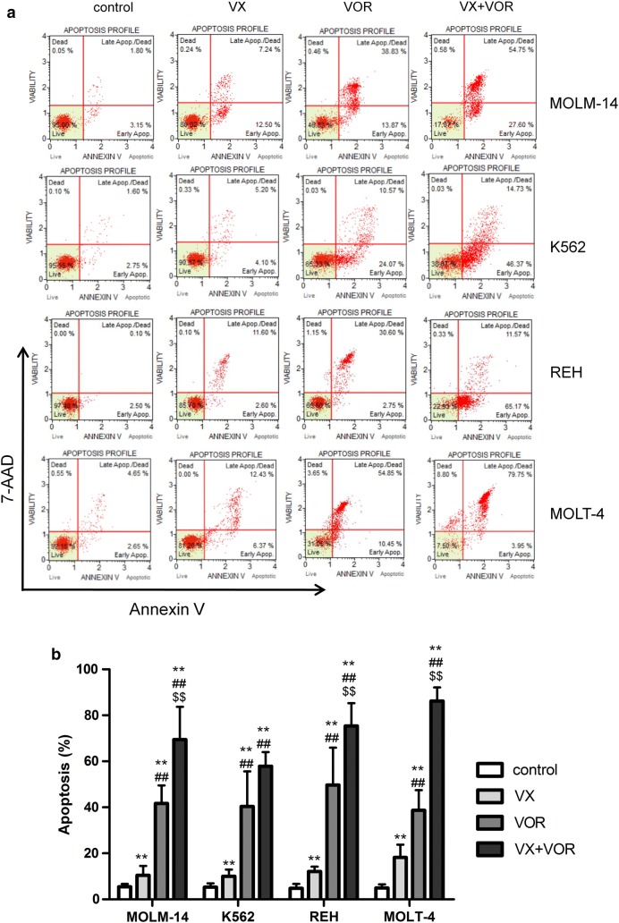
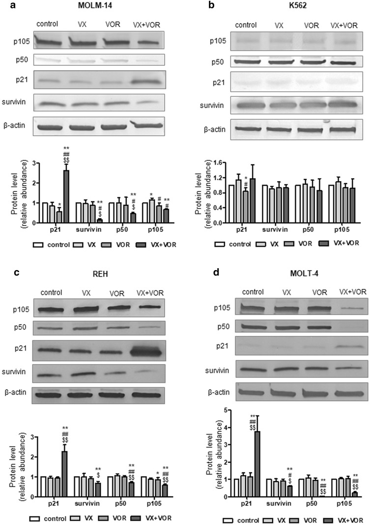
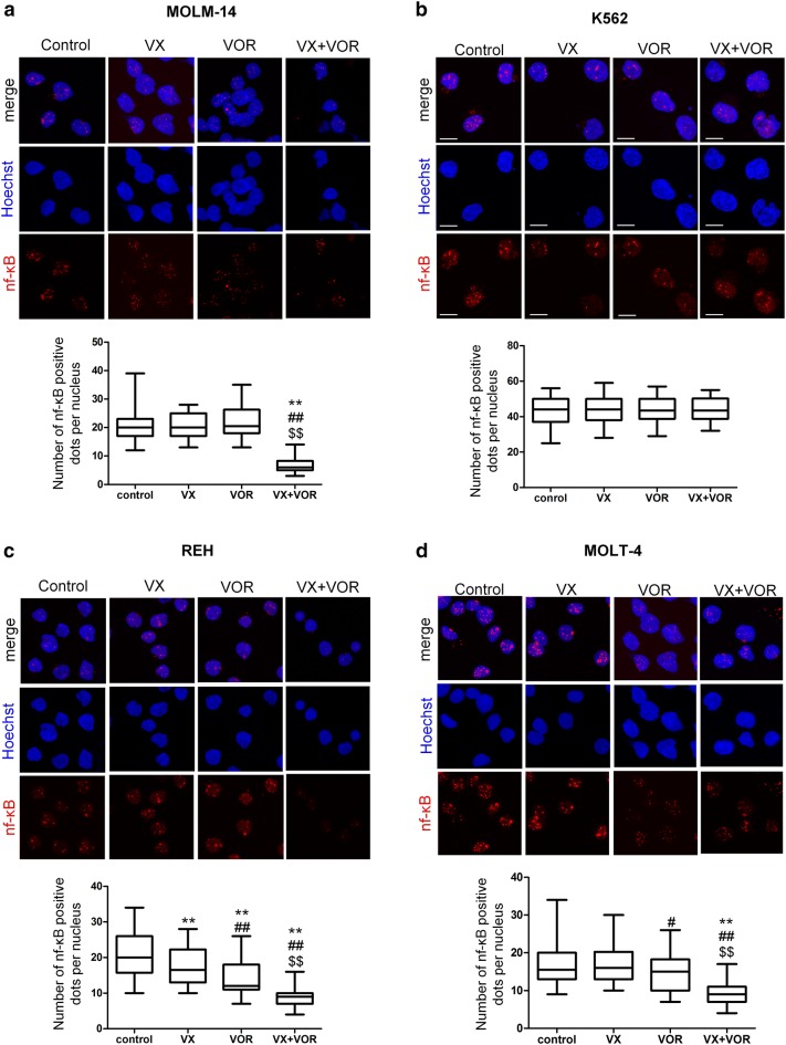
ERK1/2 inhibitors are new promising anticancer drugs. The aim of this study was to investigate the effect of the combination of ERK2 inhibitor VX-11e and voreloxin on MOLM-14, K562, REH and MOLT-4 leukemia cell lines. We found that VX-11e alone and in combination with voreloxin significantly decreased ERK activation in all cell lines tested. To evaluate the interactions of the drugs, cells were treated for 24 h with VX-11e or voreloxin alone and in combination at fixed ratios based on IC50 values. The combinatorial effects of both drugs were synergistic over a wide range of concentrations in MOLM-14, REH and MOLT-4 cell lines. In K562 cells, three effects were found to be additive, one antagonistic and only one synergistic. The results showed that incubation with both VX-11e and voreloxin inhibited the growth of leukemia cells, affected cell cycle and induced apoptosis. Furthermore, the molecular mechanism of these effects might be attributed to an increased expression of p21 and a decreased expression of survivin and NF-κB in all cell lines tested except from K562 cells. In conclusion, combination of VX-11e and voreloxin can exert a synergistic anticancer effect in leukemia cells.
Keywords: Apoptosis; Cell cycle; Leukemia cell lines; VX-11e; Voreloxin.
Conflict of interest statement
The authors declare no conflict of interest.
Figures
Similar articles
-
Combination of ERK2 and STAT3 Inhibitors Promotes Anticancer Effects on Acute Lymphoblastic Leukemia Cells.Cancer Genomics Proteomics. 2020 Sep-Oct;17(5):517-527. doi: 10.21873/cgp.20208. Cancer Genomics Proteomics. 2020. PMID: 32859630 Free PMC article.
-
TAK-733, a Selective MEK Inhibitor, Enhances Voreloxin-induced Apoptosis in Myeloid Leukemia Cells.Anticancer Res. 2018 Nov;38(11):6147-6156. doi: 10.21873/anticanres.12967. Anticancer Res. 2018. PMID: 30396931
-
The topoisomerase II inhibitor voreloxin causes cell cycle arrest and apoptosis in myeloid leukemia cells and acts in synergy with cytarabine.Haematologica. 2011 Mar;96(3):393-9. doi: 10.3324/haematol.2010.032680. Epub 2010 Dec 6. Haematologica. 2011. PMID: 21134979 Free PMC article.
-
Voreloxin, formerly SNS-595, has potent activity against a broad panel of cancer cell lines and in vivo tumor models.Cancer Chemother Pharmacol. 2009 Jun;64(1):53-65. doi: 10.1007/s00280-008-0850-3. Epub 2008 Oct 19. Cancer Chemother Pharmacol. 2009. PMID: 18931998
-
Voreloxin, a first-in-class anticancer quinolone derivative, acts synergistically with cytarabine in vitro and induces bone marrow aplasia in vivo.Cancer Chemother Pharmacol. 2010 Oct;66(5):881-8. doi: 10.1007/s00280-009-1234-z. Epub 2010 Jan 8. Cancer Chemother Pharmacol. 2010. PMID: 20058009 Free PMC article.
Cited by
-
Repurposing fluoroquinolones as cancer chemosensitizers: a way to overcome cancer therapeutic bottleneck.Naunyn Schmiedebergs Arch Pharmacol. 2025 Aug 11. doi: 10.1007/s00210-025-04508-x. Online ahead of print. Naunyn Schmiedebergs Arch Pharmacol. 2025. PMID: 40788483 Review.
-
Enhanced conversion of T cells into CAR T cells by modulation of the MAPK/ERK pathway.Cell Rep Med. 2025 Feb 18;6(2):101970. doi: 10.1016/j.xcrm.2025.101970. Epub 2025 Feb 11. Cell Rep Med. 2025. PMID: 39938523 Free PMC article.
-
ERK2-topoisomerase II regulatory axis is important for gene activation in immediate early genes.Nat Commun. 2023 Dec 14;14(1):8341. doi: 10.1038/s41467-023-44089-y. Nat Commun. 2023. PMID: 38097570 Free PMC article.
-
Multi-Omic Analysis of Glutamate Excitotoxicity in Primary Neuronal Cultures.J Neurochem. 2025 Jun;169(6):e70110. doi: 10.1111/jnc.70110. J Neurochem. 2025. PMID: 40476344 Free PMC article.
-
Targeting the MAPK/ERK and PI3K/AKT Signaling Pathways Affects NRF2, Trx and GSH Antioxidant Systems in Leukemia Cells.Antioxidants (Basel). 2020 Jul 17;9(7):633. doi: 10.3390/antiox9070633. Antioxidants (Basel). 2020. PMID: 32709140 Free PMC article.
References
-
- Enzler T, Sano Y, Choo MK, Cottam HB, Karin M, Tsao H, Park JM. Cell-selective inhibition of NF-κB signaling improves therapeutic index in a melanoma chemotherapy model. Cancer Discov. 2011;1:496–507. doi: 10.1158/2159-8290.CD-11-0143. - DOI - PMC - PubMed
-
- Huang X, Schwind S, Santhanam R, Eisfeld AK, Chiang CL, Lankenau M, Yu B, Hoellerbauer P, Jin Y, Tarighat SS, Khalife J, Walker A, Perrotti D, Bloomfield CD, Wang H, Lee RJ, Lee LJ, Marcucci G. Targeting the RAS/MAPK pathway with miR-181a in acute myeloid leukemia. Oncotarget. 2016;7:59273–59286. doi: 10.18632/oncotarget.11150. - DOI - PMC - PubMed
Publication types
MeSH terms
Substances
Grants and funding
LinkOut - more resources
Full Text Sources
Medical
Miscellaneous
