. 2019 Sep 4;101(17):1554-1562.
doi: 10.2106/JBJS.19.00034.

An Anatomical Study of the Anterosuperior Capsular Attachment Site on the Acetabulum

Affiliations

An Anatomical Study of the Anterosuperior Capsular Attachment Site on the Acetabulum

Masahiro Tsutsumi et al. J Bone Joint Surg Am. .

Abstract

Background: Despite the fact that many surgeons perform partial capsular detachment from the anterosuperior aspect of the acetabulum to correct acetabular deformities during hip arthroscopy, few studies have focused on whether these detachments influence hip joint stability. The aim of this study was to investigate the capsular attachment on the anterosuperior aspect of the acetabulum. We hypothesized that the attachment on the inferior aspect of the anterior inferior iliac spine (AIIS) is wide and fibrocartilaginous and might have a substantial role in hip joint stability.

Methods: Fifteen hips from 9 cadavers of Japanese donors were analyzed. Eleven hips were analyzed macroscopically, and the other 4 were analyzed histologically. In all specimens, the 3-dimensional morphology of the acetabulum and AIIS was examined using micro-computed tomography (micro-CT).

Results: Macroscopic analysis showed that the widths of the capsular attachments varied according to the location, and the attachment width on the inferior edge of the AIIS was significantly larger than that on the anterosuperior aspect of the acetabulum. Moreover, the capsular attachment on the inferior edge of the AIIS corresponded with the impression, which was identified by micro-CT. Histological analysis revealed that the hip joint capsule on the inferior edge of the AIIS attached to the acetabulum adjacent to the proximal margin of the labrum. In addition, the hip joint capsule attached to the inferior edge of the AIIS via the fibrocartilage.

Conclusions: The capsular attachment on the inferior edge of the AIIS was characterized by an osseous impression, large attachment width, and distributed fibrocartilage.

Clinical relevance: It appeared that the capsular attachment on the inferior edge of the AIIS was highly adaptive to mechanical stress, on the basis of its osseous impression, attachment width, and histological features. Anatomical knowledge of the capsular attachment on the inferior edge of the AIIS provides a better understanding of the pathological condition of hip joint instability.


Figures

Fig. 1
Fig. 1
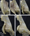
Osseous configuration of the AIIS and acetabulum shown by micro-CT. The dashed lines indicate the rough impression at the superior portion of the AIIS and anterolateral wall of the ilium. The arrowheads indicate the shallow groove at the anteromedial surface of the AIIS. The star indicates the smooth impression at the inferior portion of the AIIS. Ant = anterior, Med = medial, Post = posterior, and Sup = superior. The anteromedial (Fig. 1-A), anterior (Fig. 1-B), and anterolateral (Fig. 1-C) aspects of the right hip are shown.
Fig. 2
Fig. 2
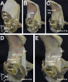
Spatial geometry of the rectus femoris and its superficial structures on the anteromedial aspect of the AIIS. Ip = iliopsoas, Sa = sartorius, Ic = iliocapsularis, RF = rectus femoris, RFd = direct head of the rectus femoris, Med = medial, Post = posterior, and Sup = superior. Fig. 2-A Anteromedial aspect of the right Ip. Fig. 2-B The Ip was partly detached from the iliac fossa and reflected to the lateral side. Fig. 2-C The origin site of the Ic is indicated by the white dashed line. Fig. 2-D The RFd after removal of the fasciae of the Ip on the surface of the RFd. Fig. 2-E Positional relationship between the origin site of the RFd and that of the Ic (indicated by the white dashed line).
Fig. 3
Fig. 3
Spatial geometry of the rectus femoris and its superficial structures on the anterolateral aspect of the AIIS. GMe = gluteus medius, GMi = gluteus minimus, TFL = tensor fasciae latae, Sa = sartorius, Cap = hip joint capsule, RF = rectus femoris, Ip = iliopsoas, RFd = direct head of the rectus femoris, RFr = reflected head of the rectus femoris, Ant = anterior, Med = medial, and Sup = superior. Fig. 3-A Anterolateral aspect of the right hip cross-section at the level of the femoral neck. Fig. 3-B Superficial muscles including the GMe, the TFL, and Sa were removed. Fig. 3-C The GMi and Ip were removed. The black dashed lines indicate deep fasciae of the GMi, Ip, and RF. Fig. 3-D The RFd and RFr after removal of the fascia of the GMi on the surface of the RFr. Fig. 3-E The origin sites of the RFd and RFr are indicated by the white dashed lines.
Fig. 4
Fig. 4
Attachment of hip joint capsule on the anterosuperior aspect of the acetabulum. Ic = iliocapsularis, RFd = direct head of the rectus femoris, Cap = hip joint capsule, RFr = reflected head of the rectus femoris, GMi = gluteus minimus, Ant = anterior, Med = medial, Post = posterior, and Sup = superior. The black dashed lines indicate the osseous attachment of the hip joint capsule. The white dashed lines indicate the origin sites of the Ic, RFd, and RFr. The white circles indicate the iliopubic ramus. The right hip joint capsule was detached from the acetabulum in the following order: anteromedial (Fig. 4-A), anterior (Fig. 4-B), and anterolateral (Fig. 4-C) aspects of the hip joint.
Fig. 5
Fig. 5
Schematic diagram indicating the measurement of the attachment of the hip joint capsule on the anterior aspect of the hip. GMi = gluteus minimus, RFd = direct head of the rectus femoris, RFr = reflected head of the rectus femoris, Ic = iliocapsularis, Med = medial, and Sup = superior. The blue region indicates the osseous attachment of the right hip joint capsule (Cap). Attachment widths were measured at the middle position between the iliopubic ramus and the anteroinferior edge of the AIIS (L1), at the anteroinferior edge of the AIIS (L2), at the posteroinferior edge of the AIIS (L3), and at the anterior edge of the origin site of the RFr (L4).
Fig. 6
Fig. 6
Histological analysis of the hip capsular attachment (Masson trichrome stain). GMi = gluteus minimus, RFd = direct head of the rectus femoris, RFr = reflected head of the rectus femoris, Ic = iliocapsularis, Med = medial, and Sup = superior. Fig. 6-A Schematic diagram of the attachment site of the hip joint capsule (Cap) on the anterior aspect of the right hip. The blue region indicates the osseous attachment of the hip joint capsule. Fig. 6-B Cross-section along line B in Fig. 6-A. Fig. 6-C Cross-section along line C in Fig. 6-A. Fig. 6-D Magnified image of the boxed region in Fig. 6-C. Four zones of tissue are clearly distinguished: fibrous connective tissue (FC), uncalcified fibrocartilage (UF), calcified fibrocartilage (CF), and bone (Bo). Fig. 6-E Cross-section along line E in Fig. 6-A. Loose connective tissue attached to the distal part of the acetabulum is indicated by the double-headed arrow. Scale: Figs. 6-B, 6-C, and Fig. 6-E = 5 mm, and Fig. 6-D = 1 mm.

Similar articles

Cited by

References

    1. Ganz R, Parvizi J, Beck M, Leunig M, Nötzli H, Siebenrock KA. Femoroacetabular impingement: a cause for osteoarthritis of the hip. Clin Orthop Relat Res. 2003. December;417:112-20. - PubMed
    1. Bedi A, Kelly BT. Femoroacetabular impingement. J Bone Joint Surg Am. 2013. January 2;95(1):82-92. - PubMed
    1. Griffin DR, Dickenson EJ, O’Donnell J, Agricola R, Awan T, Beck M, Clohisy JC, Dijkstra HP, Falvey E, Gimpel M, Hinman RS, Hölmich P, Kassarjian A, Martin HD, Martin R, Mather RC, Philippon MJ, Reiman MP, Takla A, Thorborg K, Walker S, Weir A, Bennell KL. The Warwick Agreement on femoroacetabular impingement syndrome (FAI syndrome): an international consensus statement. Br J Sports Med. 2016. October;50(19):1169-76. - PubMed
    1. Griffin DR, Dickenson EJ, Wall PDH, Achana F, Donovan JL, Griffin J, Hobson R, Hutchinson CE, Jepson M, Parsons NR, Petrou S, Realpe A, Smith J, Foster NE; FASHIoN Study Group. Hip arthroscopy versus best conservative care for the treatment of femoroacetabular impingement syndrome (UK FASHIoN): a multicentre randomised controlled trial. Lancet. 2018. June 2;391(10136):2225-35. Epub 2018 Jun 1. - PMC - PubMed
    1. Pun S, Kumar D, Lane NE. Femoroacetabular impingement. Arthritis Rheumatol. 2015. January;67(1):17-27. - PMC - PubMed