. 2019 Sep 4;2(9):e199292.
doi: 10.1001/jamanetworkopen.2019.9292.

Association of Germline Variants in Natural Killer Cells With Tumor Immune Microenvironment Subtypes, Tumor-Infiltrating Lymphocytes, Immunotherapy Response, Clinical Outcomes, and Cancer Risk

Affiliations

Association of Germline Variants in Natural Killer Cells With Tumor Immune Microenvironment Subtypes, Tumor-Infiltrating Lymphocytes, Immunotherapy Response, Clinical Outcomes, and Cancer Risk

Xue Xu et al. JAMA Netw Open. .

Erratum in

  • Error in Methods.
    [No authors listed] [No authors listed] JAMA Netw Open. 2020 Apr 1;3(4):e206708. doi: 10.1001/jamanetworkopen.2020.6708. JAMA Netw Open. 2020. PMID: 32329767 Free PMC article. No abstract available.

Abstract

Importance: Only a small fraction of patients with cancer receiving immune checkpoint therapy (ICT) respond, which is associated with tumor immune microenvironment (TIME) subtypes and tumor-infiltrating lymphocytes (TILs).

Objective: To examine whether germline variants of natural killer (NK) cells, a key component of the immune system, are associated with TIME subtypes, the abundance of TILs, response to ICT, clinical outcomes, and cancer risk.

Design, setting, and participants: This genetic association study explored TIME subtypes and examined the association of the germline genomic information of patients with cancer with TIME subtypes, abundance of TILs, response to ICT, prognosis, and cancer risk. Clinical information, tumor RNA sequencing, and whole-exome sequencing (WES) data of paired normal samples of patients with 13 common cancers (n = 5883) were obtained from the Cancer Genome Atlas. The WES data of individuals with no cancer (n = 4500) were obtained from the database of Genotypes and Phenotypes. Data collection and analysis took place in March 2017.

Main outcomes and measures: Associations between the number of germline defective genes in NK cells and survival time and the abundance of TILs.

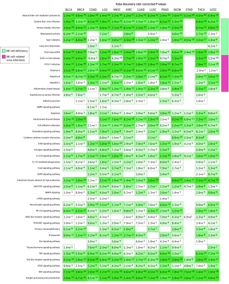
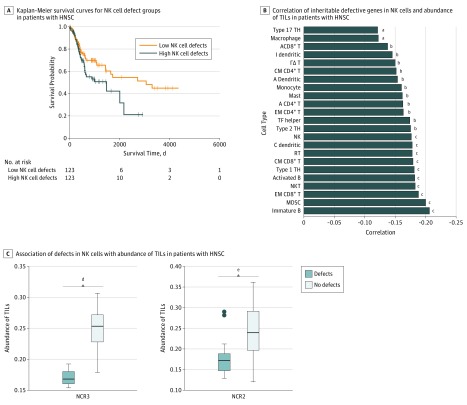
Results: Based on tumor RNA sequencing data, tumors were stratified into TIME-rich, TIME-intermediate, and TIME-poor subtypes. Tumors of TIME-rich subtype had more TILs (TIL-NK cells in TIME-rich head and neck squamous cell carcinoma [HNSC] tumors: t = 4.85; 95% CI of the difference, 0.01-0.03; P = 2.19 × 10⁻⁶) compared with TIME-intermediate HNSC tumors (t = 3.70; 95% CI of the difference, 0.01-0.03; P < .001), better prognosis (patients with HNSC: hazard ratio, 0.65; 95% CI, 0.41-1.02; P = .054) compared with TIME-intermediate and TIME-poor subtypes, and better ICT response (patients with melanoma: odds ratio [OR], 4.45; 95% CI, 0.99-27.08; P = .04). Patients with TIME-rich tumors had significantly fewer inherited defective genes in NK cells than patients with TIME-intermediate and TIME-poor tumors (patients with HNSC: OR, 0.49; 95% CI, 0.26-1.07; P = .005). Similarly, patients with cancer had significantly more inherited defective genes in NK cells than individuals with no cancer (patients with HNSC: OR, 19.09; 95% CI, 4.30-315.96; P = 6.21 × 10⁻⁴). Among 11 of 13 common cancers, the number of heritable defective genes in NK cells was significantly negatively associated with survival (patients with HNSC: hazard ratio, 1.77; 95% CI, 1.18-2.66; P = .005), abundance of TILs (patients with HNSC: R = -0.25; 95% CI, -0.65-2.17; P = 0.02), and response to ICT (patients with melanoma: OR, 4.45; 95% CI, 0.99-27.08; P = .04).

Conclusions and relevance: These results suggest that individuals who have more inherited defective genes in NK cells had a higher risk of developing cancer and that these inherited defects were associated with TIME subtypes, recruitment of TILs, ICT response, and clinical outcomes. The findings have implications for identifying individuals at risk for developing cancer of many types based on germline variants of NK cells and for improving existing ICT and chimeric antigen receptor-T cell therapy by adoptive transfer of healthy NK cells to patients with TIME-intermediate and TIME-poor tumors.


Conflict of interest statement

Conflict of Interest Disclosures: None reported.

Figures

Figure 1.
Figure 1.. Heatmaps Showing the 3 Universal Tumor Immune Microenvironment (TIME) Subtypes
A representative heatmap derived from the gene expression of immune checkpoint therapy essential genes, showing the TIME subtypes in head and neck squamous cell carcinoma. Heatmap based on log10 transformation of gene expression value. Green indicates minimum expression and red, maximum expression. Heatmaps for other cancer types are shown in eFigure 1 in the Supplement.
Figure 2.
Figure 2.. Abundance of Tumor-Infiltrating Lymphocytes (TILs) and Clinical Features of the Tumor Immune Microenvironment (TIME) Subtypes
A, Heatmaps for other cancer types are shown in eFigure 2 in the Supplement. B, Kaplan-Meier survival curves of patients with the TIME-rich subtype and the combined TIME-intermediate and TIME-poor subtypes for head and neck squamous cell carcinoma (HNSC). The survival time for the patients with TIME-rich tumors was significantly longer than those with TIME-intermediate or TIME-poor subtypes. The curves for other cancer types are shown in eFigure 3 in the Supplement. CD4 indicates cluster of differentiation 4; CD56, cluster of differentiation 56; CD8, cluster of differentiation 8; and MDSC, myeloid-derived suppressor cell.
Figure 3.
Figure 3.. Kyoto Encyclopedia of Genes and Genomes Pathway Enrichment Analysis
Heatmap shows the enriched pathways derived from the significantly differential genes of the RNA sequenced data comparing the TIME-rich subtype with the combined TIME-intermediate and TIME-poor subtypes. Columns represent Kyoto Encyclopedia of Genes and Genomes pathways, and rows represent cancer types. BLCA indicates bladder cancer; BRCA, breast cancer; COAD, colon cancer; FoxO, forkhead box protein O; GnRH, gonadotropin-releasing hormone; HNSC, head and neck squamous cell carcinoma; JAK, Janus kinases; KIRC, renal cancer; LGG, glioma cancer; LUAD, lung adenocarcinoma; LUSC, lung squamous cell cancer; NF, nuclear factor; NOD, nucleotide-binding oligomerization domain; PI3K, phosphoinositide 3-kinase; PRAD, prostate cancer; SKCM, skin cancer; STAD, stomach cancer; STAT, signal transducer and activator of transcription protein; THCA, thyroid cancer; TNF, tumor necrosis factor; UCEC, endometrial cancer; and VEGF, vascular endothelial growth factor.
Figure 4.
Figure 4.. Association of Natural Killer (NK) Cells With Defective Genes With Tumor Immune Microenvironment Subtypes and Clinical Outcomes
A, Representative Kaplan-Meier survival curve of the high-NKD gene patient group and low-NKD gene patient group in head and neck squamous cell carcinoma (HNSC). The curves for other cancer types are shown in eFigure 10 in the Supplement. B, Negative correlations between the number of inheritable defective genes in NK cells and the abundance of the tumor-infiltrating lymphocyte in tumor microenvironments for HNSC. The correlations for other cancer types are shown in eFigure 11 in the Supplement. C, Examples in HNSC showing that the abundance of tumor-infiltrating lymphocyte–NK cells was significantly lower in tumors bearing a defective gene in NK cells than the rest of the tumors. More examples are shown in eFigure 14 in the Supplement. Center horizontal line indicates mean; top and bottom borders of box, SD; whiskers, 95% CI; and circles, outliers. A indicates activated; B, B cell; cDC, conventional dendritic cell; CM, central memory; EM, effector memory; I, immature; MDSC, myeloid-derived suppressor cell; RT, regulatory T cell; T, T cell; TH, T helper cell; and TF helper, T follicular helper cell. aP < .10. bP < .05. cP < .001. dP = 2.12 × 10−3. eP = 4.81 × 10−3.
Figure 5.
Figure 5.. Heatmap Derived From Comparison of Individuals With No Cancer vs Patients With Cancer
This heatmap shows the significantly enriched pathways derived from the significantly differential germline variants between individuals with no cancer and patients with 13 cancer types (eMethods 8 in the Supplement). Columns represent APP, NK cell pathways, and NK cell–associated phenotypes, and rows represent cancer types. BLCA indicates bladder cancer; BRCA, breast cancer; COAD, colon cancer; HNSC, head and neck squamous cell carcinoma; KIRC, renal cancer; LGG, glioma cancer; LUAD, lung adenocarcinoma; LUSC, lung squamous cell cancer; PRAD, prostate cancer; SKCM, skin cancer; STAD, stomach cancer; THCA, thyroid cancer; and UCEC, endometrial cancer.

Similar articles

Cited by

References

    1. Sharma P, Allison JP. The future of immune checkpoint therapy. Science. 2015;348(6230):-. doi:10.1126/science.aaa8172 - DOI - PubMed
    1. Postow MA, Callahan MK, Wolchok JD. Immune checkpoint blockade in cancer therapy. J Clin Oncol. 2015;33(17):1974-1982. doi:10.1200/JCO.2014.59.4358 - DOI - PMC - PubMed
    1. Peng D, Kryczek I, Nagarsheth N, et al. . Epigenetic silencing of TH1-type chemokines shapes tumour immunity and immunotherapy. Nature. 2015;527(7577):249-253. doi:10.1038/nature15520 - DOI - PMC - PubMed
    1. Ribas A, Dummer R, Puzanov I, et al. . Oncolytic virotherapy promotes intratumoral T cell infiltration and improves anti-PD-1 immunotherapy. Cell. 2018;174(4):1031-1032. doi:10.1016/j.cell.2018.07.035 - DOI - PubMed
    1. Lim AI, Di Santo JP. ILC-poiesis: ensuring tissue ILC differentiation at the right place and time. Eur J Immunol. 2019;49(1):11-18. doi:10.1002/eji.201747294 - DOI - PubMed

Publication types

Grants and funding