. 2019 Sep 4;14(9):e0221394.
doi: 10.1371/journal.pone.0221394. eCollection 2019.

Display of malaria transmission-blocking antigens on chimeric duck hepatitis B virus-derived virus-like particles produced in Hansenula polymorpha

Affiliations

Display of malaria transmission-blocking antigens on chimeric duck hepatitis B virus-derived virus-like particles produced in Hansenula polymorpha

David Wetzel et al. PLoS One. .

Abstract

Background: Malaria caused by Plasmodium falciparum is one of the major threats to human health globally. Despite huge efforts in malaria control and eradication, highly effective vaccines are urgently needed, including vaccines that can block malaria transmission. Chimeric virus-like particles (VLP) have emerged as a promising strategy to develop new malaria vaccine candidates.

Methods: We developed yeast cell lines and processes for the expression of malaria transmission-blocking vaccine candidates Pfs25 and Pfs230 as VLP and VLP were analyzed for purity, size, protein incorporation rate and expression of malaria antigens.

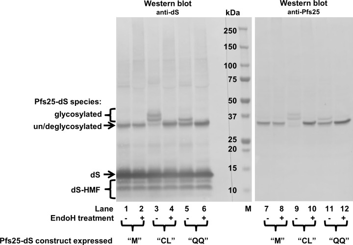
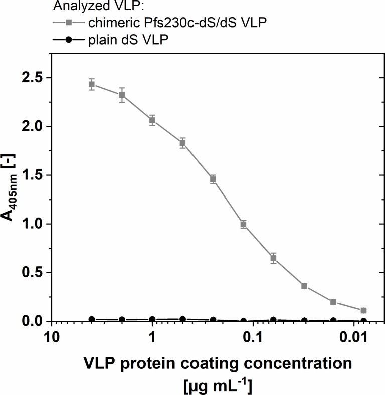
Results: In this study, a novel platform for the display of Plasmodium falciparum antigens on chimeric VLP is presented. Leading transmission-blocking vaccine candidates Pfs25 and Pfs230 were genetically fused to the small surface protein (dS) of the duck hepatitis B virus (DHBV). The resulting fusion proteins were co-expressed in recombinant Hansenula polymorpha (syn. Pichia angusta, Ogataea polymorpha) strains along with the wild-type dS as the VLP scaffold protein. Through this strategy, chimeric VLP containing Pfs25 or the Pfs230-derived fragments Pfs230c or Pfs230D1M were purified. Up to 100 mg chimeric VLP were isolated from 100 g dry cell weight with a maximum protein purity of 90% on the protein level. Expression of the Pfs230D1M construct was more efficient than Pfs230c and enabled VLP with higher purity. VLP showed reactivity with transmission-blocking antibodies and supported the surface display of the malaria antigens on the native VLP.

Conclusion: The incorporation of leading Plasmodium falciparum transmission-blocking antigens into the dS-based VLP scaffold is a promising novel strategy for their display on nano-scaled particles. Competitive processes for efficient production and purification were established in this study.


Conflict of interest statement

The authors VJ, MP, MS, DW and MW are associated with ARTES Biotechnology GmbH which owns the license for the VLP technology: Viral vectors expressing fusion of viral large envelope protein and protein of interest (No. WO2004092387A1). Recombinant proteins and virus-like particles comprising L and S polypeptides of avian hepadnaviridae and methods, nucleic acid constructs, vectors and host cells for producing same (No. WO2008025067A1). Author JM is affiliated with Evonik Technology & Infrastructure GmbH. There are no further patents, products in development or marketed products to declare. This does not alter our adherence to all the PLOS ONE policies on sharing data and materials.

Figures

Fig 1
Fig 1. Analyses of purified Pfs25-dS/dS VLP derived from strain RK#097.
(A): Reducing SDS-PAGE (left, 10 μg protein loaded) and Western blot (right, 1 μg protein loaded) comparing Pfs25-dS/dS VLP to plain dS VLP which were purified from strain A#299 and do not contain any fusion protein. Lanes 1 and 2: Coomassie stained PAA gel. Lanes 3 and 4: Western blot probed with anti-dS 7C12 mAb. Lanes 5 and 6: Western blot probed with anti-Pfs25 mAb 32F81 and analyzed on the same membrane. M: molecular weight marker. (B): TEM imaging. (C): Analysis by ELISA in comparison to plain dS VLP purified from strain A#299. The wells of the ELISA plate were coated with 1 μg mL-1 (50 μL per well) chimeric Pfs25-dS/dS VLP or same amounts of plain dS VLP. Error bars indicate standard deviation based on triplicates. (D): Size distribution determined by DLS.
Fig 2
Fig 2. Western blot analysis of different Pfs25-dS constructs in crude cell lysates before and after treatment with EndoH.
Crude cell lysates of three different recombinant H. polymorpha strains were analyzed by anti-dS (mAb 7C12) and anti-Pfs25 (mAb 32F81) Western blots. The strains co-expressed the wildtype dS and one of the three Pfs25-dS fusion protein constructs: “M” (N-terminal artificial start-methionine), “CL” (N-terminal chicken lysozyme signal sequence) or “QQ” (N-terminal chicken lysozyme signal sequence and N112Q, N187Q aa exchanges). Samples were treated with EndoH where indicated. M: molecular weight marker.
Fig 3
Fig 3. Analyses of purified Pfs230c-dS/dS VLP derived from strain RK#114.
(A): Western blot probed with anti-dS 7C12 mAb (lane 1) or probed with polyclonal anti-Pfs230 antibody (lane 2) and Coomassie stained PAA gel (lanes 3–10). Samples loaded in lanes 5–8 were treated with an EndoH. Lane 9: purified plain dS VLP. Lane 10: EndoH used. M: molecular weight marker. (B): TEM imaging at 100,000-fold magnification. (C): N-SIM analysis of purified VLP containing Pfs230c antigen showing immunolabeling of dS (green) or Pfs230c (red). Three nano-scaled spots that showed co-localization of the fluorescence markers (yellow) were representatively circled. (D): Size distribution determined by DLS.
Fig 4
Fig 4. ELISA data on purified Pfs230c-dS/dS VLP derived from strain RK#114.
The ELISA plate wells were coated with the indicated chimeric Pfs230c-dS/dS VLP or plain dS VLP. The mouse polyclonal antibody was applied at 10 μg mL-1.
Fig 5
Fig 5. Analyses of purified Pfs230D1M-dS/dS VLP derived from strain Ko#119.
(A): Coomassie stained PAA gel (lane 1, 12 μg protein loaded), Western blot probed with anti-dS 7C12 mAb (lane 2) or probed with polyclonal anti-Pfs230 antibody (lane 3). M: molecular weight marker. (B): TEM imaging at 100,000-fold magnification. (C): N-SIM analysis of purified VLP containing Pfs230D1M antigen showing immunolabeling of dS (green) or Pfs230D1M (red). Three nano-scaled spots that showed co-localization of the fluorescence markers (yellow) were representatively circled. (D): Size distribution determined by DLS.

References

    1. WHO. World Malaria Report 2017. World Health Organization; 2017. 10.1071/EC12504 - DOI
    1. Agnandji ST, Lell B, Fernandes JF, Abossolo BP, Methogo BGNO, Kabwende AL, et al. A phase 3 trial of RTS,S/AS01 malaria vaccine in African infants. N Engl J Med. 2012;367: 2284–2295. 10.1056/NEJMoa1208394 - DOI - PMC - PubMed
    1. Malaria Vaccine Funders Group. Malaria Vaccine Technology Roadmap. 2013; 1–9.
    1. Halbroth BR, Draper SJ. Recent Developments in Malaria Vaccinology. Adv Parasitol. 2015;88: 1–49. 10.1016/bs.apar.2015.03.001 - DOI - PubMed
    1. Huang DB, Wu JJ, Tyring SK. A review of licensed viral vaccines, some of their safety concerns, and the advances in the development of investigational viral vaccines. J Infect. 2004;49: 179–209. 10.1016/j.jinf.2004.05.018 - DOI - PMC - PubMed

Publication types

MeSH terms