. 2020 Feb 15;528(3):453-467.
doi: 10.1002/cne.24768. Epub 2019 Oct 2.

Projections to the putamen from neurons located in the white matter and the claustrum in the macaque

Affiliations

Projections to the putamen from neurons located in the white matter and the claustrum in the macaque

Elena Borra et al. J Comp Neurol. .

Abstract

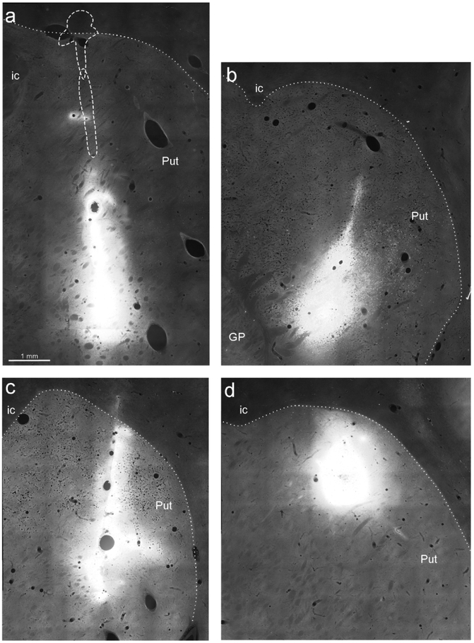
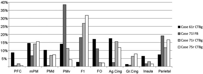
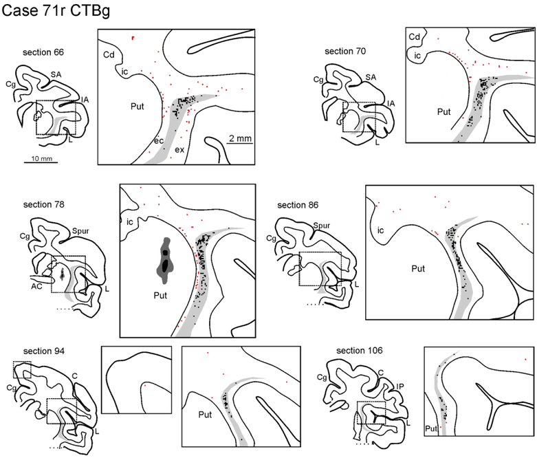
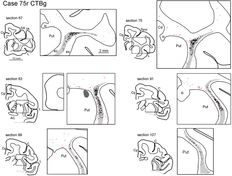
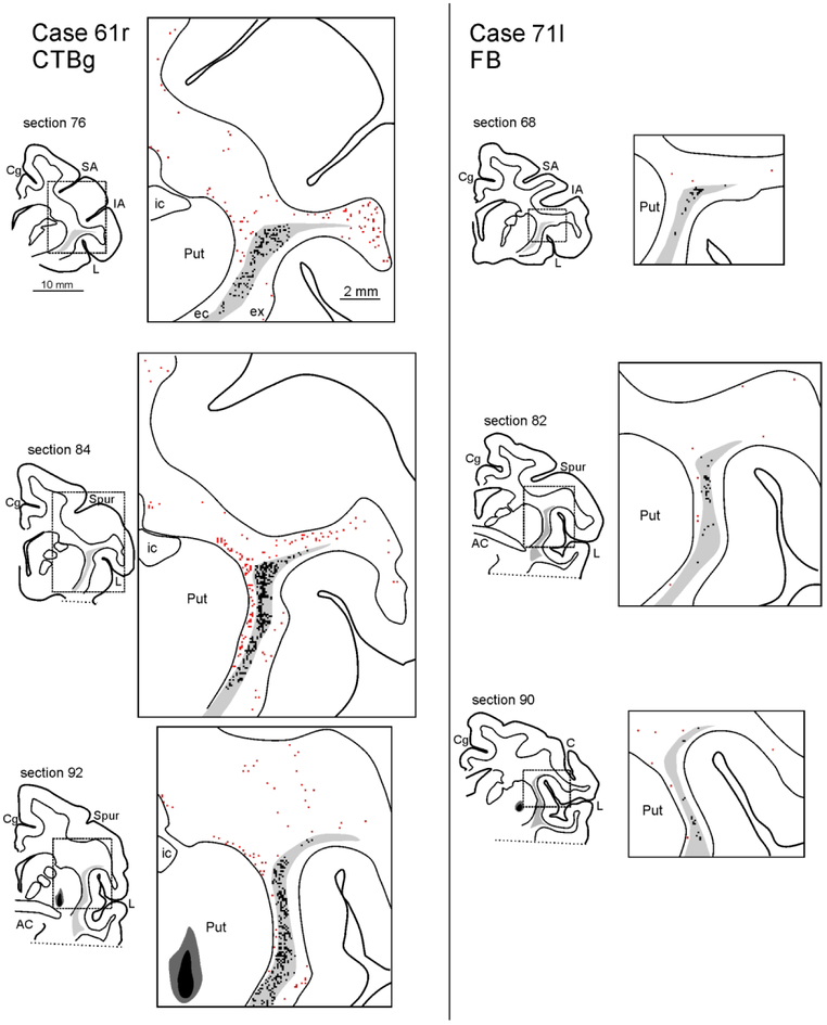
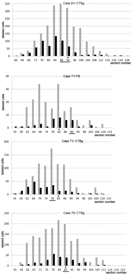
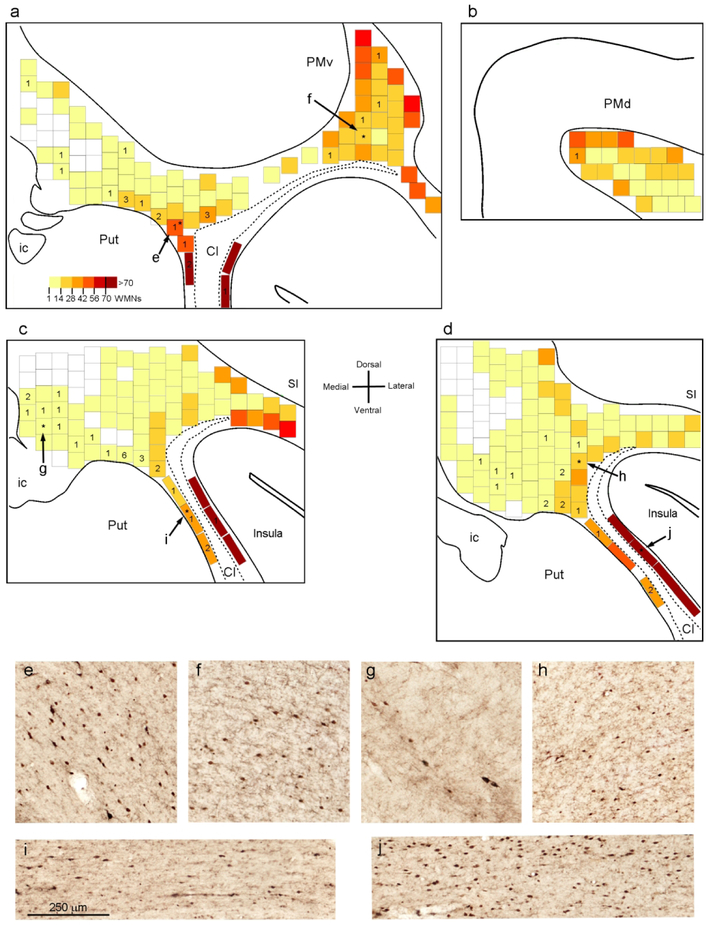
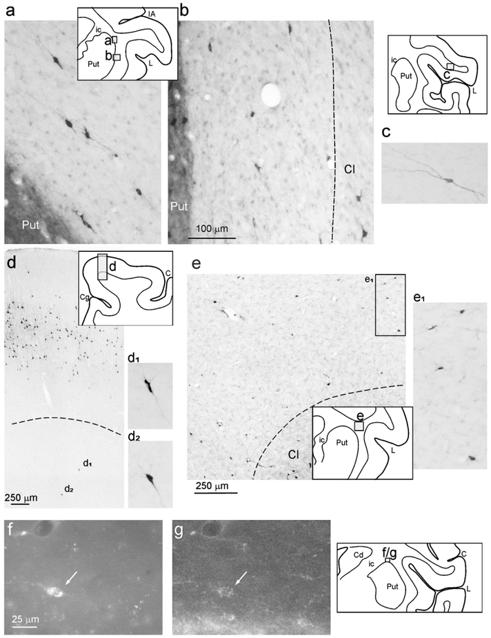
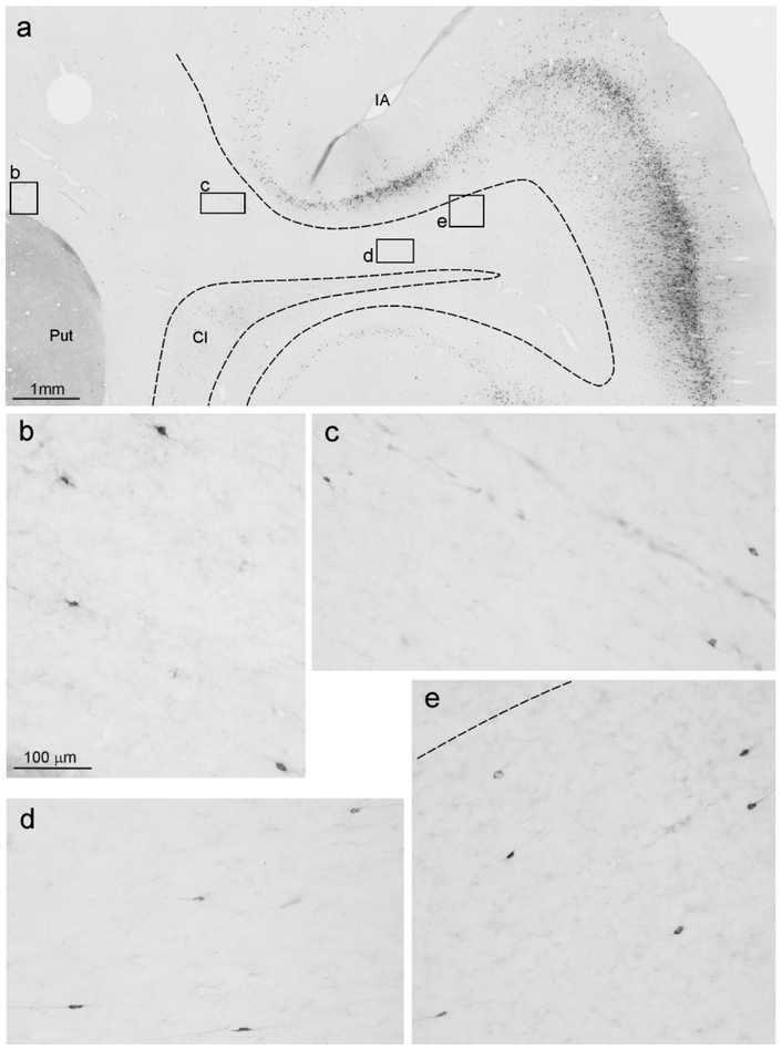
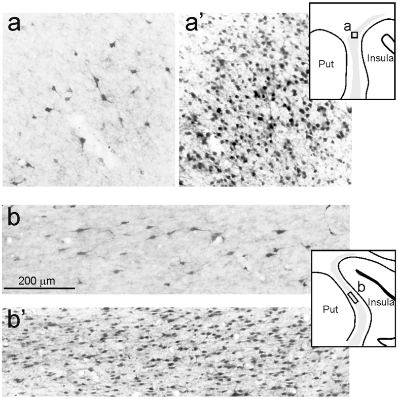
Continuing investigations of corticostriatal connections in rodents emphasize an intricate architecture where striatal projections originate from different combinations of cortical layers, include an inhibitory component, and form terminal arborizations which are cell-type dependent, extensive, or compact. Here, we report that in macaque monkeys, deep and superficial cortical white matter neurons (WMNs), peri-claustral WMNs, and the claustrum proper project to the putamen. WMNs retrogradely labeled by injections in the putamen (four injections in three macaques) were widely distributed, up to 10 mm antero-posterior from the injection site, mainly dorsal to the putamen in the external capsule, and below the premotor cortex. Striatally projecting labeled WMNs (WMNsST) were heterogeneous in size and shape, including a small GABAergic component. We compared the number of WMNsST with labeled claustral and cortical neurons and also estimated their proportion in relation to total WMNs. Since some WMNsST were located adjoining the claustrum, we wanted to compare results for density and distribution of striatally projecting claustral neurons (ClaST). ClaST neurons were morphologically heterogeneous and mainly located in the dorsal and anterior claustrum, in regions known to project to frontal, motor, and cingulate cortical areas. The ratio of ClaST to WMNsST was about 4:1 averaged across the four injections. These results provide new specifics on the connectional networks of WMNs in nonhuman primates, and delineate additional loops in the corticostriatal architecture, consisting of interconnections across cortex, claustralstriatal and striatally projecting WMNs.

Keywords: GABAergic projection neurons; RRID: AB_221544; RRID: AB_2630395; RRID: AB_477019; interstitial neurons; nonhuman primate; striatum.


Figures

Figure 1:
Figure 1:
Fluorescence photomicrographs of the injection sites in the Putamen. (a) CTBg in Case 61r (section 91), the dashed line indicates the border of the injection site observed in two sections located 600 μm and 900 μm rostral, respectively. (b) FB in Case 71l (section 90, rotated for sake of comparison). (c) CTBg in Case 71r (section 77). (d) CTBg in Case 75r (section 86). Dotted lines denote the external border of the putamen. Scale bar in a applies also to b-d. GP= globus pallidus; ic= internal capsule; Put=putamen.
Figure 2:
Figure 2:
Percent distribution of retrograde cortical labeling. Ag.Cing: agranular cingulate cortex; FO: frontal opercular cortex; Gr.Cing= granular cingulate cortex; mPM: mesial premotor cortex; PFC: prefrontal cortex; PMd: dorsal premotor cortex; PMv: ventral premotor cortex.
Figure 3:
Figure 3:
Drawings of coronal sections showing the distribution of the labeled neurons observed in the WM and the claustrum in Case 71r CTBg. The sections are shown in an anterior (section 66) to posterior (section 106) order. For each AP level, the section outline shows from where the enlargement was taken (box). In the enlargements, red dots correspond to retrogradely labeled WMNs (WMNsST) and black dots to labeled neurons within the claustrum (ClaST neurons). The claustrum is indicated with a light gray shading. In section 78 the dark grey and black regions correspond to the injection core and halo, respectively (and see Figure 1c). Because of the limited space, the location of the external (ec) and the extreme (ex) capsule are indicated only in the first section. AC: anterior commissure. C: central sulcus; Cd: caudate nucleus; Cg: cingulate sulcus; IA: inferior arcuate sulcus; IP: intraparietal sulcus; L: lateral fissure; SA: superior arcuate sulcus; Spur: spur of the arcuate sulci. Other abbreviations as in Figure 1.
Figure 4.
Figure 4.
Drawings of coronal sections showing the distribution of WMNsST and ClaST neurons in Case 75r CTBg. In section 83 the dark grey region corresponds to the injection halo (see Figure 1d). Conventions and abbreviations as in Figures 1 and 3.
Figure 5.
Figure 5.
Drawings of coronal sections showing the distribution of WMNsST and ClaST neurons in Cases 61r CTBg (at left) and 71l FB (at right). For each level, a drawing of the section shows from where the enlargement was taken. In section 92 of Case 61r and section 90 of Case 71l the dark grey and black regions correspond to the injection core and halo, respectively, drawn in fluorescence (see Figure 1a and b). Conventions and abbreviations as in Figures 1 and 3.
Figure 6:
Figure 6:
Antero-posterior distribution of labeled cells. A bar graph for each case shows the number of WMNsST (black) and ClaST neurons (grey) in coronal sections spaced 1.2 mm. The section/sections in which the injection site (see Figure 1) and the AC crossing were observed are highligted by a black line and an asterisk, respectively.
Figure 7:
Figure 7:
Distribution of WMNs in three representative sections processed for NeuN (Case 75r). (a-d) Heatmaps showing the density of WMNs in 0.25 mm2 counting boxes in sections 74 (a), 82 (b), 90 (c) and 98 (d). In counting boxes near or slightly overlapping grey matter borders, only neurons that were clearly in the WM were counted. Numbers indicate WMNsST in the adjacent sections that, by overlying the two sections, could be attributed to a specific counting box. WMNsST that could not be attributed to a specific counting box are not shown. Asterisks indicate the boxes showed in e-j. (e) WM just above the putamen and (f) deep WM below the PMv (both from a); (g) WM at the dorsal entrance of the internal capsule (from c); (h) WM near the claustrum (from d); (i) external capsule (from c); (j) extreme capsule (from d). Cl: claustrum; SI: primary somatosensory cortex. Other abbreviations as in Figures 1, 2, and 3.
Figure 8:
Figure 8:
Examples of retrogradely labeled WMNs. For each photo, a drawing of the section is shown for orientation. (a-d) are taken from Case 71r CTBg (sections 71, 94, and 112), (e) is taken from Case 61r CTBg (section 83). Dashed lines indicate the border of the WM with the claustrum (b and e) and with layer 6 (d). Scale bar in (b) applies also to (a), (c), (d1), (d2), and (e1). Note that labeled cells have a different soma area and circularity. (f) and (g) show an example (arrow) from Case 75r CTBg (section 91) of a CTBg WMNsST (f) that is GAD65/67 positive (g). Abbreviations as in Figures 1 and 3.
Figure 9:
Figure 9:
Examples of WMNsST from Case 61r CTBg (section 76). Scale bar in (b) applies also to (c-e). Conventions and abbreviations as in Figures 1, 3, and 8.
Figure 10:
Figure 10:
Examples of ClaST neurons vs total neurons in counting boxes of 0.25 mm2 in the claustrum, from Case 75r. For each level, a drawing of the section is shown for orientation. (a) and (b) are from sections 79 and 91 processed for CTBg; (a’) and (b’) are from adjacent sections processed for NeuN.

Similar articles

Cited by

References

    1. Alloway KD, Smith JB, Mowery TM, Watson GDR. 2017. Sensory Processing in the Dorsolateral Striatum: The Contribution of Thalamostriatal Pathways. Front Syst Neurosci. 11:53. doi: 10.3389/fnsys.2017.00053. - DOI - PMC - PubMed
    1. Arikuni T, Kubota K. 1985. Claustral and amygdaloid afferents to the head of the caudate nucleus in macaque monkeys. Neurosci Res. 2:239–254. - PubMed
    1. Benavides-Piccione R, DeFelipe J. 2003. Different populations of tyrosine-hydroxylase-immunoreactive neurons defined by differential expression of nitric oxide synthase in the human temporal cortex. Cereb Cortex. 13:297–307. - PubMed
    1. Benavides-Piccione R, DeFelipe J. 2007. Distribution of neurons expressing tyrosine hydroxylase in the human cerebral cortex. J Anat. 211:212–22. - PMC - PubMed
    1. Case L, Broberger C. 2018. Neurotensin broadly recruits inhibition via white matter neurons in the mouse cerebral cortex: synaptic mechanisms for decorrelation. Cereb Cortex. 28:2711–2724. doi: 10.1093/cercor/bhx149. - DOI - PubMed

Publication types