. 2019 Nov;112(5):1453-1470.
doi: 10.1111/mmi.14371. Epub 2019 Sep 13.

DdlR, an essential transcriptional regulator of peptidoglycan biosynthesis in Clostridioides difficile

Affiliations

DdlR, an essential transcriptional regulator of peptidoglycan biosynthesis in Clostridioides difficile

Laurent Bouillaut et al. Mol Microbiol. 2019 Nov.

Abstract

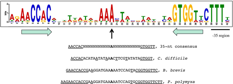
D-Ala-D-Ala ligase, encoded by ddl genes, is responsible for the synthesis of a dipeptide, D-Ala-D-Ala, an essential precursor of bacterial peptidoglycan. In Clostridioides difficile, the single ddl gene is located upstream of the ddlR gene, which encodes a putative transcriptional regulator. Using mutational and transcriptional analysis and DNA-binding assays, DdlR was found to be a direct activator of the ddl ddlR operon. DdlR is a member of the MocR/GabR-type proteins that have aminotransferase-like, pyridoxal 5'-phosphate-binding domains. A DdlR mutation that prevented covalent binding of pyridoxal 5'-phosphate abolished the ability of DdlR to activate transcription. Addition of D-Ala-D-Ala to the medium inactivated DdlR, reducing dipeptide biosynthesis. In contrast, D-Ala-D-Ala limitation caused a dramatic increase in expression from the ddl promoter. Though uncommon for transcription regulators, C. difficile DdlR is essential, as the ddlR null mutant cells could not grow even in complex laboratory media in the absence of D-Ala-D-Ala. A dyad symmetry sequence, which is located immediately upstream of the -35 region of the ddl promoter, serves as an important element of the DdlR-binding site. This sequence is conserved upstream of putative DdlR targets in other bacteria of classes Clostridia and Bacilli, indicating a similar mode of regulation of these genes.


Figures

Fig. 1.
Fig. 1.
Organization of the ddl ddlR operon and the sequence of the ddl and ddlR regulatory regions. The likely initiation codons, the ddl termination codon, the 10 and 35 promoter regions, and the +1 gene positions are in boldface. The directions of transcription and translation are indicated by the arrows. The sequence protected by DdlR on the template strand in DNase I footprinting experiments is underlined. The 33-bp dyad-symmetry sequence is in boldface and italicized. The coordinates of the 5’ and 3’ ends of the sequence with respect to the +1 position of the ddl gene and the locations of the p1 and p2 mutations are indicated.
Fig. 2.
Fig. 2.
Effect of D-Ala-D-Ala on growth of the C. difficile parent strain and ddlR and ddl mutants. Cells were grown overnight in TY complex medium with or without D-Ala-D-Ala (DADA) (20 µg/ml) and diluted 100-fold in the same medium without D-Ala-D-Ala or with indicated concentrations of D-Ala-D-Ala. 630 - strain 630∆erm; ddlR - strain LB-CD28, ddl - strain BCD27.
Fig. 3.
Fig. 3.
Binding of DdlR to the ddl regulatory region as detected by a gel-shift assay. A radioactively labeled ddlp+ DNA fragment was incubated with increasing amounts of purified DdlR without or with 2 mM D-Ala-D-Ala and 100 µM PLP. DdlR monomer concentrations used (nM) are indicated below each lane, and the concentrations needed to shift ~50% of DNA fragments, are underlined. The arrows indicate the bands corresponding to unbound ddl DNA and the complex of DdlR with ddl DNA. Each gel-shift assay was repeated at least two times.
Fig. 4.
Fig. 4.
Binding of DdlR or DdlR1 to various regulatory regions as detected by a gel-shift assay. Radioactively labeled CD630_23440 (A), ddlR (B), ddlp1 (C), ddlp2 (D), and ddlp+ (E, F) DNA fragments were incubated with increasing amounts of purified DdlR (A-D) or DdlR1 (E, F). 100 µM PLP was added for the experiment shown in panel F. DdlR or DdlR1 monomer concentrations used (nM) are indicated below each lane, and the concentrations, which are needed to shift ~50% of DNA fragments, are underlined.
Fig. 5.
Fig. 5.
DNase I footprinting analysis of DdlR binding to the ddl regulatory region. ddlp+ (A) or ddlp2 (B) DNA fragments, radioactively labeled on the template strand, were incubated with increasing concentrations of purified DdlR in the absence or presence of 100 µM PLP or 100 µM PLP and 2 mM D-Ala-D-Ala. DdlR monomer concentrations used (nM) are indicated below each lane. The corresponding A + G sequencing ladders is shown in the left lane. The protected area is shown by a vertical line. The positions of hypersensitive bands are indicated by arrows.
Fig. 6.
Fig. 6.
UV-visible spectra of DdlR, DdlR1, and PLP. 40 µM DdlR (A) or DdlR1 (B) were in 50 mM HEPES - 500 mM NaCl - 92.5 mM imidazole (pH 7.5) buffer. The inset in panel A compares the spectra of DdlR and of free 40 µM PLP in 50 mM HEPES (pH 7.5) buffer. The intensity of the absorption peak at A430 manifested by wild-type DdlR indicates that more than half of DdlR molecules are bound to PLP.
Fig. 7.
Fig. 7.
Motif logo for the conserved sequence in the putative DdlR-binding sites of different bacteria. The logo was generated by the MEME function of the MEME suite (Bailey and Elkan, 1994). The dyad symmetry region is indicated by horizontal arrows and the conserved central nucleotide by a vertical arrow. The partial, putative −35 region is underlined. The nucleotide sequences of the regulatory regions of genes from 11 Clostridia and Bacilli strains were analyzed: ddl from C. difficile, Clostridium tetani, Clostridium butyricum, Clostridium acetobutylicum, Clostridium pasteurianum, and Clostridium ljungdahlii, ddlR from Bacillus anthracis, Lysinibacillus sphaericus, and Paenibacillus polymyxa, and alr from Clostridium botulinum and C. butyricum. The simplified consensus sequence and the corresponding actual sequences from three genomes are shown below the logo. The elements of dyad symmetry are underlined.

Similar articles

Cited by

References

    1. Al-Zyoud WA, Hynson RM, Ganuelas LA, Coster AC, Duff AP, Baker MA et al. (2016) Binding of transcription factor GabR to DNA requires recognition of DNA shape at a location distinct from its cognate binding site. Nucl. Acids Res, 44, 1411–1420. - PMC - PubMed
    1. Bailey TL and Elkan C (1994) Fitting a mixture model by expectation maximization to discover motifs in biopolymers. Proc. Int. Conf. Intell. Syst. Mol. Biol, 2, 28–36. - PubMed
    1. Barbe V, Cruveiller S, Kunst F, Lenoble P, Meurice G, Sekowska A et al. (2009) From a consortium sequence to a unified sequence: the Bacillus subtilis 168 reference genome a decade later. Microbiology, 155, 1758–1775. - PMC - PubMed
    1. Barreteau H, Kovac A, Boniface A, Sova M, Gobec S and Blanot D (2008) Cytoplasmic steps of peptidoglycan biosynthesis. FEMS Microbiol. Rev, 32, 168–207. - PubMed
    1. Belitsky BR (2004) Bacillus subtilis GabR, a protein with DNA-binding and aminotransferase domains, is a PLP-dependent transcriptional regulator. J. Mol. Biol, 340, 655–664. - PubMed

Publication types

MeSH terms