. 2019 Sep 3;11(9):2062.
doi: 10.3390/nu11092062.

Indole-3-Acetic Acid Alleviates Nonalcoholic Fatty Liver Disease in Mice via Attenuation of Hepatic Lipogenesis, and Oxidative and Inflammatory Stress

Affiliations

Indole-3-Acetic Acid Alleviates Nonalcoholic Fatty Liver Disease in Mice via Attenuation of Hepatic Lipogenesis, and Oxidative and Inflammatory Stress

Yun Ji et al. Nutrients. .

Abstract

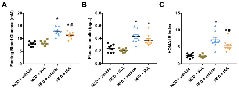
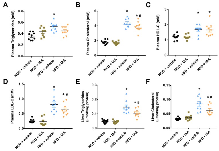
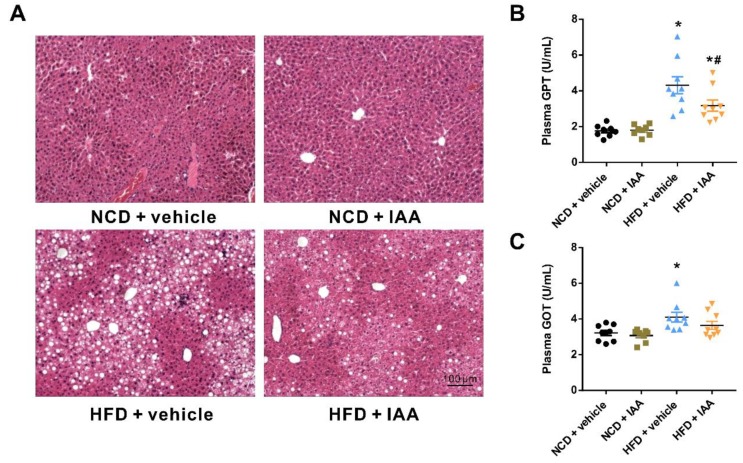
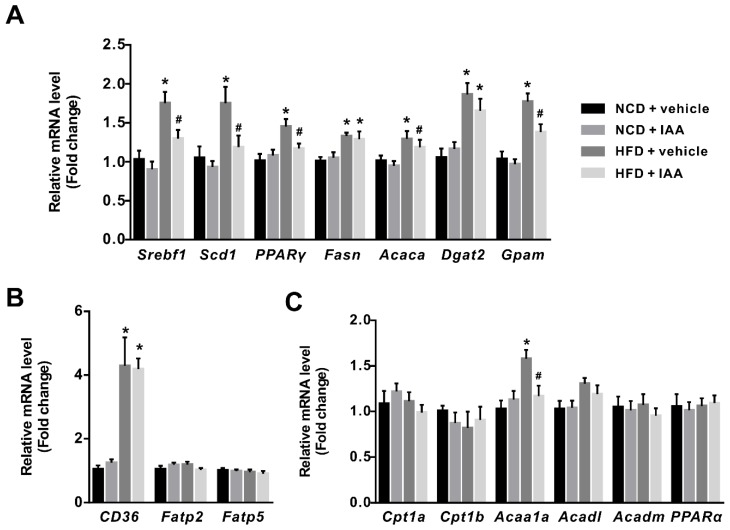
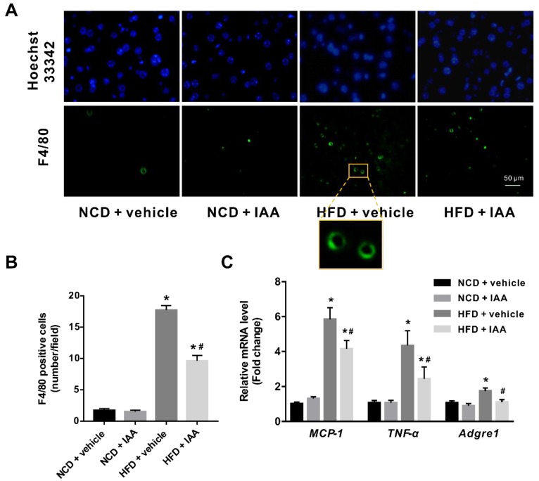
Recent evidences have linked indole-3-acetic acid (IAA), a gut microbiota-derived metabolite from dietary tryptophan, with the resistance to liver diseases. However, data supporting IAA-mediated protection against nonalcoholic fatty liver disease (NAFLD) from an in vivo study is lacking. In this study, we assessed the role of IAA in attenuating high-fat diet (HFD)-induced NAFLD in male C57BL/6 mice. Administration of IAA (50 mg/kg body weight) by intraperitoneal injection was found to alleviate HFD-induced elevation in fasting blood glucose and homeostasis model assessment of insulin resistance (HOMA-IR) index as well as plasma total cholesterol, low-density lipoprotein cholesterol (LDL-C), and glutamic-pyruvic transaminase (GPT) activity. Histological examination further presented the protective effect of IAA on liver damage induced by HFD feeding. HFD-induced an increase in liver total triglycerides and cholesterol, together with the upregulation of genes related to lipogenesis including sterol regulatory element binding-protein 1 (Srebf1), steraroyl coenzyme decarboxylase 1 (Scd1), peroxisome proliferator-activated receptor gamma (PPARγ), acetyl-CoA carboxylase 1 (Acaca), and glycerol-3-phosphate acyltransferase, mitochondrial (Gpam), which were mitigated by IAA treatment. The results of reactive oxygen species (ROS) and malonaldehyde (MDA) level along with superoxide dismutase (SOD) activity and glutathione (GSH) content in liver tissue evidenced the protection of IAA against HFD-induced oxidative stress. Additionally, IAA attenuated the inflammatory response of liver in mice exposed to HFD as shown by the reduction in the F4/80-positive macrophage infiltration and the expression of monocyte chemoattractant protein-1 (MCP-1) and tumor necrosis factor-α (TNF-α). In conclusion, our findings uncover that IAA alleviates HFD-induced hepatotoxicity in mice, which proves to be associated with the amelioration in insulin resistance, lipid metabolism, and oxidative and inflammatory stress.

Keywords: NAFLD; indole-3-acetic acid; inflammation; lipid metabolism; oxidative stress; steatosis.


Conflict of interest statement

The authors declare no conflicts of interest.

Figures

Figure 1
Figure 1
Indole-3-acetic acid (IAA) protected against high-fat-diet (HFD)-induced insulin resistance in mice. (A) Fasting blood glucose levels determined by a glucometer in mice fasted overnight. (B) Results from ELISA for plasma insulin. (C) HOMA-IR calculated as described in the Materials and Methods section. Data are expressed as the mean ± standard error of the mean. n = 8–9. * p < 0.05 vs. NCD + vehicle; # p < 0.05 vs. HFD + vehicle.
Figure 2
Figure 2
Lipid contents in the plasma and liver of vehicle or indole-3-acetic acid (IAA)-treated mice subjected to normal chow diet (NCD) or high-fat diet (HFD). (AD) display plasma total triglycerides, total cholesterol, HDL-C, and LDL-C, respectively. (E,F) show the levels of liver triglyceride and cholesterol. Results are described as the mean ± standard error of the mean. n = 8–9. * p < 0.05 vs. NCD + vehicle; # p < 0.05 vs. HFD + vehicle.
Figure 3
Figure 3
Protective effects of indole-3-acetic acid (IAA) on high-fat diet (HFD)-induced liver injury. (A) Representative images of hematoxylin & eosin staining results for liver tissue sections. Scale bar represents 100 µm. (B) Plasma glutamic-pyruvic transaminase (GPT) and (C) glutamic oxalacetic transaminase (GOT) levels. Data are presented as the mean ± standard error of the mean. n = 8–9. * p < 0.05 vs. NCD + vehicle; # p < 0.05 vs. HFD + vehicle.
Figure 4
Figure 4
Effect of indole-3-acetic acid (IAA) on genes involved in lipid metabolism in liver of mice fed with normal chow diet (NCD) or high-fat diet (HFD). The relative expression levels of genes related to (A) lipogenesis, (B) fatty acid uptake, and (C) β-oxidation of fatty acids were quantified by RT-qPCR. The bar chart presents mean ± standard error of the mean of the fold change values acquired from at least 6 biologically replicates for each gene. * p < 0.05 vs. NCD + vehicle; # p < 0.05 vs. HFD + vehicle.
Figure 5
Figure 5
Changes in oxidative stress markers in mice exposed to high-fat diet (HFD) and indole-3-acetic acid (IAA) treatment. (A) plasma total antioxidant capacity (T-AOC), (B) and (C) liver reactive oxygen species (ROS) and malondialdehyde (MDA) level, F, fluorescence intensity. (D,E) liver superoxide dismutase (SOD) and catalase (CAT) activity. (F,G) the content of glutathione (GSH) and oxidized glutathione (GSSG), and (H) the ratio of GSH/GSSG. Values are shown as the mean ± standard error of the mean. * p < 0.05 vs. NCD + vehicle; # p < 0.05 vs. HFD + vehicle.
Figure 6
Figure 6
Inflammatory stress in liver of mice induced by high-fat diet (HFD) was ameliorated by indole-3-acetic acid (IAA) treatment. (A) Immunofluorescence analysis for F4/80 antigen in liver tissue section. Scale bar represents 50 µm. (B) Count of F4/80 positive cells per field (at least six field per mice) (C) Relative mRNA abundance of MCP-1, TNF-α, and Adgre1. Values are presented as the mean ± standard error of the mean. n = 6. * p < 0.05 vs. NCD + vehicle; # p < 0.05 vs. HFD + vehicle.

References

    1. Norheim F., Hui S.T., Kulahcioglu E., Mehrabian M., Cantor R.M., Pan C., Parks B.W., Lusis A.J. Genetic and hormonal control of hepatic steatosis in female and male mice. J. Lipid Res. 2017;58:178–187. doi: 10.1194/jlr.M071522. - DOI - PMC - PubMed
    1. Abd El-Kader S.M., El-Den Ashmawy E.M. Non-alcoholic fatty liver disease: The diagnosis and management. World J. Hepatol. 2015;7:846–858. doi: 10.4254/wjh.v7.i6.846. - DOI - PMC - PubMed
    1. Younossi Z., Anstee Q.M., Marietti M., Hardy T., Henry L., Eslam M., George J., Bugianesi E. Global burden of NAFLD and NASH: Trends, predictions, risk factors and prevention. Nat. Rev. Gastroenterol. Hepatol. 2018;15:11–20. doi: 10.1038/nrgastro.2017.109. - DOI - PubMed
    1. Rau M., Schilling A.K., Meertens J., Hering I., Weiss J., Jurowich C., Kudlich T., Hermanns H.M., Bantel H., Beyersdorf N., et al. Progression from Nonalcoholic Fatty Liver to Nonalcoholic Steatohepatitis Is Marked by a Higher Frequency of Th17 Cells in the Liver and an Increased Th17/Resting Regulatory T Cell Ratio in Peripheral Blood and in the Liver. J. Immunol. 2016;196:97–105. doi: 10.4049/jimmunol.1501175. - DOI - PubMed
    1. Takahashi Y., Fukusato T. Histopathology of nonalcoholic fatty liver disease/nonalcoholic steatohepatitis. World J. Gastroenterol. 2014;20:15539–15548. doi: 10.3748/wjg.v20.i42.15539. - DOI - PMC - PubMed