. 2019 Oct 25;294(43):15733-15742.
doi: 10.1074/jbc.RA119.010123. Epub 2019 Sep 4.

REV7 has a dynamic adaptor region to accommodate small GTPase RAN/ Shigella IpaB ligands, and its activity is regulated by the RanGTP/GDP switch

Affiliations

REV7 has a dynamic adaptor region to accommodate small GTPase RAN/ Shigella IpaB ligands, and its activity is regulated by the RanGTP/GDP switch

Xin Wang et al. J Biol Chem. .

Abstract

REV7, also termed mitotic arrest-deficient 2-like 2 (MAD2L2 or MAD2B), acts as an interaction module in a broad array of cellular pathways, including translesion DNA synthesis, cell cycle control, and nonhomologous end joining. Numerous REV7 binding partners have been identified, including the human small GTPase Ras-associated nuclear protein (RAN), which acts as a potential upstream regulator of REV7. Notably, the Shigella invasin IpaB hijacks REV7 to disrupt cell cycle control to prevent intestinal epithelial cell renewal and facilitate bacterial colonization. However, the structural details of the REV7-RAN and REV7-IpaB interactions are mostly unknown. Here, using fusion protein and rigid maltose-binding protein tagging strategies, we determined the crystal structures of these two complexes at 2.00-2.35 Å resolutions. The structures revealed that both RAN and IpaB fragments bind the "safety belt" region of REV7, inducing rearrangement of the C-terminal β-sheet region of REV7, conserved among REV7-related complexes. Of note, the REV7-binding motifs of RAN and IpaB each displayed some unique interactions with REV7 despite sharing consensus residues. Structural alignments revealed that REV7 has an adaptor region within the safety belt region that can rearrange secondary structures to fit a variety of different ligands. Our structural and biochemical results further indicated that REV7 preferentially binds GTP-bound RAN, implying that a GTP/GDP-bound transition of RAN may serve as the molecular switch that controls REV7's activity. These results provide insights into the regulatory mechanism of REV7 in cell cycle control, which may help with the development of small-molecule inhibitors that target REV7 activity.

Keywords: IpaB; RAN; REV7; Shigella invasin; cell cycle; crystal structure; host–pathogen interaction; mitotic arrest–deficient 2-like 2 (MAD2L2); protein complex; small GTPase; structural biology.


Conflict of interest statement

The authors declare that they have no conflicts of interest with the contents of this article

Figures

Figure 1.
Figure 1.
Design and overall structure of the REV7–RAN complex. A, sequence alignment of REV7-binding motifs. Conserved residues are colored white on a red background; similar residues are shown in red boxes. B, GST pulldown of MBP-tagged REV7 by GST-RAN (174–185). Top, Coomassie Blue stain of the pulldown results (arrows). Bottom, Coomassie Blue stain of input MBP-REV7 and MBP alone (arrows). C, schematic of the REV7.RAN3 fusion protein. The HORMA (Hop1p, Rev7, and MAD2 proteins) domain is colored light blue. RANRBM (167–185) is colored magenta, and REV3α-helix is colored gray. D, overall structure of the REV7–RANRBM complex. REV7 is colored light blue and green (C-terminal safety belt), and the RANRBM fragment is shown in magenta. The termini (yellow circles) and secondary structure elements (white boxes) are labeled. The disordered region is indicated by dots. E, 2Fo-Fc electron density map of the RANRBM fragment contoured at 1.2 σ level.
Figure 2.
Figure 2.
Interaction details of the REV7–RAN complex. A, structural details of the interactions between REV7 (light blue and green) and RANRBM (magenta). The key residues are shown as sticks and labeled. Water molecules are shown as red spheres. Electrostatic interactions are shown as yellow dots. B and C, Phe-176 and Pro-180 (magenta sticks) of RANRBM bind into the hydrophobic pockets of REV7 (electrostatic representation). D, GST pulldown of MBP-tagged REV7 variants by GST-RANRBM. Top, Coomassie Blue stain of 30% input MBP-REV7 variants (arrow). Bottom, Coomassie Blue stain of the pulldown results (arrows). E, MBP pulldown of His-tagged, full-length RAN variants by MBP-REV7. Top, Coomassie Blue stain of input His-RAN variants (arrow). Bottom, Coomassie Blue stain of the pulldown results (arrows).
Figure 3.
Figure 3.
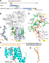
Crystal structure of the MBP-tagged REV7–IpaB complex. A, schematic of the MBP-REV7.IpaB3 fusion protein. The HORMA domain is colored light blue. IpaBRBM is colored orange, and REV3α-helix is colored gray. B, overall structure of the MBP-tagged REV7–IpaB complex. REV7 is colored light blue and green. The C-terminal safety belt and the IpaBRBM fragment are shown in orange, and the MBP tag is shown in white. The termini (yellow circles) and secondary structure elements (white boxes) are labeled. C, 2Fo-Fc electron density map of IpaBRBM fragment contoured at 1.2 σ level. D, structural details of the interactions between REV7 (light blue and green) and IpaBRBM (orange). The key residues are shown as sticks and labeled. E, the chaperone-binding domain (CBD) and RBM of IpaB have an identical sequence. F, crystal structure of the Shigella IpaB–IpgC complex (PDB code 3GZ1). IpgC is colored cyan, and IpaBCBD is shown as wheat-colored sticks. G, the CBD and RBM of Shigella IpaB present different conformations despite the identical sequence.
Figure 4.
Figure 4.
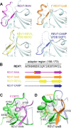
Structural definition of the REV7 adaptor region to fit the variance of RBMs. A, structures of the adaptor region (156–173) of REV7 in the REV7–RAN, REV7–IpaB, REV7–REV3RBM2 (PDB code 6BC8), and REV7-CAMP (PDB code 5XPT) complexes. The adaptor regions are highlighted by the indicated colors. B, schematic of the secondary structure elements of the REV7 adaptor region upon binding various partners. Disordered regions are indicated by dashed lines. C, hydrophobic interactions between Phe-176 of RANRBM (magenta) and the residues lining the adaptor region of REV7 (green). D, hydrophobic interactions between Leu-63 of Shigella IpaB (orange) and the residues lining the adaptor region of REV7 (green).
Figure 5.
Figure 5.
RanGTP shows higher affinity for REV7 than RanGDP. A, MBP pulldown of His-tagged, full-length RAN variants by MBP-tagged REV7. Mutant RAN (T24N) is a mimetic of RanGDP, and mutant RAN (L43E) is a mimetic of RanGTP. Top, Western blot (WB) of input His-RAN variants using anti-His antibodies. Bottom, Western blot of the pulldown results using anti-His and anti-MBP antibodies. B, HEK293 cells were double-transfected with yellow fluorescent protein–REV7 and HA-tagged RAN WT or HA-tagged RanGDP (T24N), and in vivo co-IP against REV7 was performed. C, HEK293 cells were transfected with myc-CDH1 or double-transfected with myc-CDH1 and HA-tagged RAN WT. In vivo co-IP against Cdc27 was performed. D, SPR analyses of REV7–RAN interactions. Purified MBP-REV7 was immobilized on a CM5 chip. The affinities were evaluated over a concentration range from 0.625 μm to 20 μm of His-RAN variants in 2-fold increments. The concentration of 0.625 μm was repeated as an internal control. The calculated Kd values are indicated. RU, resonance units.
Figure 6.
Figure 6.
RanGTP exposes its C-terminal moiety to bind REV7. A, structure comparison of RanGDP and RanGTP. The cartoon models of RanGDP (PDB code 3GJ0) and RanGTP (PDB code 1K5D) are colored wheat and orange (the C-terminal moiety), and their REV7-binding motifs are colored magenta and labeled. B, model of the conformation changes in REV7–RAN binding. The conformation change from RanGDP to RanGTP exposes its C-terminal moiety. Free REV7 adopts an open-form conformation. Upon binding RanGTP, the C-terminal moiety of REV7 also undergoes structural rearrangement to form safety belt architecture in the REV7–RanGTP complex.

References

    1. Lawrence C. W., Das G., and Christensen R. B. (1985) REV7, a new gene concerned with UV mutagenesis in yeast. Mol. Gen. Genet. 200, 80–85 10.1007/BF00383316 - DOI - PubMed
    1. Murakumo Y., Roth T., Ishii H., Rasio D., Numata S., Croce C. M., and Fishel R. (2000) A human REV7 homolog that interacts with the polymerase ζ catalytic subunit hREV3 and the spindle assembly checkpoint protein hMAD2. J. Biol. Chem. 275, 4391–4397 10.1074/jbc.275.6.4391 - DOI - PubMed
    1. Murakumo Y., Ogura Y., Ishii H., Numata S., Ichihara M., Croce C. M., Fishel R., and Takahashi M. (2001) Interactions in the error-prone postreplication repair proteins hREV1, hREV3, and hREV7. J. Biol. Chem. 276, 35644–35651 10.1074/jbc.M102051200 - DOI - PubMed
    1. Hara K., Hashimoto H., Murakumo Y., Kobayashi S., Kogame T., Unzai S., Akashi S., Takeda S., Shimizu T., and Sato M. (2010) Crystal structure of human REV7 in complex with a human REV3 fragment and structural implication of the interaction between DNA polymerase ζ and REV1. J. Biol. Chem. 285, 12299–12307 10.1074/jbc.M109.092403 - DOI - PMC - PubMed
    1. Bomar M. G., D'Souza S., Bienko M., Dikic I., Walker G. C., and Zhou P. (2010) Unconventional ubiquitin recognition by the ubiquitin-binding motif within the Y family DNA polymerases ι and Rev1. Mol. Cell 37, 408–417 10.1016/j.molcel.2009.12.038 - DOI - PMC - PubMed

Publication types

LinkOut - more resources