. 2019 Sep 4;4(5):e00439-19.
doi: 10.1128/mSphere.00439-19.

Potential Role of Extracellular ATP Released by Bacteria in Bladder Infection and Contractility

Affiliations

Potential Role of Extracellular ATP Released by Bacteria in Bladder Infection and Contractility

Behnam Abbasian et al. mSphere. .

Abstract

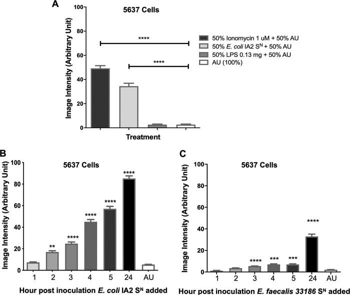
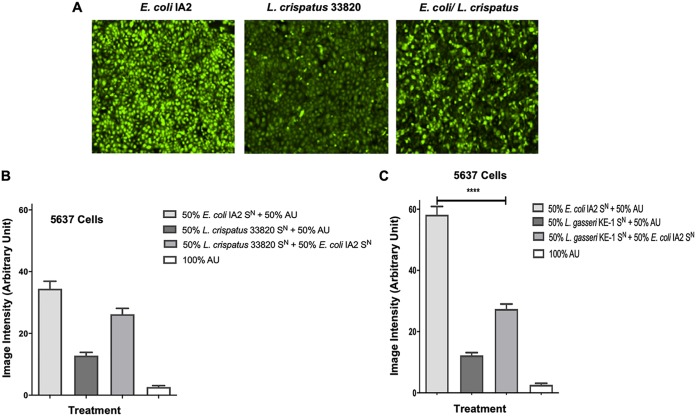
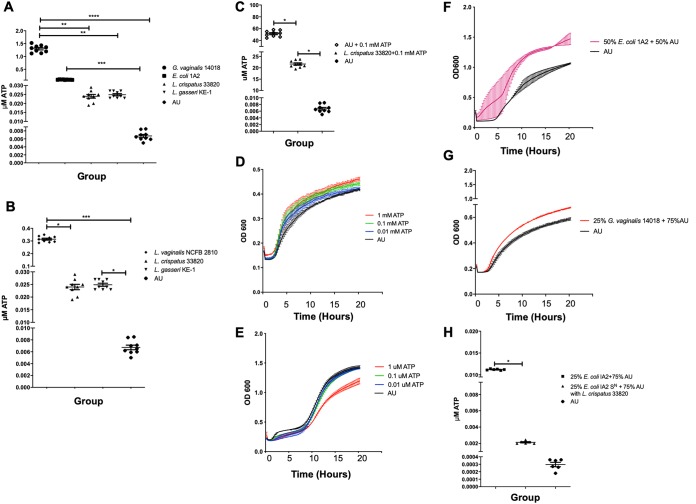
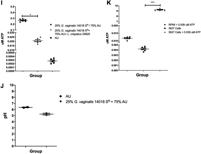
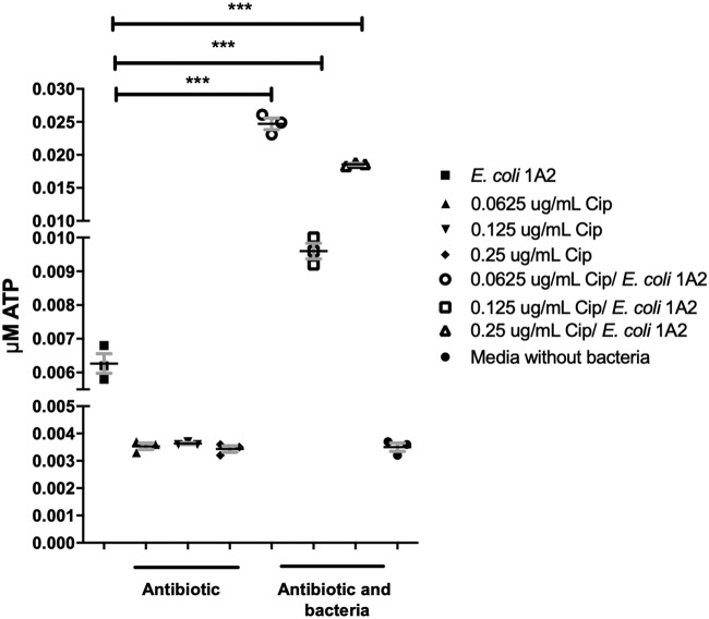
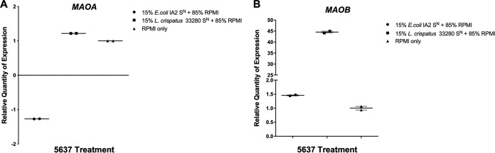
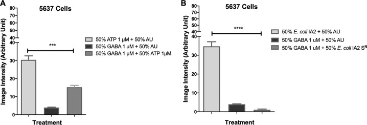
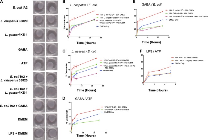
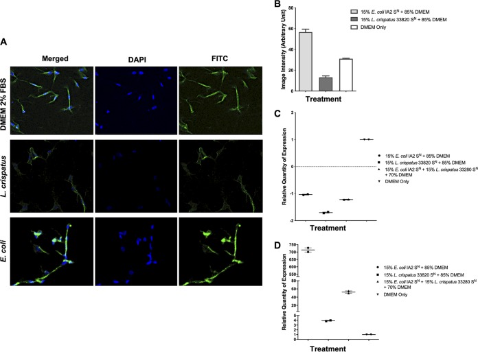
Urgency urinary incontinence (UUI) and overactive bladder (OAB) can both potentially be influenced by commensal and urinary tract infection-associated bacteria. The sensing of bladder filling involves interplay between various components of the nervous system, eventually resulting in contraction of the detrusor muscle during micturition. This study models host responses to various urogenital bacteria, first by using urothelial bladder cell lines and then with myofibroblast contraction assays. To measure responses, we examined Ca2+ influx, gene expression, and alpha smooth muscle actin deposition assays. Organisms such as Escherichia coli and Gardnerella vaginalis were found to strongly induce Ca2+ influx and contraction, whereas Lactobacillus crispatus and L. gasseri did not induce this response. Additionally, supernatants from lactobacilli impeded Ca2+ influx and contraction induced by uropathogens. Upon further investigation of factors associated with purinergic signaling pathways, the Ca2+ influx and contraction of cells correlated with the amount of extracellular ATP produced by E. coli Certain lactobacilli appear to mitigate this response by utilizing extracellular ATP or producing inhibitory compounds that may act as a receptor agonist or Ca2+ channel blocker. These findings suggest that members of the urinary microbiota may be influencing UUI or OAB.IMPORTANCE The ability of uropathogenic bacteria to release excitatory compounds, such as ATP, may act as a virulence factor to stimulate signaling pathways that could have profound effects on the urothelium, perhaps extending to the vagina. This may be countered by the ability of certain commensal urinary microbiota constituents, such as lactobacilli. Further understanding of these interactions is important for the treatment and prevention of UUI and OAB. The clinical implications may require a more targeted approach to enhance the commensal bacteria and reduce ATP release by pathogens.

Keywords: ATP; Escherichia coli; Gardnerella; Lactobacillus; extracellular.


Figures

FIG 1
FIG 1
Bacterial supernatant induces Ca2+ influx in 5637 uroepithelial cells. Bacterial supernatant (SN) of E. coli IA2 after overnight culture was added to 5637 cells at a 50:50 ratio with artificial urine (AU) to assess its ability to induce influx of Ca2+ into the cell (A). Supernatants from either E. coli IA2 (B) or E. faecalis 33186 (C) were taken from cultures at 1, 2, 3, 4, 5, and 24 h postinoculum and tested for their ability to induce Ca2+ influx in the 5637 cells relative to the AU control. Each bar represents the total average image intensity over 60 s following treatment of a sample. Statistical significance was determined using Dunn’s multiple-comparison test. **, P < 0.01; ***, P < 0.001; ****, P < 0.0001.
FIG 2
FIG 2
Effect of Lactobacillus supernatant on Ca2+ influx in 5637 urothelial cells induced by uropathogenic supernatant. (A) Fluorescent microscopy images of Ca2+ influx caused by supernatants (SN) from E. coli IA2, L. crispatus 33820, and a mixture of supernatants from the two bacteria. Bacterial supernatant from either E. coli IA2, L. crispatus 33820 (B) or L. gasseri KE-1 (C) overnight cultures was mixed 50:50 with either AU or bacterial supernatants to measure Ca2+ influx in 5637 urothelial cells (B and C). Each bar represents the total average image intensity over 60 s following treatment of a sample. Statistical significance was determined using Dunn’s multiple-comparison test. ****, P < 0.0001.
FIG 3
FIG 3
Release and utilization of extracellular and supplemented ATP by bacteria. E. coli, L. crispatus, L. gasseri, G. vaginalis, and L. vaginalis supernatants (SN) were collected from overnight cultures grown in AU and measured for ATP (A and B). Statistical significance was determined using Tukey’s test (P ≤ 0.05). L. crispatus was grown in AU supplemented with 0.1 mM ATP overnight, and the amount of ATP was evaluated by luminometer (C). Growth of L. crispatus and E. coli was measured in the presence of different concentrations of ATP in AU (D and E) and additionally for L. crispatus supplemented with E. coli or G. vaginalis supernatants (F and G). The ability of L. crispatus to reduce the amount of ATP in AU supplemented with 25% E. coli supernatant (H) and 25% G. vaginalis supernatant (I) individually was also examined. L. crispatus 33820 was grown in media supplemented with G. vaginalis 14018 to assess the change in pH (J). 5637 urothelial cells were incubated in RPMI, supplemented with small quantities of ATP, and incubated for 2 min to assess the amount of ATP released (K). Statistical significance was determined using Dunn’s multiple-comparison test. *, P < 0.05; **, P < 0.01; ***, P < 0.001; ****, P < 0.0001.
FIG 3
FIG 3
Release and utilization of extracellular and supplemented ATP by bacteria. E. coli, L. crispatus, L. gasseri, G. vaginalis, and L. vaginalis supernatants (SN) were collected from overnight cultures grown in AU and measured for ATP (A and B). Statistical significance was determined using Tukey’s test (P ≤ 0.05). L. crispatus was grown in AU supplemented with 0.1 mM ATP overnight, and the amount of ATP was evaluated by luminometer (C). Growth of L. crispatus and E. coli was measured in the presence of different concentrations of ATP in AU (D and E) and additionally for L. crispatus supplemented with E. coli or G. vaginalis supernatants (F and G). The ability of L. crispatus to reduce the amount of ATP in AU supplemented with 25% E. coli supernatant (H) and 25% G. vaginalis supernatant (I) individually was also examined. L. crispatus 33820 was grown in media supplemented with G. vaginalis 14018 to assess the change in pH (J). 5637 urothelial cells were incubated in RPMI, supplemented with small quantities of ATP, and incubated for 2 min to assess the amount of ATP released (K). Statistical significance was determined using Dunn’s multiple-comparison test. *, P < 0.05; **, P < 0.01; ***, P < 0.001; ****, P < 0.0001.
FIG 4
FIG 4
The ability of subtherapeutic concentrations of ciprofloxacin to induce E. coli to release more ATP. Data represent a single biological experiment. This was to determine the minimum inhibitory and subtherapeutic concentrations of exposure to this antibiotic (Cip). E. coli cells were grown in an overnight culture in various sub-MICs of ciprofloxacin and released significant quantities of ATP at different sub-MIC antibiotic concentrations.
FIG 5
FIG 5
MAOA and MAOB expression in 5637 urothelial cells following stimulation with bacterial supernatant. Supernatants (SN) from overnight cultures E. coli IA2 and L. crispatus 33820 were added to 5637 cell cultures for 3 h, after which cells were lysed and RNA collected. Expression of the genes encoding monamine oxidases (MAOA/MAOB) was measured by quantitative PCR using GAPDH as the reference gene. Samples were normalized to the unstimulated (RPMI) control (A and B). Data are representative of two biological experiments.
FIG 6
FIG 6
Effect of GABA and ATP on Ca2+ influx in 5637 urothelial cells. To evaluate the ability of GABA to inhibit the stimulation of Ca2+ influx caused by ATP, AU containing 1 μM GABA was mixed with 1 μM ATP in AU (A). Similarly, to test the ability of GABA to reduce the stimulation of Ca2+ influx caused by bacterial supernatant (SN), 1 μM GABA was mixed with E. coli IA2 supernatant (B). Statistical significance was determined using Dunn’s multiple-comparison test. ***, P < 0.001; ****, P < 0.0001.
FIG 7
FIG 7
Bacterial supernatants can cause contraction of a myofibroblast-populated collagen matrix. (A) Images of myofibroblast-populated collagen matrix when treated with bacterial supernatants (SN) from E. coli IA2, L. crispatus 33820, and L. gasseri KE-1 mixed with DMEM. GABA, ATP, and LPS were included as controls. (B and C) Contraction of myofibroblasts over time when treated with bacterial supernatants from overnight cultures of either E. coli IA2 alone or in combination with L. crispatus 33820 (B) or L. gasseri KE-1 (C) in DMEM. (D and E) Contraction of myofibroblasts when treated with 1 μM ATP or GABA (D) or supernatants from an overnight culture of E. coli IA2 in DMEM (E). (F) Contraction of myofibroblasts when treated with 1 μM ATP or 0.13 mg/ml LPS. DMEM alone was used as a control for all experiments.
FIG 8
FIG 8
Induction of α-SMA by E. coli IA2 supernatant and mitigation by L. crispatus 33280 supernatant. (A and B) Overnight culture supernatants (SN) from E. coli IA2 and L. crispatus 33280 cultures either alone or in combination were cocultured with myofibroblasts for 1 h to determine levels of α-SMA. Image intensity was measured by confocal microscopy with DAPI and fluorescein isothiocyanate (FITC) to show staining of α-SMA. (C and D) Overnight culture supernatants from E. coli IA2 and L. crispatus 33280 cultures either alone or in combination were cocultured with myofibroblasts that were grown in the collagen matrix and then incubated at 37°C with 5% CO2 for 3 h, and following this, RNA was extracted. Expression of the genes encoding α-SMA (ACTA2) and TNF-α (TNF) was measured by quantitative PCR using GAPDH as a reference gene. Samples were normalized to the unstimulated control (DMEM) (C and D).

References

    1. Stewart WF, Van Rooyen JB, Cundiff GW, Abrams P, Herzog AR, Corey R, Hunt TL, Wein AJ. 2003. Prevalence and burden of overactive bladder in the United States. World J Urol 20:327–336. doi:10.1007/s00345-002-0301-4. - DOI - PubMed
    1. Norton P, Brubaker L. 2006. Urinary incontinence in women. Lancet 367:57–67. doi:10.1016/S0140-6736(06)67925-7. - DOI - PubMed
    1. de Groat WC, Yoshimura N. 2015. Anatomy and physiology of the lower urinary tract. Handb Clin Neurol 130:61–108. doi:10.1016/B978-0-444-63247-0.00005-5. - DOI - PubMed
    1. Fowler C, Griffiths D, de Groat W. 2008. The neural control of micturition. Nat Rev Neurosci 9:453–466. doi:10.1038/nrn2401. - DOI - PMC - PubMed
    1. Foxman B. 2002. Epidemiology of urinary tract infections: incidence, morbidity, and economic costs. Am J Med 113(Suppl 1A):5S–13S. doi:10.1016/S0002-9343(02)01054-9. - DOI - PubMed

Publication types

LinkOut - more resources