. 2019 Sep 4;11(508):eaav5908.
doi: 10.1126/scitranslmed.aav5908.

Neutrophil extracellular traps, B cells, and type I interferons contribute to immune dysregulation in hidradenitis suppurativa

Affiliations

Neutrophil extracellular traps, B cells, and type I interferons contribute to immune dysregulation in hidradenitis suppurativa

Angel S Byrd et al. Sci Transl Med. .

Abstract

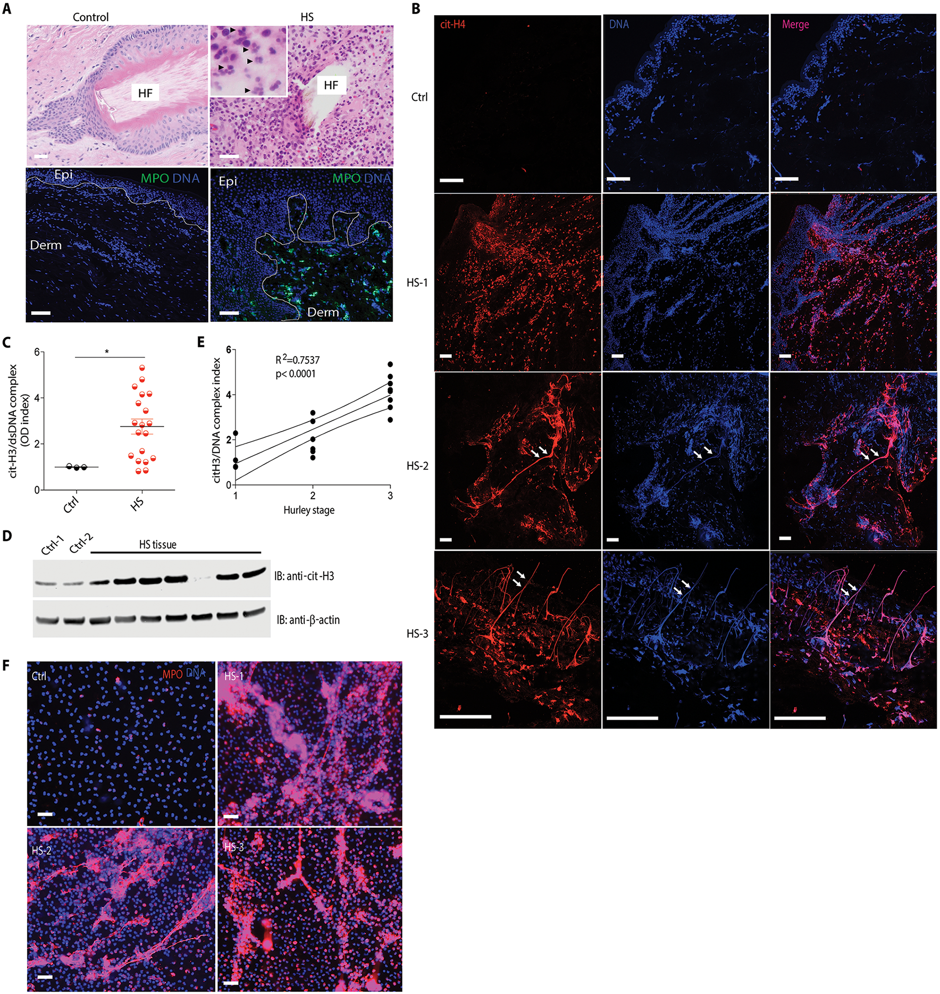
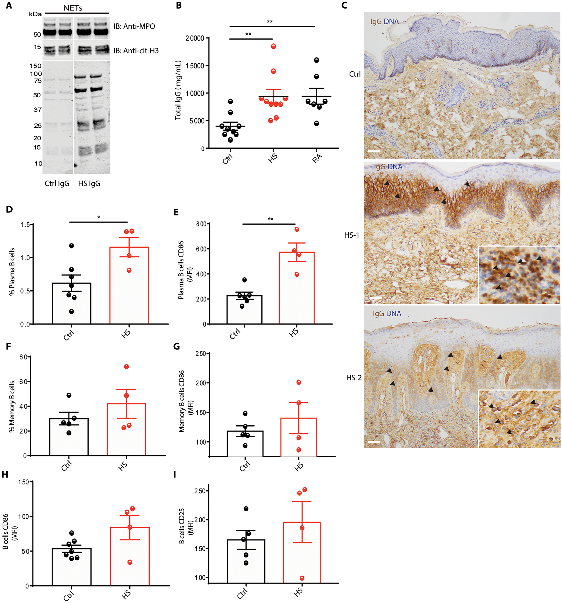
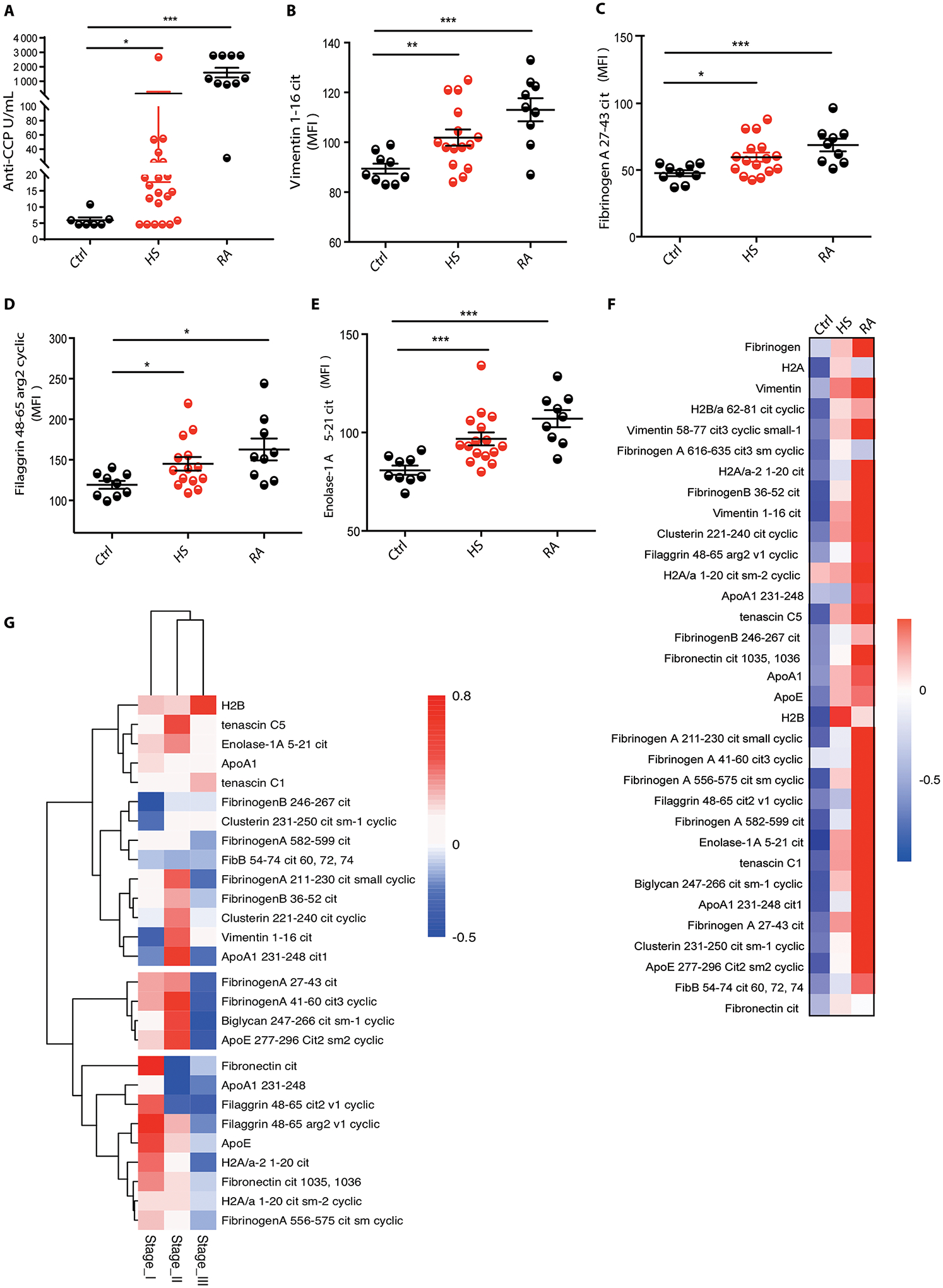
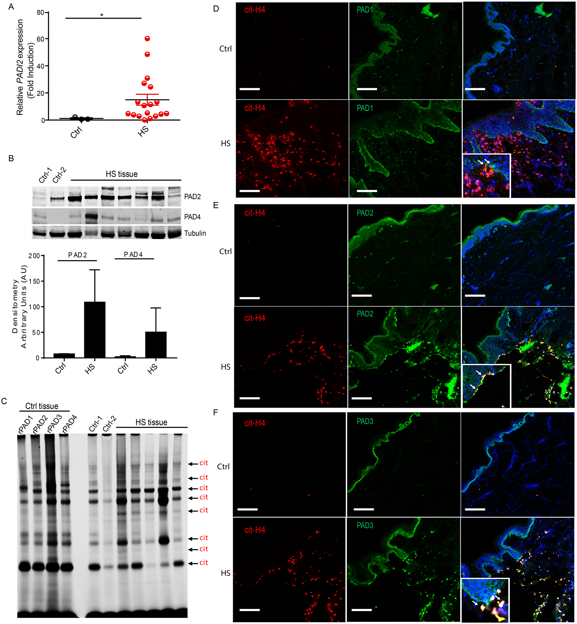
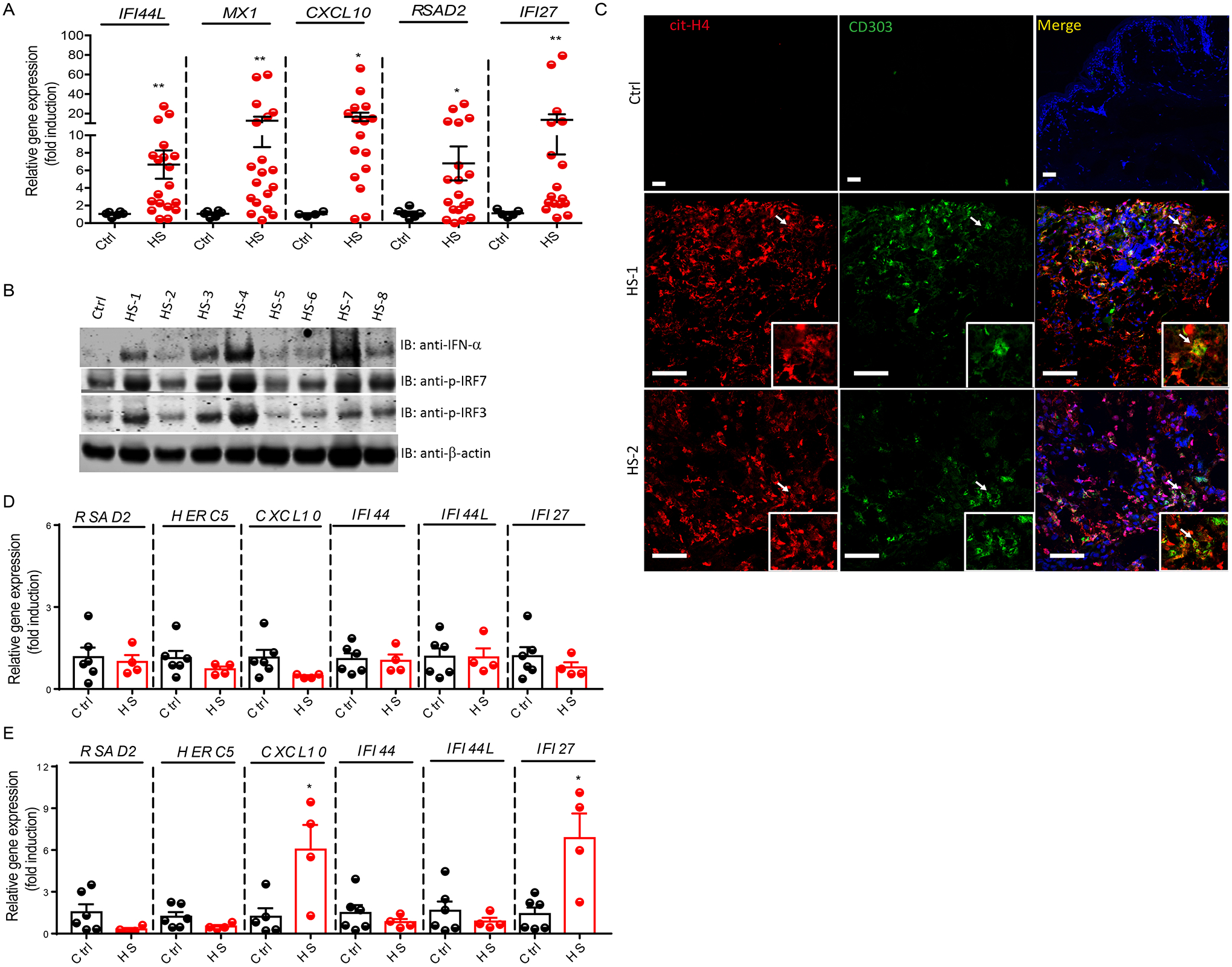
Hidradenitis suppurativa (HS), also known as acne inversa, is an incapacitating skin disorder of unknown etiology manifested as abscess-like nodules and boils resulting in fistulas and tissue scarring as it progresses. Given that neutrophils are the predominant leukocyte infiltrate in HS lesions, the role of neutrophil extracellular traps (NETs) in the induction of local and systemic immune dysregulation in this disease was examined. Immunofluorescence microscopy was performed in HS lesions and detected the prominent presence of NETs. NET complexes correlated with disease severity, as measured by Hurley staging. Neutrophils from the peripheral blood of patients with HS peripheral also displayed enhanced spontaneous NET formation when compared to healthy control neutrophils. Sera from patients recognized antigens present in NETs and harbored increased antibodies reactive to citrullinated peptides. B cell dysregulation, as evidenced by elevated plasma cells and IgG, was observed in the circulation and skin from patients with HS. Peptidylarginine deiminases (PADs) 1 to 4, enzymes involved in citrullination, were differentially expressed in HS skin, when compared to controls, in association with enhanced tissue citrullination. NETs in HS skin coexisted with plasmacytoid dendritic cells, in association with a type I interferon (IFN) gene signature. Enhanced NET formation and immune responses to neutrophil and NET-related antigens may promote immune dysregulation and contribute to inflammation. This, along with evidence of up-regulation of the type I IFN pathway in HS skin, suggests that the innate immune system may play important pathogenic roles in this disease.


Conflict of interest statement

Competing interests: All unrelated to the work reported and had no impact or conflicts related to this manuscript: A.S.B. and G.A.O. are investigators for Eli Lilly. J.M.S. is a Consultant/Speaker for Allergen and Co-Founder of LifeSprout. L.S.M. is a paid consultant for AstraZeneca, Janssen Research & Development and Armirall and has received stock from Noveome Biotherapeutics, which are developing therapeutics against infections, inflammatory and autoimmune diseases. L.S.M. serves on the scientific advisory board for Integrated Biotherapeutics, which is developing vaccines against pathogens. A.Z.R. has NDAs with Pliant Therapeutics and xMD Diagnostics and is on the Advisory Board for Escala Therapeutics. L.S.M. has received grants from AstraZeneca, MedImmune, Pfizer, Regeneron Pharmaceuticals, Boehringer Ingelheim, and Moderna Therapeutics, which are developing therapeutics against infections, inflammatory and autoimmune diseases. A.S.B. received honorarium for speaking at the 77th Annual Society for Investigative Dermatology meeting – AbbVie Sponsored Symposium. All other authors declare no potential conflicts of interest.

Figures

Figure 1.
Figure 1.. NETs are present in HS lesions and correlate with disease severity.
(A) Biopsies from HS patients stained with H&E (upper panel, neutrophils indicated with arrows) and immunofluorescence staining against myeloperoxidase (MPO, green) and Hoechst (DNA, blue) (lower panel). Epi= epidermis, Derm= dermis, HF=hair follicle. Scale bars, 50 μm. (B) Representative confocal images of lesions from HS patients stained for citrullinated histone H4 (cit-H4, red) and Hoechst (DNA, blue), arrows show co-localization of cit-H4 and DNA; Scale bars, 100 μm. (C) Homogenized lysates from resected tissues from various Hurley stages were tested for citrullinated histone H3 and DNA complexes. Results are the mean +/− SEM, *p<0.05, Mann-Whitney U test analysis was used. (D) Cit-H3 protein abundance was evaluated in tissue from HS patients and assessed by Western blot. (E) Correlation of citrullinated histone H3 and DNA complexes with HS severity as determined by the Hurley stage. One-way ANOVA Brown-Forsythe test analysis was used. (F) NETosis of peripheral blood neutrophils from HS patients and control neutrophils. MPO is red and DNA is blue. Scale bars, 50 μm.
Figure 2.
Figure 2.. Antibodies recognizing NET antigens and citrullinated peptides are present in HS serum.
(A) NETs were resolved in a SDS-PAGE gel and transferred onto a nitrocellulose membrane. Membrane was probed incubated with serum from control or HS patients. Autoantibodies were detected using anti-IgG secondary antibody. Detection of MPO and cit-H3 were used as loading controls for NETs. (B) ELISA of total IgG in HS, RA, and control (Ctrl) samples. (C) Representative images of control and HS tissue stained for intercellular IgG IHC within the epidermis and IgG-positive plasma cells arrows; Nuclei/DNA were stained with hematoxylin (blue). Scale bars, 50 μm. Peripheral blood from control and HS patients were analyzed for the frequency and activation status of (D,E) plasma cells, (F,G) memory B cells, and (H, I) CD19+ B cells by flow cytometry. Results are the mean +/− SEM, *p<0.05, **p<0.01, Mann-Whitney U test analysis was used.
Figure 3.
Figure 3.. Autoantibodies against citrullinated antigens are present in HS patients.
(A) Ctrl (n=7), HS (n=21), and RA (n=10) sera were tested for the presence of autoantibodies against cyclic citrullinated peptides (anti-CCP1). Control (Ctrl) sera were used as negative control and Rheumatoid arthritis (RA) sera were used as positive control. Results are the mean +/− SEM, *p<0.05, ***p<0.001, Mann-Whitney U test analysis was used. (B) Ctrl (n=9), HS (n=16) and RA (n=9) sera were analyzed for the presence of antibodies recognizing citrullinated vimentin, (C) citrullinated fibrinogen, (D) citrullinated filaggrin, and (E) citrullinated enolase using a bead-based assay. Results are the mean +/− SEM, *p<0.05, **p<0.01, ***p<0.001, Mann-Whitney U test analysis was used. (F) Heat map of the elevated antibodies recognizing citrullinated peptides present in HS sera (n=16) when compared to control sera (n=10). RA sera (n=9) were used as control. (G) Heat map of unsupervised clustering of citrullinated autoantibodies stratified by Hurley stage (Stage I n=4; Stage II n=5; Stage III n=8).
Figure 4.
Figure 4.. Peptidylarginine deiminase expression and activity are increased in HS lesions.
(A) Padi2 qPCR analysis of lesions from HS patients and control tissue. *p<0.05; Mann-Whitney U test analysis was used. (B) PAD2 and PAD4 protein analysis in tissue from HS patients as assessed by Western blot. Tubulin was used as a loading control. Densitometry of PAD2/Tub and PAD4/Tub (lower panel) (C) PAD activity was measured using Rh-PG probe against citrulline in HS and control tissue. Ctrl tissue with recombinant PADs 1–4 were used as a positive control for substrates for citrullination pattern detection. Representative confocal images of NETs (cit-H4, red), and (D) PAD1 (green), (E) PAD2 (green), (F) PAD3 (green) in HS and control tissues. Arrows indicate co-existence of NETs and PADs. Hoechst was used to counterstain nuclei in blue. Scale bars, 100 μm.
Figure 5.
Figure 5.. An enhanced type I interferon signature is detected in HS lesions.
RNA was isolated from homogenized HS lesional tissue or control tissue. (A) Quantitative PCR analysis of HS lesions (n=16) and control tissue (n=5) for type I interferon genes IFI44L, MX1, CXCL10, RSAD2, and IFI27. (B) Protein analysis of key interferon regulators, IRF3 and IRF7, in HS tissue and control tissue. Beta-actin was used as a loading control. (C) NETs and pDCs were visualized in HS tissue by staining cit-H4 (red) and CD303/BDCA-2 (green), respectively, showing co-existence of NETs and CD303 positive cells, arrows. Nuclei were stained blue. Scale bars, 100 μm. (D) PBMC and (E) neutrophils from HS patients (n=4) and control (n=6) were analyzed for type I IFN regulated genes. Results are the mean +/− SEM, *p<0.05, **p<0.01, Mann-Whitney U test analysis was used.

References

    1. B Jemec G, Kimball AB, Hidradenitis suppurativa: Epidemiology and scope of the problem. J Am Acad Dermatol. 73, S4–7 (2015). - PubMed
    1. Revuz J, Hidradenitis suppurativa. J Eur Acad Dermatol Venereol. 23, 985–998 (2009). - PubMed
    1. Vlassova N, Kuhn D, Okoye GA, Hidradenitis suppurativa disproportionately affects African Americans: a single-center retrospective analysis. Acta Derm Venereol. 95, 990–991(2015). - PubMed
    1. Lee DE, Clark AK, Shi VY, Hidradenitis Suppurativa: Disease Burden and Etiology in Skin of Color. Dermatology. 233, 456–61 (2017). - PubMed
    1. Deckers IE, Kimball AB, The Handicap of Hidradenitis Suppurativa. Dermatol Clin.34,17–22 (2016). - PubMed

Publication types

MeSH terms