. 2019 Sep 4;11(508):eaaw8828.
doi: 10.1126/scitranslmed.aaw8828.

Overcoming adaptive therapy resistance in AML by targeting immune response pathways

Affiliations

Overcoming adaptive therapy resistance in AML by targeting immune response pathways

Katelyn Melgar et al. Sci Transl Med. .

Abstract

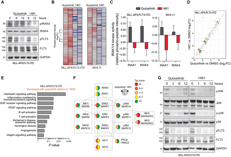
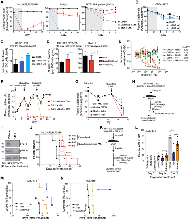
Targeted inhibitors to oncogenic kinases demonstrate encouraging clinical responses early in the treatment course; however, most patients will relapse because of target-dependent mechanisms that mitigate enzyme-inhibitor binding or through target-independent mechanisms, such as alternate activation of survival and proliferation pathways, known as adaptive resistance. Here, we describe mechanisms of adaptive resistance in FMS-like receptor tyrosine kinase (FLT3)-mutant acute myeloid leukemia (AML) by examining integrative in-cell kinase and gene regulatory network responses after oncogenic signaling blockade by FLT3 inhibitors (FLT3i). We identified activation of innate immune stress response pathways after treatment of FLT3-mutant AML cells with FLT3i and showed that innate immune pathway activation via the interleukin-1 receptor-associated kinase 1 and 4 (IRAK1/4) complex contributes to adaptive resistance in FLT3-mutant AML cells. To overcome this adaptive resistance mechanism, we developed a small molecule that simultaneously inhibits FLT3 and IRAK1/4 kinases. The multikinase FLT3-IRAK1/4 inhibitor eliminated adaptively resistant FLT3-mutant AML cells in vitro and in vivo and displayed superior efficacy as compared to current targeted FLT3 therapies. These findings uncover a polypharmacologic strategy for overcoming adaptive resistance to therapy in AML by targeting immune stress response pathways.


Conflict of interest statement

Competing interests: C.J.T. M.W., J.-K.J., K.M., and D.T.S. are inventors on the following patent PCT/US2017/047088: Compounds, compositions, methods for treating disease, and methods for preparing compounds. K.M., M.M.W., J.-K.J., C.J.T., and D.T.S. are inventors on a patent application (WO/2018/038988) jointly held by the CCHMC and the U.S. National Institutes of Health covering chemical composition, methods of preparation and methods of treatment for small molecules reported in this manuscript including NCGC1481. D.T.S. has received support from Celgene and honoraria from Curis Inc. R.L.L. is on the supervisory board of Qiagen and is a scientific advisor to Loxo, Imago, C4 Therapeutics, and IsoPlexis, which each include an equity interest. R.L.L. also received research support from and consulted for Celgene and Roche, received research support from Prelude Therapeutics, consulted for Incyte, Novartis, MorphoSys, and Janssen, and received honoraria from Lilly and Amgen for invited lectures and from Gilead for grant reviews. All other authors declare that they have no competing interests.

Figures

Fig. 1.
Fig. 1.. FLT3-ITD AML develops adaptive resistance and activates innate immune pathways after FLT3i treatment.
(A) Overview of experimental design to evaluate adaptive resistance. CFC, colony-forming cell assay. (B) MLL-AF9;FLT3-ITD or MV4;11 cells were cultured with quizartinib for 3 days and then replated in fresh medium, and cell viability was measured by AnnexinV staining. Values are expressed as means ± SEM from three biological replicates. (C) After 10 days in liquid culture [from (B)], the remaining viable cells were plated in methylcellulose, and colony formation was determined after 7 days. Values are expressed as means ± SD from two biological replicates. (D) Serine-threonine kinase (STK) PamChip analysis was performed on protein lysates isolated from MLL-AF9;FLT3-ITD and MV4;11 cells treated with quizartinib (0.3 nM) for 6 and 12 hours. Hierarchical clustering analysis was performed on differentially phosphorylated peptides in the indicated groups relative to DMSO (two biological replicates). (E) In-cell active kinases inferred from the phosphorylated peptides (STK PamChip) are shown for each of the indicated conditions. (F) Pathway enrichment of differential in-cell kinase activity in MLL-AF9;FLT3-ITD and MV4;11 cells treated with quizartinib for 6 and 12 hours was determined using Panther. (G) Pathway enrichment of differentially expressed genes (>2-fold, P < 0.05) in MLL-AF9;FLT3-ITD cells treated with quizartinib for 12 hours was determined using ToppGene (n = 3 per group).
Fig. 2.
Fig. 2.. Innate immune signaling via IRAK1/4 mediates adaptive resistance to FLT3i.
(A) IRAK1 and IRAK4 activity in MLL-AF9;FLT3-ITD cells treated with quizartinib for 6 and 12 hours is shown on the basis of the relative phosphorylation of the indicated peptides on the STK PamChip array (summary of four IRAK1- or three IRAK4-independent peptides from two biological replicates). (B) Immunoblotting of pFLT3 and pIRAK4 in MLL-AF9;FLT3-ITD cells treated with quizartinib for the indicated times. GAPDH, glyceraldehyde-3-phosphate dehydrogenase. (C) Immunoblotting of pFLT3 and pIRAK4 in MV4;11 cells treated with quizartinib for the indicated times. (D) Immunoblotting of pIRAK4 and total IRAK4 in MLL-AF9;FLT3-ITD cells treated in vitro with gilteritinib (10 nM) or from the peripheral blood of patients with FLT3-ITD AML treated with gilteritinib for the indicated number of days. (E) Delta Bliss score of MLL-AF9;FLT3-ITD cells treated with the indicated concentrations of quizartinib and IRAK-Inh for 48 hours was calculated on the basis of the cellular metabolic activity using CellTiter-Glo. (F) Viability of MLL-AF9;FLT3-ITD cells treated for 3 days with DMSO (vehicle control), quizartinib (0.5 μM), IRAK-Inh (10 μM), or quizartinib and IRAK-Inh. Individual data points are shown along with the mean from two biological replicates. (G) After 10 days in liquid culture [from (F)], the remaining viable cells were plated in methylcellulose, and colony formation was determined after 7 days. Values are expressed as means ± SEM from four biological replicates. (H) MV4;11 cells expressing control shRNA (shControl-GFP) or shRNA targeting IRAK4 (shIRAK4-GFP) were treated with DMSO or quizartinib (1 or 50 nM) for 3 days. The proportion of GFP+ cells over 10 days in culture is shown relative to day 0. Individual data points are shown along with the mean from two biological replicates. *P < 0.05 (unpaired, two-tailed t test). ns, not significant. (I) MV4;11 cells expressing an empty GFP vector (vector) or a GFP vector with IRAK4 (IRAK4) were treated with DMSO or quizartinib (1 or 50 nM) for 3 days. The proportion of GFP+ cells over 10 days in culture is shown relative to day 0. Values are expressed as means ± SEM from four biological replicates. *P = 0.029 (Mann-Whitney).
Fig. 3.
Fig. 3.. Structure activity relationship of small-molecule inhibitors reveals the importance of targeting IRAK1/4 and FLT3 in FLT3+ AML.
(A) Chemical structures of NCGC1410, NCGC2376, and NCGC2327. (B) The half maximal inhibitory concentration (IC50) for NCGC1410, NCGC2376, and NCGC2327 on IRAK1, IRAK4, and FLT3 activity (Reaction Biology). (C) Metabolic activity of MLL-AF9;FLT3-ITD cells treated with NCGC1410, NCGC2376, or NCGC2327 for 72 hours as measured by CellTiter-Glo. Values are expressed as means ± SEM from three biological replicates. (D) Viability of MLL-AF9;FLT3-ITD cells treated for 3 days with DMSO (vehicle control), NCGC1410, NCGC2376, or NCGC2327. Individual data points are shown along with the mean from two biological replicates. (E) Immunoblotting of MLL-AF9;FLT3-ITD cells treated with quizartinib (50 nM), quizartinib and IRAK-Inh (10 μM), NCGC2327 (50 nM), or IRAK-Inh.
Fig. 4.
Fig. 4.. NCGC1481 is a potent small-molecule inhibitor of FLT3 and IRAK1/4.
(A) Chemical structure of NCGC1481 (1481). The IC50 and equilibrium dissociation constant (Kd) for NCGC1481 with IRAK1, IRAK4, and FLT3 is shown below the structure. (B) NCGC1481-binding pocket of IRAK4 from NCGC1481-IRAK4 crystal structure. IRAK4 is shown as a ribbon structure along with contact residues. NCGC1481 is shown in green and blue. (C) Reaction Biology kinome map showing selectivity of NCGC1481 across 369 purified and active kinases. (D) KiNativ in situ kinome profile of NCGC1481 in MV4;11 cells showing the expressed and active kinases (left dendrogram) and these inhibited by NCGC1481 (right dendrogram). (E) Top kinases (listed in alphabetical order) inhibited by NCGC1481 as determined by the KiNativ in situ kinome profile and the corresponding IC50 value for NCGC1481 as determined versus purified active kinases at Reaction Biology (R.B.). N.D., not determined. (F) Kinase inhibitory activity (Reaction Biology, IC50 values) for top kinase targets for selected NCGC1481 analogs with variable cytotoxicity (far right column) versus MV4;11 cells. (G) Metabolic activity of MLL-AF9, MLL-AF9;FLT3-ITD, and MLL-AF9;NRAS cells treated with the indicated concentration of NCGC1481 for 72 hours as measured by CellTiter-Glo. Values are expressed as means ± SEM from three biological replicates. (H) Viability of primary AML cells from 13 patients was determined in the presence of NCGC1481 for 48 hours by trypan blue exclusion from two biological replicates. The heat map indicates the mean value. The FLT3-ITD status of the patients is indicated below the heat map and in table S7.
Fig. 5.
Fig. 5.. NCGC1481 inhibits compensatory IRAK1/4 signaling in FLT3-ITD AML cells.
(A) Immunoblotting of pFLT3 and pIRAK4 in MLL-AF9;FLT3-ITD and MV4;11 cells treated with IC10 of quizartinib or NCGC1481 for 6 and 12 hours. (B) STK PamChip analysis was performed on protein lysates isolated from MLL-AF9;FLT3-ITD and MV4;11 cells treated with IC10 of quizartinib or NCGC1481 for 12 hours. (C) IRAK1 and IRAK4 activity in MLL-AF9;FLT3-ITD and MV4;11 cells treated with IC10 of quizartinib or NCGC1481 for 12 hours (summary of four IRAK1- or three IRAK4-independent peptides from two biological replicates). (D) Differential gene expression of MLL-AF9;FLT3-ITD treated with IC10 of quizartinib or NCGC1481 for 12 hours. Individual data points represent means from biological triplicate samples. (E) Pathway enrichment of differential gene expression (RNA-seq) in MLL-AF9;FLT3-ITD cells treated with quizartinib for 12 hours was determined using ToppGene. EGF, epidermal growth factor; PDGF, platelet-derived growth factor. (F) Network map of active kinases in MLL-AF9;FLT3-ITD cells treated with quizartinib for 12 hours. Tau (τ) scores indicate activity inferred from the phosphorylated peptides (STK PamChip). The left half of the circle represents data from quizartinib-treated cells. The right half of the circle represents data from NCGC1481-treated cells. (G) Immunoblotting of MLL-AF9;FLT3-ITD cells treated with 1 nM quizartinib or NCGC1481 for the indicated times.
Fig. 6.
Fig. 6.. NCGC1481 prevents adaptive resistance in FLT3-ITD AML cells in vitro and in vivo.
(A) MLL-AF9;FLT3-ITD, MV4;11, or FLT3-ITD AML patient-derived cells were cultured with quizartinib or NCGC1481 for 3 days and replated in fresh medium, and cell viability was measured by AnnexinV staining. Values are expressed as means ± SEM from three to four biological replicates for cell lines and two replicates for the patient sample. *P < 0.05 (unpaired, two-tailed t test). (B) Healthy human CD34+ umbilical cord blood (UCB) cells were treated with 1481 (1 or 5 μM) for 3 days and replated in fresh medium. Cell viability was measured by AnnexinV staining. Individual data points are shown along with the mean from two biological replicates. (C) After 10 days in liquid culture [from (B)], the remaining viable CD34+ cells were plated in methylcellulose, and colony formation was determined after 14 days. Values are expressed as means ± SEM from four biological replicates. (D) After 10 days in liquid culture [from (A)], the remaining viable cells were plated in methylcellulose, and colony formation was determined after 7 days. Values are expressed as means ± SEM from four biological replicates. *P < 0.05 (unpaired, two-tailed t test). (E) MV4;11 cells were cultured with quizartinib (5 μM) for 3 days and replated in fresh medium, and cell proliferation was determined after treatment with the indicated concentration of NCGC1481 or quizartinib (Qztnb) for 72 hours. Values are expressed as means ± SEM from three biological replicates. (F) MLL-AF9;FLT3-ITD cells were cultured with quizartinib (5 μM) for 3 days, plated in fresh medium (days 0 and 7), and then replated in medium containing quizartinib (5 μM) or NCGC1481 (5 μM) at days 7 and 17. Cell viability was measured by AnnexinV staining. Individual data points are shown along with the mean from two to three biological replicates. (G) FLT3-ITD AML patient-derived cells were cultured with quizartinib (5 μM) for 3 days, plated in fresh medium, and then replated in medium containing DMSO, quizartinib (5 μM), or NCGC1481 (5 μM) at day 7. Cell viability was measured by AnnexinV staining. Individual data points are shown along with the mean from two biological replicates. (H) Overview of experimental design of xenograft studies. Parental or quizartinib refractory [from (E)] MLL-AF9;FLT3-ITD cells were intravenously injected into NRGS mice. On day 10 after transplant, the mice were intraperitoneally (IP) treated with NCGC1481 (30 mg/kg) or vehicle control daily (n = 5 mice per condition). (I) After 48 hours of treatment with NCGC1481, MLL-AF9;FLT3-ITD (GFP+) cells were isolated from the BM for immunoblot (IB) analysis. (J) Disease-free survival of NRGS mice xenografted with parental or quizartinib refractory MLL-AF9;FLT3-ITD cells and treated with NCGC1481 or vehicle (n = 5 mice per condition). *P < 0.05 and **P < 0.01 (Mantel-Cox test). (K) Overview of experimental design of xenograft studies using FLT3-ITD AML cells obtained from a patient (FLT3+ AML-174). FLT3-ITD AML cells were intravenously injected into NSGS mice. Two weeks after transplant, mice were treated with vehicle control, quizartinib (15 mg/kg), or NCGC1481 (30 mg/kg) intraperitoneally daily. (L) BM aspirates were analyzed for leukemic burden on days 0, 14, and 28 after treatment (n = 6 mice per condition). Values are expressed as means ± SEM from six individual mice. *P < 0.05 (unpaired, two-tailed t test). (M) Leukemia-free survival of NRGS mice xenografted with AML-174 patient cells and treated with quizartinib, NCGC1481, or vehicle (n = 5 to 7 mice per group). **P < 0.005 (Mantel-Cox test). (N) Leukemia-free survival of NRGS mice xenografted with AML-019 patient cells and treated with quizartinib, NCGC1481, or vehicle (n = 4 to 5 mice per group). **P < 0.005 (Mantel-Cox test).

Comment in

Similar articles

Cited by

References

    1. Chu SH, Small D, Mechanisms of resistance to FLT3 inhibitors. Drug Resist. Updat 12, 8–16 (2009). - PMC - PubMed
    1. Larrosa-Garcia M, Baer MR, FLT3 inhibitors in acute myeloid leukemia: Current status and future directions. Mol. Cancer Ther 16, 991–1001 (2017). - PMC - PubMed
    1. Griffith J, Black J, Faerman C, Swenson L, Wynn M, Lu F, Lippke J, Saxena K, The structural basis for autoinhibition of FLT3 by the juxtamembrane domain. Mol. Cell 13, 169–178 (2004). - PubMed
    1. Gilliland DG, Griffin JD, The roles of FLT3 in hematopoiesis and leukemia. Blood 100, 1532–1542 (2002). - PubMed
    1. Grafone T, Palmisano M, Nicci C, Storti S, An overview on the role of FLT3-tyrosine kinase receptor in acute myeloid leukemia: Biology and treatment. Oncol. Rev 6, e8 (2012). - PMC - PubMed

Publication types

MeSH terms

Substances