. 2020 Aug 3;19(3):195-206.
doi: 10.2463/mrms.mp.2019-0018. Epub 2019 Sep 4.

Deep Learning Based Noise Reduction for Brain MR Imaging: Tests on Phantoms and Healthy Volunteers

Affiliations

Deep Learning Based Noise Reduction for Brain MR Imaging: Tests on Phantoms and Healthy Volunteers

Masafumi Kidoh et al. Magn Reson Med Sci. .

Abstract

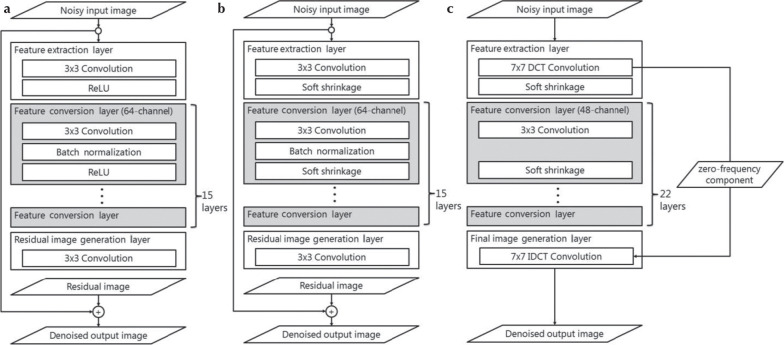
Purpose: To test whether our proposed denoising approach with deep learning-based reconstruction (dDLR) can effectively denoise brain MR images.

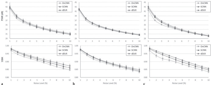
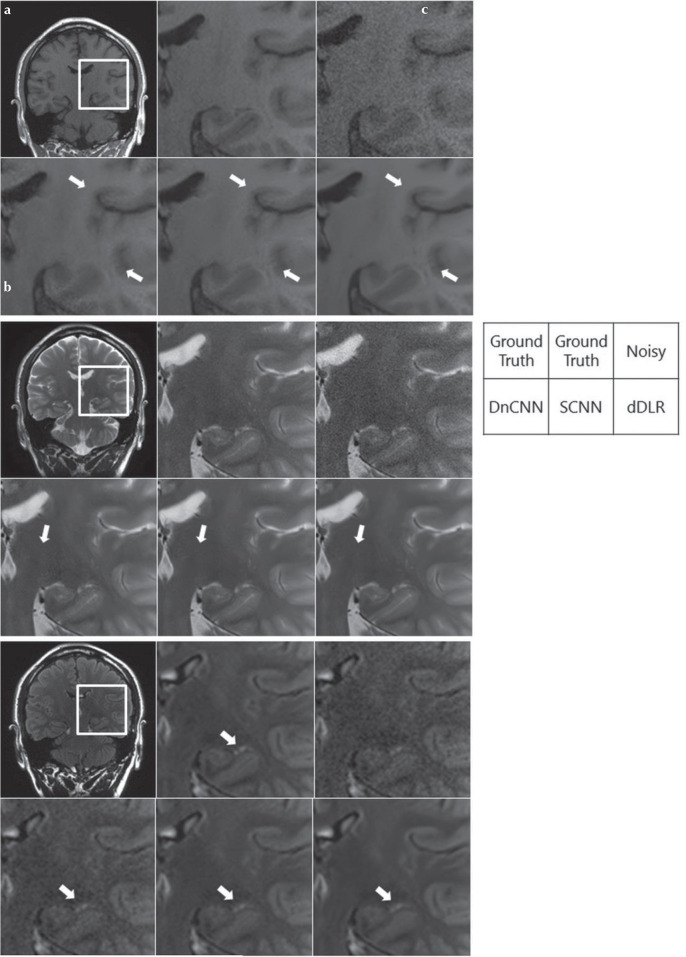
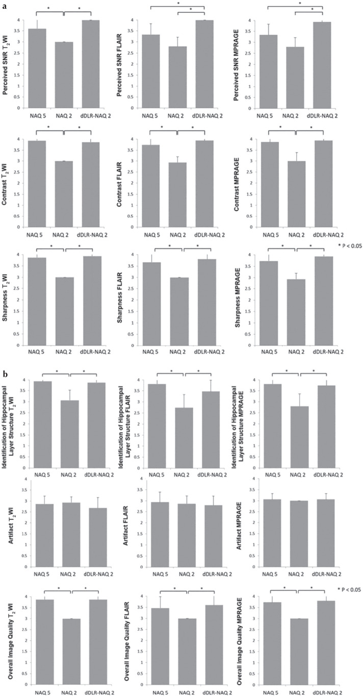
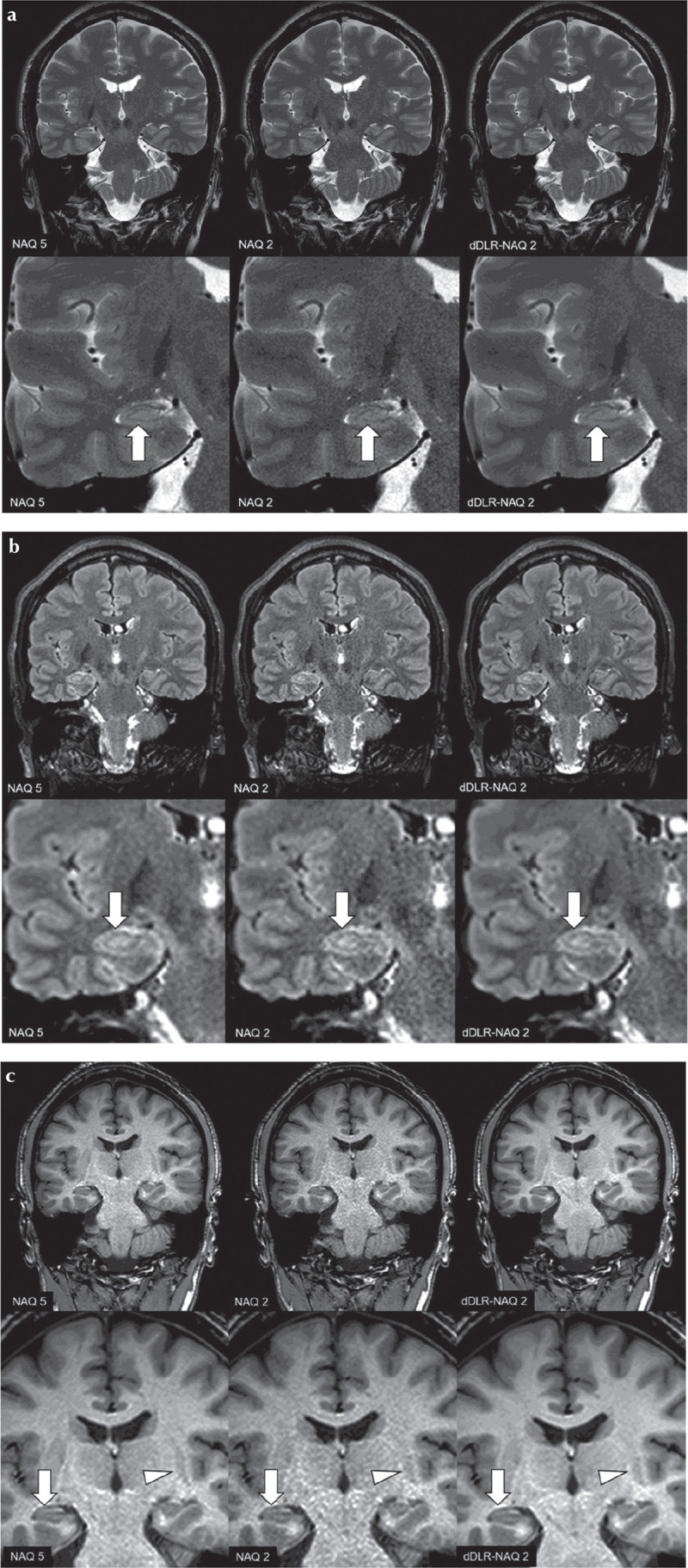
Methods: In an initial experimental study, we obtained brain images from five volunteers and added different artificial noise levels. Denoising was applied to the modified images using a denoising convolutional neural network (DnCNN), a shrinkage convolutional neural network (SCNN), and dDLR. Using these brain MR images, we compared the structural similarity (SSIM) index and peak signal-to-noise ratio (PSNR) between the three denoising methods. Two neuroradiologists assessed the image quality of the three types of images. In the clinical study, we evaluated the denoising effect of dDLR in brain images with different levels of actual noise such as thermal noise. Specifically, we obtained 2D-T2-weighted image, 2D-fluid-attenuated inversion recovery (FLAIR) and 3D-magnetization-prepared rapid acquisition with gradient echo (MPRAGE) from 15 healthy volunteers at two different settings for the number of image acquisitions (NAQ): NAQ2 and NAQ5. We reconstructed dDLR-processed NAQ2 from NAQ2, then compared with SSIM and PSNR. Two neuroradiologists separately assessed the image quality of NAQ5, NAQ2 and dDLR-NAQ2. Statistical analysis was performed in the experimental and clinical study. In the clinical study, the inter-observer agreement was also assessed.

Results: In the experimental study, PSNR and SSIM for dDLR were statistically higher than those of DnCNN and SCNN (P < 0.001). The image quality of dDLR was also superior to DnCNN and SCNN. In the clinical study, dDLR-NAQ2 was significantly better than NAQ2 images for SSIM and PSNR in all three sequences (P < 0.05), except for PSNR in FLAIR. For all qualitative items, dDLR-NAQ2 had equivalent or better image quality than NAQ5, and superior quality to that of NAQ2 (P < 0.05), for all criteria except artifact. The inter-observer agreement ranged from substantial to near perfect.

Conclusion: dDLR reduces image noise while preserving image quality on brain MR images.

Keywords: brain magnetic resonance imaging; deep learning convolutional neural network; image reconstruction; noise reduction.


Conflict of interest statement

Conflicts of Interest

Kensuke Shinoda, Masahiro Nambu, and Yuichi Yamashita are employees of Canon Medical Systems Corporation. Kenzo Isogawa is an employee of Corporate Research and Development Center, Toshiba Corporation. The other authors declare that they have no conflicts of interest.

Figures

Fig. 1
Fig. 1
Convolutional neural network (CNN) architecture of (a) denoising convolutional neural network (DnCNN), (b) shrinkage convolutional neural network (SCNN) and (c) deep learning-based reconstruction (dDLR). (a) DnCNN is a conventional denoising method featuring residual learning and batch normalization in hidden layers. (b) SCNN differs from DnCNN in that the activation function is a soft-shrinkage function. (c) dDLR is a plain CNN, not a residual neural network. dDLR uses discrete cosine transform (DCT) convolution to divide the data into a zero-frequency component path and a path with 48 high frequency components for denoising. A soft-shrinkage activation function is applied in both SCNN and dDLR to provide adaptive denoising at various noise levels using a single CNN without a requirement to train a unique CNN at each level. IDCT, inverse discrete cosine transform; ReLU, Rectified Linear Unit.
Fig. 2
Fig. 2
Activation function using soft-shrinkage. T is the threshold of the soft-shrinkage activation function. T is calculated by multiplying the noise level σ of the input noisy image and a coefficient α which is one of training parameters in our convolutional neural network. ReLU, Rectified Linear Unit.
Fig. 3
Fig. 3
Training, validation, and test data sets. (a) Training data sets were generated from eight examinations (T1WI, T2WI, PDWI, etc.) from the brain and knee of five of the volunteers. After noise addition and data augmentation, 32400 patches of training pairs were obtained. (b) Validation data sets were created from six examinations such as T1WI, T2WI, PDWI, etc. of the brain and knee from four of the volunteers. After noise addition, 660 validation pairs were obtained. (c) Test data sets were obtained from three examinations of five volunteers. Oblique-coronal T1WI, T2WI and FLAIR images were acquired. Fat Sat., fat saturation; Brain ToF, time-of-flight images from brain MR angiography; FLAIR, fluid-attenuated inversion recovery; PDW, proton-density-weighted; T1WI, T1-weighted image; T2WI, T2-weighted image.
Fig. 4
Fig. 4
Peak signal-to-noise ratio (PSNR) and structure similarity (SSIM) index values at different noise levels (1–10%) for DnCNN, SCNN, and dDLR on (a) T1WI, (b) T2WI, and (c) fluid-attenuated inversion recovery (FLAIR) images. Across all noise levels for all three types of images, dDLR was superior to DnCNN and SCNN with regard to both PSNR and SSIM (P < 0.01). dDLR, deep learning-based reconstruction; DnCNN, denoising convolutional neural network; SCNN, shrinkage convolutional neural network.
Fig. 5
Fig. 5
Visual comparison of noise reduction performance between DnCNN, SCNN and dDLR in a 38-year old male volunteer. Top-left is a ground-truth (10-number of image acquisition [NAQ]-like) image, and the others are magnified images of the rectangular annotated area in the ground-truth image. Top-center: magnified ground-truth image, top-right: magnified noise-added image without denoising, bottom-left: magnified image denoised with DnCNN, bottom-center: denoised with SCNN, and bottom-right: denoised with dDLR. Denoising was applied to the artificially noise-added images as follows: (a) T1WI with 3% noise, (b) T2WI with 4% noise, and (c) fluid-attenuated inversion recovery (FLAIR) with 2% noise. dDLR unambiguously reduced image noise while preserving intrinsic structures and structural boundaries (arrows) compared with DnCNN and SCNN images. dDLR, deep learning-based reconstruction; DnCNN, denoising convolutional neural network; SCNN, shrinkage convolutional neural network; T1WI, T1-weighted image; T2WI, T2-weighted image.
Fig. 6
Fig. 6
(a) Results of qualitative assessments: perceived SNR, image contrast and image sharpness. Perceived SNR of dDLR-NAQ2 was significantly higher than that of NAQ2 in all sequences (P < 0.05). Perceived SNR of NAQ5 was significantly higher than that of NAQ2 in T2WI (P < 0.05). In FLAIR and MPRAGE, perceived SNR of dDLR-NAQ2 was significantly higher than that of NAQ5 (both P < 0.05). Image contrast for both dDLR-NAQ2 and NAQ5 was significantly higher than that of NAQ2 in all sequences (P < 0.05), and there was no significant difference between dDLR-NAQ2 and NAQ5 (T2WI: P = 0.95; FLAIR: P = 0.64; MPRAGE: P = 0.95). For image sharpness, dDLR-NAQ2 and NAQ5 were both significantly superior to NAQ2 in all sequences (P < 0.05), and there was no significant difference between dDLR-NAQ2 and NAQ5 (T2WI: P = 0.95; FLAIR: P = 0.79; MPRAGE: P = 0.64). (b) Results of qualitative assessments: identification of hippocampal layer structure, artifact and overall image quality. For identification of hippocampal layer structure, dDLR-NAQ2 and NAQ5 were both significantly superior to NAQ2 in all sequences (P < 0.05), and there was no significant difference between dDLR-NAQ2 and NAQ5 (T2WI: P = 0.94; FLAIR: P = 0.38; MPRAGE: P = 0.91). There were no significant differences in artifacts between NAQ5, NAQ2 and dDLR-NAQ2 (T2WI: P = 0.20; FLAIR: P = 0.47; MPRAGE: P = 0.37). For overall image quality, dDLR-NAQ2 and NAQ5 were both significantly superior to NAQ2 for all sequences (P < 0.05), and there was no significant difference between dDLR-NAQ2 and NAQ5 (T2WI: P = 1.00; FLAIR: P = 0.72; MPRAGE: P = 0.94). FLAIR, fluid-attenuated inversion recovery; dDLR, deep learning-based reconstruction; NAQ, number of image acquisition; MPRAGE, magnetization-prepared rapid acquisition with gradient echo; T2WI, T2-weighted image.
Fig. 7
Fig. 7
A 38-year-old male healthy volunteer. Upper row: original magnification, lower row: magnified image. (a) T2-weighted image (T2WI): A NAQ2 has higher image noise than NAQ5 and dDLR-NAQ2. Identification of the hippocampal layer structure is superior in both NAQ5 and dDLR-NAQ2 compared with NAQ2 (arrows). (b) FLAIR: NAQ2 demonstrates higher image noise than NAQ5 and dDLR-NAQ2. Identification of the hippocampal layer structure is again superior in NAQ5 and dDLR-NAQ2 compared with NAQ2 (arrows). (c) magnetization-prepared rapid acquisition with gradient echo (MPRAGE) NAQ2 demonstrates higher image noise than NAQ5 and dDLR-NAQ2. Identification of the hippocampal layer structure is superior in NAQ5 and dDLR-NAQ2 compared with NAQ2 (arrows). Contrast between the left putamen and its surrounding white matter is also superior in NAQ5 and dDLR-NAQ2 compared with NAQ2 (arrowheads). dDLR, deep learning-based reconstruction; NAQ, number of image acquisition.

References

    1. Malmgren K, Thom M. Hippocampal sclerosis—origins and imaging. Epilepsia 2012; 53:19–33. - PubMed
    1. Jonkman LE, Klaver R, Fleysher L, Inglese M, Geurts JJ. Ultra-high-field MRI visualization of cortical multiple sclerosis lesions with T2 and T2*: a postmortem MRI and histopathology study. AJNR Am J Neuroradiol 2015; 36:2062–2067. - PMC - PubMed
    1. Thom M. Review: Hippocampal sclerosis in epilepsy: a neuropathology review. Neuropathol Appl Neurobiol 2014; 40:520–543. - PMC - PubMed
    1. McDonnell MJ. Box-filtering techniques. Comput Graph Image Process 1981; 17:65–70.
    1. Buades A, Coll B, Morel JM. A review of image denoising algorithms, with a new one. Multiscale Model Simul 2005; 4:490–530.