. 2019 Sep 4;10(1):3975.
doi: 10.1038/s41467-019-11606-x.

Protein prenylation restrains innate immunity by inhibiting Rac1 effector interactions

Affiliations

Protein prenylation restrains innate immunity by inhibiting Rac1 effector interactions

Murali K Akula et al. Nat Commun. .

Abstract

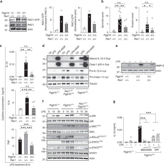
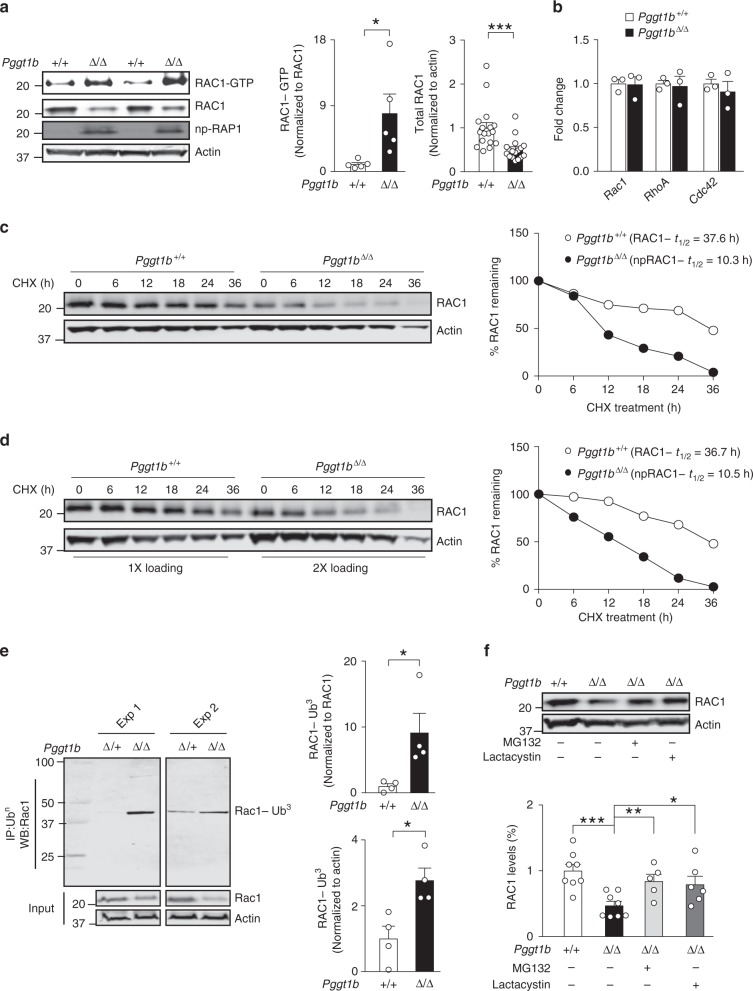
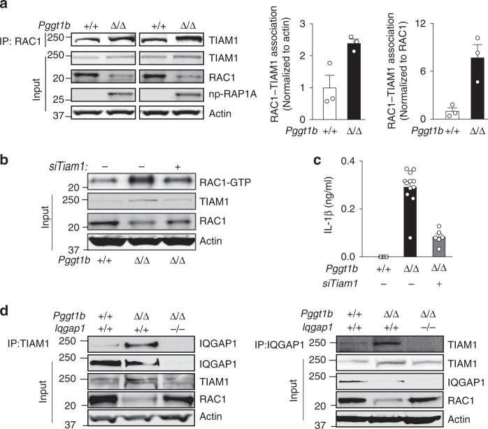
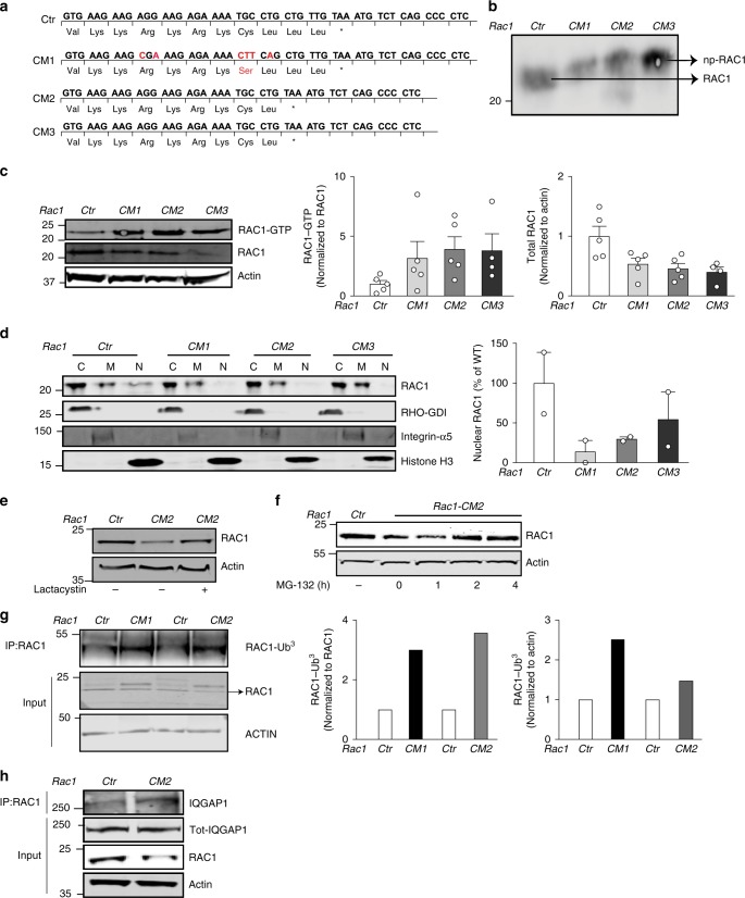
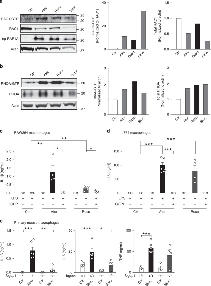
Rho family proteins are prenylated by geranylgeranyltransferase type I (GGTase-I), which normally target proteins to membranes for GTP-loading. However, conditional deletion of GGTase-I in mouse macrophages increases GTP-loading of Rho proteins, leading to enhanced inflammatory responses and severe rheumatoid arthritis. Here we show that heterozygous deletion of the Rho family gene Rac1, but not Rhoa and Cdc42, reverses inflammation and arthritis in GGTase-I-deficient mice. Non-prenylated Rac1 has a high affinity for the adaptor protein Ras GTPase-activating-like protein 1 (Iqgap1), which facilitates both GTP exchange and ubiquitination-mediated degradation of Rac1. Consistently, inactivating Iqgap1 normalizes Rac1 GTP-loading, and reduces inflammation and arthritis in GGTase-I-deficient mice, as well as prevents statins from increasing Rac1 GTP-loading and cytokine production in macrophages. We conclude that blocking prenylation stimulates Rac1 effector interactions and unleashes proinflammatory signaling. Our results thus suggest that prenylation normally restrains innate immune responses by preventing Rac1 effector interactions.


Conflict of interest statement

The authors declare no competing interests.

Figures

Fig. 1
Fig. 1
Rac1 haploinsufficiency rescues arthritis and inflammatory signaling in Pggt1bΔ/Δ mice. a Left, Western blots showing steady-state levels of GTP-bound and total Rac1 in BM macrophages isolated from Pggt1bΔ/+, Pggt1bΔ/Δ, and littermate Rac1Δ/+Pggt1bΔ/Δ mice. Actin was used as a loading control. Right, Bar graphs showing mean Rac1-GTP levels determined by densitometry (n = 2 per genotype). b Synovitis and erosion score in joints of 12-week-old Pggt1b+/+ (n = 4), Pggt1bΔ/Δ (n = 12), and Rac1Δ/+Pggt1bΔ/Δ (n = 9) mice. c Cytokine concentrations, 8 h after LPS (10 ng/ml) stimulation, in medium of primary bone marrow (BM) macrophages isolated from Pggt1b+/+ (n = 3), Pggt1bΔ/Δ (n = 4), and Rac1Δ/+Pggt1bΔ/Δ (n = 3) mice. d Western blots showing levels of mature Il-1β and caspase-1 in supernatants (Sup), and pro-Il-1β and pro-caspase-1 in lysates (Lys) of LPS (200 ng/ml) stimulated BM macrophages; tubulin in lysates was used as a loading control. The antibiotic nigericin (28 mM) was used as a positive control for inflammasome-mediated caspase-1 activation and Il-1β production. e Western blot showing levels of Mmp13 in medium of LPS-stimulated BM macrophages; Actin in lysates was used as a loading control. f Western blots showing phosphorylated (p) and total levels of intracellular signaling mediators in lysates of BM macrophages isolated 0, 15, and 30 min after LPS stimulation. g Concentration of Il-1β in medium of LPS-stimulated BM macrophages (n = 3/genotype) that had been pre-incubated for 1 h with inhibitors of p38 (SB203580; 1 and 5 µM) and ROS (DPI; 500 nM and 5 µM). For ce, g, similar results were observed in two to three independent experiments. Error bars presented as s.e.m. when n is equal to or more than three. Significance between groups were calculated with two tailed Student’s t test (c, g) and one-way ANOVA with Tukey’s post hoc test (b). n.s. not significant, *P < 0.05, **P < 0.01, ***P < 0.001
Fig. 2
Fig. 2
GGTase-I knockout increases Rac1-GTP loading, but reduces Rac1 total levels. a Left, western blots showing steady-state levels of GTP-bound and total Rac1 in BM macrophages. Nonprenylated RAP1A was used as marker of GGTase-I-deficient cells; actin was used as a loading control. Middle and right, amount of GTP-bound (n = 5/genotype) and total Rac1 (n = 17/genotype) in BM macrophages determined by densitometry of protein bands. b Quantitative polymerase chain reaction (QPCR) data showing levels of Rac1, Rhoa, and Cdc42 expression in cDNA of BM macrophages (n = 3 per genotype). c Left, western blots showing levels of Rac1 and Actin that remain in BM macrophages at various time points after incubation with cycloheximide (20 µg/ml) to stop protein synthesis. Equal amounts of total proteins were loaded. Right, densitometry of protein bands normalized to time-point 0 within each genotype. d Similar experiment as in (c) except twice the amount of total proteins from the Pggt1bΔ/Δ lysates were loaded compared to Pggt1bΔ/+ to obtain similar Rac1 levels at time-point 0. e Left, immunoprecipitation (IP) of Ubiquitin (Ub) followed by western blots for Rac1. Direct western blots were performed on the same lysates (input) to quantify total levels of Rac1 and Actin. The molecular weight of the main band was ~45 kDa which corresponds to Rac1 conjugated with three Ubs. Two independent experiments are shown. Right, amount of ubiquitin-bound Rac1 determined by densitometry of protein bands (n = 4/genotype). f Upper panel, western blots showing total Rac1 levels in lysates of BM macrophages incubated for 10 h with proteasome inhibitors MG-132 (15 µM) and lactacystin (15 µM). Lower panel, quantification of protein bands normalized to Actin and expressed as percent of control (Pggt1bΔ/+). Similar results were obtained three times. Error bars represent s.e.m. Significance between groups were calculated with two tailed Student’s t test. *P < 0.05, **P < 0.01, ***P < 0.001
Fig. 3
Fig. 3
Iqgap1 binds nonprenylated Rac1 and mediates GTP loading, cytokine production, and arthritis. a Left, immunoprecipitation (IP) of Rac1 in BM macrophage lysates followed by western blots for Iqgap1. Direct western blots were performed on the same lysates (Input) to quantify total Iqgap1 and Rac1 levels. Nonprenylated Rap1A was used as marker of GGTase-I-deficient cells; actin was used as a loading control. Middle and right, levels of Rac1-bound Iqgap1 was determined by densitometry of IP-western blots of BM macrophages from six mice/genotype. b Synovitis and erosion scores in joints of 12-week-old Pggt1bΔ/+ (n = 4), Pggt1bΔ/Δ (n = 9), and littermate Iqgap1–/–Pggt1bΔ/Δ (n = 7) mice. c Cytokine concentrations, 8 h after LPS stimulation, in medium of BM macrophages isolated from Pggt1bΔ/+, Pggt1bΔ/Δ, and Iqgap1–/–Pggt1bΔ/Δ mice (n = 3/genotype). d Left panel, Western blots showing levels of GTP-bound Rac1, total Rac1, and Iqgap1 in BM macrophages isolated from Pggt1bΔ/+, Pggt1bΔ/Δ, and Iqgap1−/−Pggt1bΔ/Δ mice. Right panels, levels of GTP-bound and total Rac1 determined by densitometry (n = 5/genotype). e Western blots of macrophage lysates isolated at base-line and 15 and 30 min after LPS stimulation. f Confocal microscopy images showing F-Actin staining of BM macrophages. Scale bars: 10 μm. Error bars represent s.e.m. The graph shows adhesive area of 20–30 macrophages in ten randomly selected fields analyzed in cells from two mice/genotype. Significance between groups were calculated with two tailed Student’s t test (a, c, d, f) and one-way ANOVA with Tukey’s post hoc test (b). n.s. not significant, *P < 0.05, **P < 0.01, ***P < 0.001
Fig. 4
Fig. 4
Tiam1 binds nonprenylated Rac1 and supports GTP loading and Il-1β production. a Left, immunoprecipitation (IP) of Rac1 in BM macrophage lysates followed by western blots for Tiam1. Direct western blots were performed on the same lysates (Input) to quantify total Tiam1 and Rac1 levels. Middle and right, levels of Rac1-bound Tiam1 was determined by densitometry of IP-western blots (n = 3/genotype). Actin was the loading control. b Western blot showing levels of GTP-bound Rac1 in lysates of BM macrophages pre-incubated for 24 h with scrambled or Tiam1-targeted siRNAs. Direct western blots were performed on the same lysates (Input) to quantify total Tiam1 and Rac1 levels. c Concentration of Il-1β, 8 h after LPS stimulation, in medium of BM macrophages pre-incubated for 24 h with scrambled or Tiam1-targeted siRNAs (n = 2 genotype). d Left, IP of Tiam1 followed by western blot for Iqgap1. Right, IP of Iqgap1 followed by western blot for Tiam1. Direct western blots were performed on the same lysates (Input) to quantify total Iqgap1, Tiam1, and Actin levels. Error bars represent s.e.m. Significance between groups were calculated with two tailed Student’s t test. *P < 0.05
Fig. 5
Fig. 5
Editing the endogenous Rac1 CAAX-motif increases GTP loading and reduces total Rac1 levels. a Predicted amino acid sequence from sanger sequence results of PCR-amplified Rac1 DNA and cDNA fragments. The DNA/cDNA was from HEK-293 clones whose Rac1 gene had been edited at the CAAX sequence by CRISPR/Cas9-facilitated homologous recombination to prevent geranylgeranylation by GGTase-I. CM1–3, CAAX mutant clones; Ctr, un-edited control clone. b Left, western blot showing electrophoretic mobility of wild-type Rac1 in Rac1Ctr lysates, and nonprenylated (np) Rac1 in CM1–3 lysates. c Left, western blots showing amounts of GTP-bound Rac1, total Rac1, and the loading control Actin in lysates of the gene-edited cells. Middle and right, levels of GTP-bound and total Rac1 determined by densitometry; data are mean of three independent experiments for each cell line. d Left, western blots showing the distribution of Rac1 in cytosol, membrane, and nuclear fractions (designated C, M, and N, respectively) of the gene-edited cells. Rho-GDI1, integrin-α5, and histone H3 were used as markers for cytosol, membrane, and nuclear fractions, respectively. Right, levels of nuclear Rac1 determined by densitometry data of three independent experiments and expressed as percent of Rac1Ctr. e, f Western blots showing levels of total Rac1 and the loading control Actin in lysates of gene-edited cells isolated after incubation with 15 µM lactacystin for 12 h (e); and 50 µM MG-132 at different time points (f). g Left, immunoprecipitation (IP) of Rac1 in gene-edited cells followed by western blot for Ubiquitin. Direct western blots were performed on the same lysates (Input) to quantify total Rac1 levels. Actin was used as a loading control. Right, Bar graphs showing levels of Rac1-Ub3. h Immunoprecipitation (IP) of Rac1 in gene-edited cells followed by western blot for Iqgap1. Direct western blots were performed on the same lysates (Input) to quantify Iqgap1 and Rac1 levels. Actin was used as a loading control. Error bars represent s.e.m
Fig. 6
Fig. 6
Statins increase Rac1-GTP and cytokine production in a GGPP- and Iqgap1-dependent fashion. a Left, western blots showing levels of GTP-bound and total Rac1 in lysates of RAW 264.7 macrophages incubated for 3 weeks with Atorvastatin (5 µM), Rosuvastatin (2.5 µM), and Simvastatin (1 µM). Np-Rap1A was used as a marker of GGTase-I-deficient cells and Actin as a loading control. Middle and right, amounts of GTP-bound and total Rac1 determined by densitometry. b Left, western blots showing levels of GTP-bound and total RhoA in the same cells as in (a). Middle and right, amounts of GTP-bound and total RhoA determined by densitometry. c Il-1β concentration, before and 8 h after LPS stimulation, in medium of RAW 264.7 macrophages incubated with Atorvastatin (5 µM) and Rosuvastatin (2.5 µM) for 21 days. GGPP (10 µM) was added to the cells 3 days before LPS stimulation. d Similar experiment as in c performed with J774 macrophages. e Cytokine concentration in medium of LPS-stimulated Iqgap1+/+ and Iqgap1−/− macrophages incubated with Simvastatin (5 µM) for 60 h. Error bars represent s.e.m. Significance between groups were calculated with two tailed Student’s t test. *P < 0.05, **P < 0.01, ***P < 0.001

Similar articles

Cited by

References

    1. Zhang FL, Casey PJ. Influence of metal ions on substrate binding and catalytic activity of mammalian protein geranylgeranyltransferase type-I. Biochem. J. 1996;320(Pt 3):925–932. - PMC - PubMed
    1. Hori Y, et al. Post-translational modifications of the C-terminal region of the rho protein are important for its interaction with membranes and the stimulatory and inhibitory GDP/GTP exchange proteins. Oncogene. 1991;6:515–522. - PubMed
    1. Solski PA, Helms W, Keely PJ, Su L, Der CJ. RhoA biological activity is dependent on prenylation but independent of specific isoprenoid modification. Cell Growth Differ. 2002;13:363–373. - PMC - PubMed
    1. Sahai E, Marshall CJ. Rho-GTPases and cancer. Nat. Rev. Cancer. 2002;2:133–142. - PubMed
    1. Kusama T, et al. Selective inhibition of cancer cell invasion by a geranylgeranyltransferase-I inhibitor. Clin. Exp. Metastasis. 2003;20:561–567. - PubMed

Publication types

MeSH terms

Substances