. 2019 Sep 4;9(1):12725.
doi: 10.1038/s41598-019-49192-z.

HilD induces expression of a novel Salmonella Typhimurium invasion factor, YobH, through a regulatory cascade involving SprB

Affiliations

HilD induces expression of a novel Salmonella Typhimurium invasion factor, YobH, through a regulatory cascade involving SprB

María M Banda et al. Sci Rep. .

Abstract

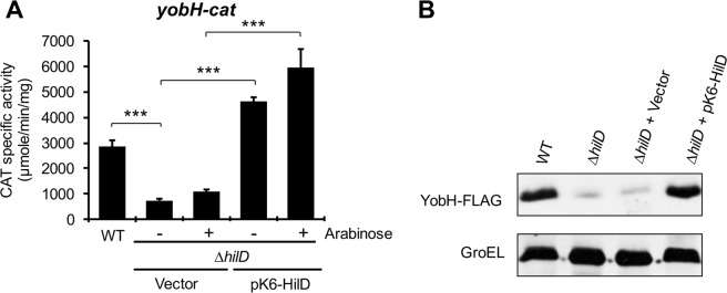
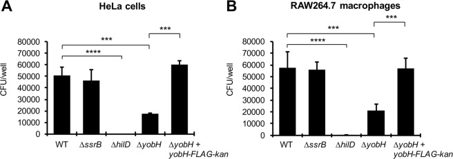
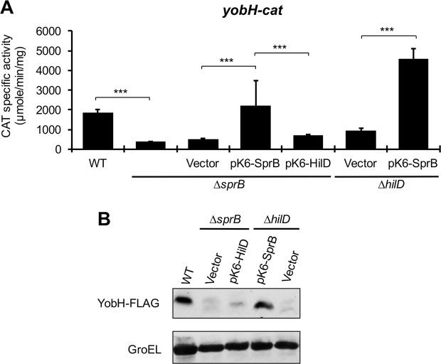
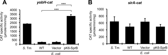
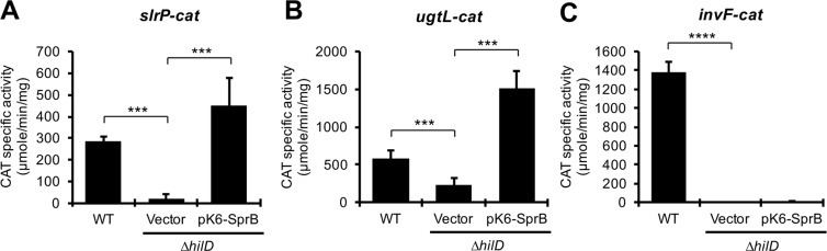
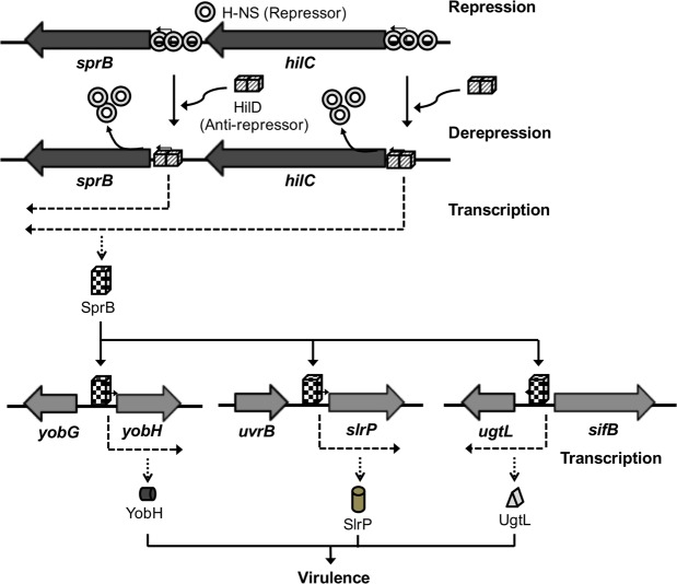
HilD is an AraC-like transcriptional regulator encoded in the Salmonella pathogenicity island 1 (SPI-1), which actives transcription of many genes within and outside SPI-1 that are mainly required for invasion of Salmonella into host cells. HilD controls expression of target genes directly or by acting through distinct regulators; three different regulatory cascades headed by HilD have been described to date. Here, by analyzing the effect of HilD on the yobH gene in Salmonella enterica serovar Typhimurium (S. Typhimurium), we further define an additional regulatory cascade mediated by HilD, which was revealed by previous genome-wide analyses. In this regulatory cascade, HilD acts through SprB, a LuxR-like regulator encoded in SPI-1, to induce expression of virulence genes. Our data show that HilD induces expression of sprB by directly counteracting H-NS-mediated repression on the promoter region upstream of this gene. Then, SprB directly activates expression of several genes including yobH, slrP and ugtL. Interestingly, we found that YobH, a protein of only 79 amino acids, is required for invasion of S. Typhimurium into HeLa cells and mouse macrophages. Thus, our results reveal a novel S. Typhimurium invasion factor and provide more evidence supporting the HilD-SprB regulatory cascade.


Conflict of interest statement

The authors declare no competing interests.

Figures

Figure 1
Figure 1
HilD positively regulates the expression of yobH (SL1344_1770) in LB. (A) Activity of the yobH-cat transcriptional fusion from the pyobH-cat plasmid, was determined in the WT S. Typhimurium SL1344 strain and its isogenic ∆hilD mutant containing the pMPM-K6Ω vector or the pK6-HilD plasmid, with (+) and without (−) induction (0.001% L-arabinose). Means and standard deviations from three independent experiments performed in duplicate are shown. Statistically different values are indicated (***p < 0.001). (B) Expression of YobH-FLAG in WT S. Typhimurium SL1344 strain and its isogenic ∆hilD mutant containing the pMPM-K6Ω vector or the pK6-HilD plasmid, was analyzed by Western blotting by using an anti-FLAG monoclonal antibody. GroEL was detected as a loading control with an anti-GroEL polyclonal antibody. Blots were cropped from different parts of the same gel. CAT specific activity and YobH-FLAG expression were determined from samples of bacterial cultures grown for 9 h in LB at 37 °C.
Figure 2
Figure 2
YobH is involved in the S. Typhimurium invasion of HeLa cells and macrophages. Epithelial HeLa cells (A) and murine RAW 264.7 macrophages (B) were infected with the WT S. Typhimurium SL1344 strain and its isogenic ∆ssrB, ∆hilD, ∆yobH and ∆yobH + yobH-FLAG-kan (∆yobH complemented) mutants. Invasion was quantified by enumerating the intracellular CFUs at 1 h post-infection, using a gentamicin protection assay. Means and standard deviations from three independent experiments performed in duplicate are shown. Statistically different values are indicated (***p < 0.001; ****p < 0.0001).
Figure 3
Figure 3
SprB is required for the expression of yobH in LB. Activity of the yobH-cat transcriptional fusion from the pyobH-cat plasmid, was determined in the WT S. Typhimurium SL1344 strain and its isogenic ∆SPI-1, ∆hilD, ∆sprB, ∆hilC, ∆hilA, ∆invF, ∆rtsA, ∆ssrB, ∆sinR and ∆flhDC mutants. CAT specific activity was quantified from samples of bacterial cultures grown for 9 h in LB at 37 °C. Means and standard deviations from three independent experiments performed in duplicate are shown. Statistically different values are indicated (***p < 0.001).
Figure 4
Figure 4
HilD induces the expression of yobH through SprB. (A) Activity of the yobH-cat transcriptional fusion from the pyobH-cat plasmid, was determined in the WT S. Typhimurium SL1344 strain and its isogenic ∆sprB mutant containing or not the pMPM-K6Ω vector, or the pK6-SprB or pK6-HilD plasmids, as well as in the ∆hilD mutant containing the pMPM-K6Ω vector or the pK6-SprB plasmid. Means and standard deviations from three independent experiments performed in duplicate are shown. Statistically different values are indicated (***p < 0.001). (B) Expression of YobH-FLAG in WT S. Typhimurium SL1344 strain and its isogenic ∆sprB mutant containing the pMPM-K6Ω vector or the pK6-HilD plasmid, as well as in the ∆hilD mutant containing the pMPM-K6Ω vector or the pK6-SprB plasmid, was analyzed by Western blotting by using an anti-FLAG monoclonal antibody. GroEL was detected as a loading control with an anti-GroEL polyclonal antibody. Blots were cropped from different parts of the same gel. CAT specific activity and YobH-FLAG expression were quantified from samples of bacterial cultures grown for 9 h in LB at 37 °C.
Figure 5
Figure 5
SprB induces expression of yobH in the absence of Salmonella-specific regulators. Activity of the yobH-cat (A) and sirA-cat (B) transcriptional fusions from the pyobH-cat and psirA-cat plasmids, respectively, was determined in the WT S. Typhimurium SL1344 strain and in the WT E. coli MC4100 strain containing or not the pMPM-K6Ω vector or the pK6-SprB plasmid expressing SprB under an arabinose inducible promoter. CAT specific activity was quantified from samples of bacterial cultures grown for 9 h in LB containing 0.001% L-arabinose, at 37 °C. Means and standard deviations from three independent experiments performed in duplicate are shown. Statistically different values are indicated (***p < 0.001).
Figure 6
Figure 6
HilD positively controls expression of sprB. Activity of the sprB-cat transcriptional fusion from the psprB-cat plasmid, was determined in the WT S. Typhimurium SL1344 strain (A,B) and its isogenic ∆hilD mutant containing or not the pMPM-K6Ω vector or the pK6-HilD plasmid (A), as well as in the WT E. coli MC4100 strain containing or not the pMPM-K6Ω vector or the pK6-HilD plasmid (B). CAT specific activity was quantified from samples of bacterial cultures grown for 9 h in LB at 37 °C. Means and standard deviations from three independent experiments performed in duplicate are shown. Statistically different values are indicated (***p < 0.001). EMSAs were performed with purified MBP-HilD (0, 0.1, 0.5 and 1 µM) and a DNA fragment containing the regulatory region of sprB (C). A DNA fragment containing the regulatory region of ppK was used as a negative internal control. The DNA-protein complexes, indicated by an asterisk, were resolved in a nondenaturing 6% polyacrylamide gel and stained with ethidium bromide.
Figure 7
Figure 7
HilD directly displaces H-NS-mediated repression on sprB. (A) Activity of the sprB-cat transcriptional fusion from the psprB-cat plasmid, was determined in the WT S. Typhimurium SL1344 strain and its isogenic ∆hilD mutant containing or not the pMPM-T6Ω vector, or the pT6-HNS-WT or pT6-HNS-G113D plasmids, with (+) and without (−) induction (0.1% L-arabinose). CAT specific activity was quantified from samples of bacterial cultures grown for 9 h in LB at 37 °C. Means and standard deviations from three independent experiments performed in duplicate are shown. Statistically different values are indicated (***p < 0.001). (B) EMSAs were performed with purified H-NS-FH (0, 0.2, 0.45 and 0.7 µM) and a DNA fragment containing the regulatory region of sprB. A DNA fragment containing the regulatory region of ppK was used as a negative internal control. The DNA-protein complexes, indicated by an asterisk, were resolved in a nondenaturing 6% polyacrylamide gel and stained with ethidium bromide. (C) Competitive nonradioactive EMSAs between H-NS and HilD on the regulatory region of sprB. Purified H-NS-FH protein was added at 0.6 µM (lanes 3 to 8) and purified MBP-HilD protein was added at 0.2, 0.4, 0.6, 0.8 and 1 µM (lanes 4 to 8, respectively). No proteins were added in lane 1 and MBP-HilD was added at 1 µM in lane 2. The DNA-protein complexes were resolved in a nondenaturing 6% polyacrylamide gel. The upper panel shows the protein-DNA complexes stained with ethidium bromide and the lower panel shows the immunoblot detection of H-NS-FH from the DNA-protein complexes. Blots for DNA or protein detection were cropped from different gels.
Figure 8
Figure 8
The HilD-SprB regulatory cascade induces expression of the slrP and ugtL genes. Activity of the slrP-cat, ugtL-cat and invF-cat transcriptional fusions from the pslrP-cat, pugtL-cat and pinvF-cat plasmids, was determined in the WT S. Typhimurium SL1344 strain and its isogenic ∆hilD mutant containing the pMPM-K6Ω vector or the pK6-SprB plasmid. CAT specific activity was quantified from samples of bacterial cultures grown for 9 h in LB at 37 °C. Means and standard deviations from three independent experiments performed in duplicate are shown. Statistically different values are indicated (***p < 0.001; ****p < 0.0001).
Figure 9
Figure 9
Model for the expression of YobH, SlrP and UgtL mediated by the HilD-SprB regulatory cascade. H-NS represses expression of sprB by binding the two promoter regions transcribing this gene. HilD binds to and thus displaces the H-NS repressor complex from these promoter regions, which allows expression of SprB that finally activates transcription of the yobH, slrP and ugtL virulence genes. Transcription of sprB from the promoter upstream of hilC and the effect of HilD and H-NS on this promoter were reported previously,,,,. The previously defined regulation of ugtL and slrP by SlyA and/or PhoP is not depicted in the model but it is described in text.

Similar articles

Cited by

References

    1. Haraga A, Ohlson MB, Miller SI. Salmonellae interplay with host cells. Nat. Rev. Microbiol. 2008;6:53–66. doi: 10.1038/nrmicro1788. - DOI - PubMed
    1. Fabrega A, Vila J. Salmonella enterica Serovar Typhimurium Skills To Succeed in the Host: Virulence and Regulation. Clin. Microbiol. Rev. 2013;26:308–341. doi: 10.1128/CMR.00066-12. - DOI - PMC - PubMed
    1. Haselbeck AH, et al. Current perspectives on invasive nontyphoidal Salmonella disease. Curr. Opin. Infect. Dis. 2017;30:498–503. doi: 10.1097/QCO.0000000000000398. - DOI - PMC - PubMed
    1. Eng S-K, et al. Salmonella: A review on pathogenesis, epidemiology and antibiotic resistance. Front. Life Sci. 2015;8:284–293. doi: 10.1080/21553769.2015.1051243. - DOI
    1. Porwollik S, McClelland M. Lateral gene transfer in. Salmonella. Microbes Infect. 2003;5:977–989. doi: 10.1016/S1286-4579(03)00186-2. - DOI - PubMed

Publication types

Substances

LinkOut - more resources