. 2019 Sep;573(7773):287-290.
doi: 10.1038/s41586-019-1530-7. Epub 2019 Sep 4.

Structures of influenza A virus RNA polymerase offer insight into viral genome replication

Affiliations

Structures of influenza A virus RNA polymerase offer insight into viral genome replication

Haitian Fan et al. Nature. 2019 Sep.

Abstract

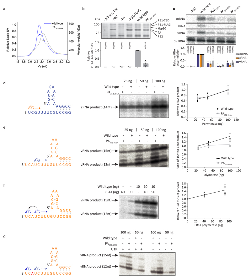
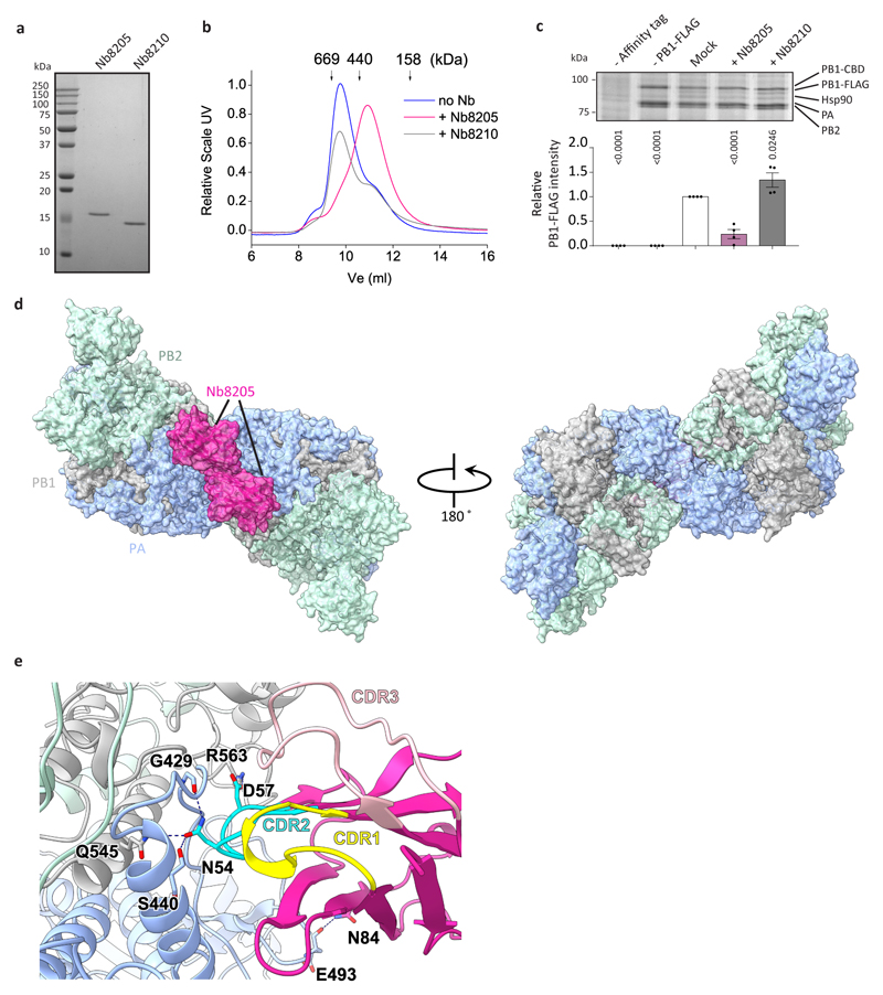
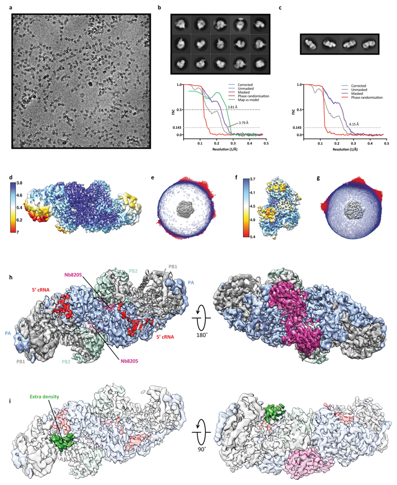
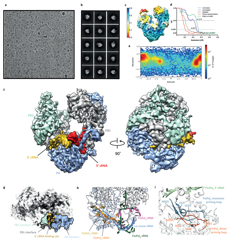
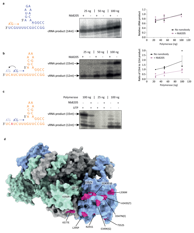
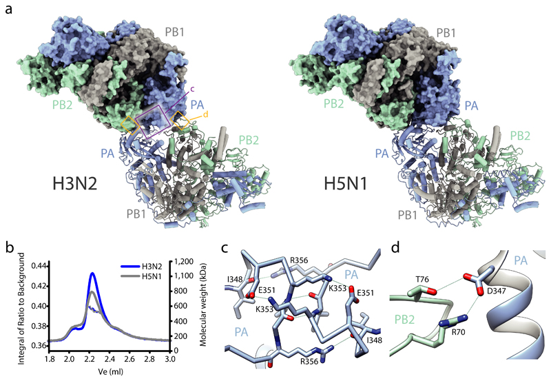
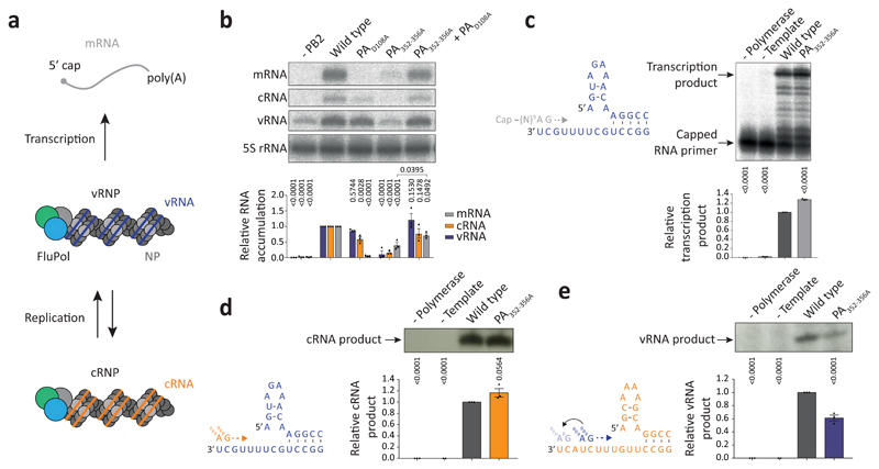
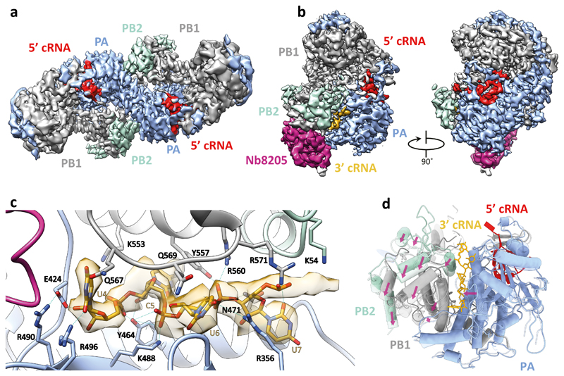
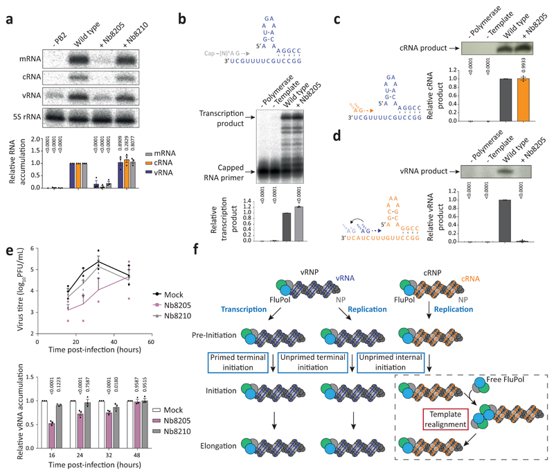
Influenza A viruses are responsible for seasonal epidemics, and pandemics can arise from the transmission of novel zoonotic influenza A viruses to humans1,2. Influenza A viruses contain a segmented negative-sense RNA genome, which is transcribed and replicated by the viral-RNA-dependent RNA polymerase (FluPolA) composed of PB1, PB2 and PA subunits3-5. Although the high-resolution crystal structure of FluPolA of bat influenza A virus has previously been reported6, there are no complete structures available for human and avian FluPolA. Furthermore, the molecular mechanisms of genomic viral RNA (vRNA) replication-which proceeds through a complementary RNA (cRNA) replicative intermediate, and requires oligomerization of the polymerase7-10-remain largely unknown. Here, using crystallography and cryo-electron microscopy, we determine the structures of FluPolA from human influenza A/NT/60/1968 (H3N2) and avian influenza A/duck/Fujian/01/2002 (H5N1) viruses at a resolution of 3.0-4.3 Å, in the presence or absence of a cRNA or vRNA template. In solution, FluPolA forms dimers of heterotrimers through the C-terminal domain of the PA subunit, the thumb subdomain of PB1 and the N1 subdomain of PB2. The cryo-electron microscopy structure of monomeric FluPolA bound to the cRNA template reveals a binding site for the 3' cRNA at the dimer interface. We use a combination of cell-based and in vitro assays to show that the interface of the FluPolA dimer is required for vRNA synthesis during replication of the viral genome. We also show that a nanobody (a single-domain antibody) that interferes with FluPolA dimerization inhibits the synthesis of vRNA and, consequently, inhibits virus replication in infected cells. Our study provides high-resolution structures of medically relevant FluPolA, as well as insights into the replication mechanisms of the viral RNA genome. In addition, our work identifies sites in FluPolA that could be targeted in the development of antiviral drugs.


Conflict of interest statement

Competing interests The authors declare no competing interests.

Figures

Extended Data Fig. 1
Extended Data Fig. 1. Subunit organisation of FluPolA heterotrimers.
a, b, Views of the structure of human H3N2 (a) and avian H5N1 (b) FluPolA heterotrimers, coloured according to subunit. c–e, Structures of human H3N2 FluPolA subunits PA (c), PB1 (d) and PB2 (e), coloured and labelled by domain. f, Domain maps of each H3N2 FluPolA subunit. g-i, The 2D Fo – mFc electron density maps of FluPolA dimer interface as shown in Fig. 1c (g, stereo view), and 1d (h, stereo view), as well as of the complete FluPolA dimer (i), are shown in gray mesh (contoured at 1.5 σ, all from the H3N2 FluPolA structure model).
Extended Data Fig. 2
Extended Data Fig. 2. Effect of mutations at the dimer interface on FluPolA dimerisation and activity.
a, SEC-MALS analysis of wild type and PA352-356A mutant H3N2 FluPolA (n=1 independent experiment). Smooth lines reflect the relative UV signal of SEC and dotted lines indicate estimated molecular weight for each frame. Note that monomeric FluPolA heterotrimer has an approximate molecular weight of 255 kDa. b, Effect of PA352-356A mutation on FluPolA dimerisation in HEK-293T cells. Data are mean ± s.e.m., n=3 independent transfections. One-way ANOVA. P < 0.05 is considered significant. c, Effect of mutations designed to destabilise PB2 and PA loops at the FluPolA dimer interface on FluPolA activity in a vRNP reconstitution assay. Data are mean ± s.e.m., n=3 independent transfections. Two-way ANOVA. P < 0.05 is considered significant. d, e, Effect of PA352-356A mutation on in vitro ApG-primer replication by FluPolA on a vRNA (d) and cRNA (e) template. f, Effect of an active site polymerase mutant (PB1a) on in vitro ApG-primer replication by FluPolA on a cRNA template. Data are mean ± s.e.m., n=4 independent reactions. g, Omitting UTP from in vitro ApG-primer replication by FluPolA on a cRNA template affects the synthesis of the 15 nucleotide full-length vRNA but not of the 12 nucleotide short vRNA indicating that the 12 nucleotide product is derived from internal initiation by the ApG dinucleotide at positions 4 and 5 of the cRNA template. The position in the template at which UTP is required is indicated in red. Representative data from n=2 independent reactions. For gel source data, see Supplementary Fig. 2.
Extended Data Fig. 3
Extended Data Fig. 3. Single-particle cryo-EM analysis of human H3N2 FluPolA bound to cRNA promoter.
a, Representative micrograph of cRNA-bound FluPolA heterotrimer particles embedded in vitreous ice. b, Representative 2D class averages. c, FSC curves for 3D reconstruction using gold-standard refinement in RELION, indicating overall map resolution of 4.07 Å and the model-to-map FSC. Curves are shown for phase randomisation, unmasked, masked and phase-randomisation-corrected masked maps. d, 3D reconstruction locally filtered and coloured according to RELION local resolution. e, Angular distribution of particle projections with the cryo-EM map shown in grey. f, Cryo-EM density of the PA loop 352-356 at the dimer interface. g, Cryo-EM map of cRNA-bound FluPolA dimer refined without symmetry imposed (C1), revealing an extra density (green) located next to the 3ʹ end of the 5ʹ cRNA close to the template entry channel. h, Close-up views highlighting cryo-EM extra density (dark green) with the 3′ vRNA strand from the superimposed FluPolB structure (PDB: 5MSG, light green) inserting into the polymerase active site. Localisation of the 3ʹ vRNA shows that bases are positioned in the extra density facing the density corresponding to the 3ʹ end of the 5ʹ cRNA, suggesting the presence of a promoter RNA duplex region as observed in vRNA-bound FluPolB . The extra density is consistent with the presence of a 3ʹ cRNA in one of the heterotrimers of the cRNA-bound FluPolA dimer, oriented towards the polymerase active site.
Extended Data Fig. 4
Extended Data Fig. 4. The effect of Nb8205 on FluPolA dimerisation.
a, SDS-PAGE of purified nanobodies (n=1 independent experiment). B, Analytical SEC of FluPolA in complex with nanobodies (n=4 for Nb8205 and n=2 for Nb8210, with similar results). c, Effect of nanobodies on FluPolA dimerisation in HEK-293T cells. Data are mean ± s.e.m., n=4 independent transfections. One-way ANOVA. P < 0.05 is considered significant. For gel source data, see Supplementary Fig. 2. d, Crystal structure of H3N2 FluPolA in complex with Nb8205. e, Close-up view of FluPolA-Nb8205 interactions. Residues involved in hydrogen bonding interactions are labelled and hydrogen bonds are indicated with dashed lines. The complementarity determining regions (CDRs) are coloured individually and labelled.
Extended Data Fig. 5
Extended Data Fig. 5. Single-particle cryo-EM analysis of monomeric and dimeric cRNA-bound human H3N2 FluPolA heterotrimer in complex with Nb8205.
a, Representative micrograph of cRNA-bound FluPolA in complex with Nb8205 embedded in vitreous ice. b, Representative 2D class averages. c, FSC curves for the 3D reconstruction using gold-standard refinement in RELION, indicating overall map resolution of 3.79 Å and 4.15 Å for the monomeric and dimeric FluPolA form, respectively, and the model-to-map FSC. Curves are shown for phase randomisation, unmasked, masked and phase-randomisation-corrected masked maps. d, f, 3D reconstruction locally filtered and coloured according to RELION local resolution for the dimeric (d) and monomeric (f) form. e, g, Angular distribution of particle projections for the dimeric (e) and monomeric (g) form with the cryo-EM map shown in grey. h, Dimer of FluPolA heterotrimers bound to cRNA promoter and Nb8205 rigid body fitted into the cryo-EM map of dimeric cRNA-bound FluPolA heterotrimer in complex with Nb8205. i, Cryo-EM map of the dimeric cRNA-bound FluPolA heterotrimer in complex with Nb8205 revealing an extra density (green) located next to the 3ʹ end of the 5ʹ cRNA, as observed for the cRNA-bound FluPolA dimer (Extended Data Fig. 3g, h).
Extended Data Fig. 6
Extended Data Fig. 6. Single-particle cryo-EM analysis of cRNA-bound FluPolB.
a, Representative micrograph of cRNA-bound FluPolB heterotrimer particles embedded in vitreous ice b, Representative 2D class averages. c, 3D reconstruction locally filtered and coloured according to RELION local resolution. d, FSC curves for the 3D reconstruction using gold-standard refinement in RELION, indicating overall map resolution of 4.18 Å and the model-to-map FSC. Curves are shown for the phase randomisation, unmasked, masked, phase-randomisation-corrected masked maps. e, Angular distribution of particle projections according to cryoSPARC v2.5 non-uniform refinement. f, Cryo-EM map of cRNA-bound FluPolB. g, Comparison of the dimerisation interface and the 3ʹ cRNA binding site in H3N2 FluPolA (PDB: 6QNW and 6QPG). h, 3ʹ cRNA binding site in FluPolA and FluPolB overlaps with the previously identified 3′ vRNA binding site in the La Crosse orthobunyavirus polymerase (PDB: 5AMQ). Sites of 3ʹ vRNA binding at surface of the polymerase in FluPolB (PDB: 4WRT) and in the polymerase active site for FluPolB (PDB: 5MSG) are shown for comparison,. i, Comparison of the structure of dimeric FluPolA to monomeric FluPolB (PDB: 5MSG) reveals a movement of the priming loop that protrudes from the PB1 thumb subdomain into the polymerase active site. Resolved PB1 residues closest to the tip of the priming loop, E638 and M656, move away from the corresponding E637 and M655 residues in FluPolB and the polymerase active site, indicated by the end of the 3ʹ vRNA, by approximately 7 Å.
Extended Data Fig. 7
Extended Data Fig. 7. Single-particle cryo-EM analysis of human H3N2 FluPolA bound to vRNA promoter.
a, Representative micrograph of vRNA-bound FluPolA heterotrimer particles embedded in vitreous ice. b, Representative 2D class averages. c, FSC curves for 3D reconstruction using gold-standard refinement in RELION, indicating overall map resolution of 3.01 Å and the model-to-map FSC. Curves are shown for phase randomisation, unmasked, masked and phase-randomisation-corrected masked maps. d, 3D reconstruction locally filtered and coloured according to RELION local resolution. e, Angular distribution of particle projections with the cryo-EM map shown in grey. f, Cryo-EM map of vRNA-bound FluPolA heterotrimer revealing the presence of a fully resolved priming loop. g, Close-up views highlighting the stacking of the 3′ vRNA by the priming loop. h, Cartoon illustration of the role of polymerase dimerisation in template realignment during replication initiation on a cRNA template. Base-pairing between the 5′ and 3′ cRNA positions bases 4 and 5 of the 3′ cRNA next to the catalytic aspartates (PB1 amino acid residues D445 and D446) in the active site to allow internal replication initiation by the synthesis of a pppApG dinucleotide. The priming loop stacks the cRNA template through PB1 amino acid P651 (left panel). Rotation of the PB1 thumb/PB2-N1 domain triggered by polymerase dimerisation results in a movement of the priming loop and backtracking of the stacked template (arrows). Backtracking is also facilitated by an interaction of PB2 amino acid residue R46 with the 3′ cRNA introducing a ‘kink’ in the template. Backtracking positions bases 1 and 2 of the cRNA template opposite the pppApG dinucleotide that remains coordinated by the catalytic aspartates. The resulting replication complex is ready to extend the pppApG dinucleotide by incorporating the next incoming NTP (right panel).
Extended Data Fig. 8
Extended Data Fig. 8. Effect of Nb8205 on FluPolA activity and mapping of host adaptive mutations at the FluPolA dimer interface.
a, b, Effect of Nb8205 on in vitro ApG-primer replication by FluPolA on a vRNA (a) and cRNA (b) template. Data are mean ± s.e.m., n=3 independent reactions. c, Omitting UTP from in vitro ApG-primer replication by FluPolA on a cRNA template affects the synthesis of the 15 nucleotide full-length vRNA but not of the 12 nucleotide short vRNA. The position in the template at which UTP is required is indicated in red. Representative data from n=2 independent reactions. For gel source data, see Supplementary Fig. 2. d, Crystal structure of H3N2 FluPolA with amino acid residues implicated in avian to mammalian host adaptation of influenza A viruses indicated.
Fig. 1
Fig. 1. Structures of human H3N2 and avian H5N1 FluPolA.
a, Crystal structures of dimers of FluPolA heterotrimers from human H3N2 (left) and avian H5N1 (right) influenza A viruses. Regions at the dimer interface (shown in close-up in panels c and d) are boxed. b, SEC-SAXS analysis of human H3N2 and avian H5N1 FluPolA (n=3 independent experiments for H3N2 with similar results and n=1 for H5N1). Smooth lines reflect the relative UV signal of SEC and dotted lines indicate estimated molecular weight for each frame. Note that monomeric FluPolA heterotrimer has an approximate molecular weight of 255 kDa. c, d, Interactions between loops 352-356 of the PA C-terminal domains (c) and the PA C-terminal domain and PB2 N1 subdomain (d) at the FluPolA dimer interface. Dashed lines indicate hydrogen bonds.
Fig. 2
Fig. 2. Mutations at the FluPolA dimer interface inhibit cRNA to vRNA replication.
a, Scheme of transcription and replication by FluPolA in the context of viral ribonucleoproteins (vRNPs). b, vRNP reconstitution assay with the PA352-356A dimer mutant and complementation with the transcription-deficient PAD108A mutant. Data are mean ± s.e.m., n=3 independent transfections. Two-way ANOVA. P < 0.05 is considered significant. mRNA signals for PA352-356A with and without PAD108A were compared by two-tailed unpaired t-test. P < 0.05 is considered significant. c, Effect of the PA352-356A mutation on in vitro transcription by FluPolA primed with a capped RNA primer. Data are mean ± s.e.m., n=3 independent reactions. One-way ANOVA. P<0.05 is considered significant. d, e, Effect of the PA352-356A mutation on in vitro primer-independent replication by FluPolA on a vRNA (d) and cRNA (e) template. Data are mean ± s.e.m., n=3 independent reactions. One-way ANOVA. P < 0.05 is considered significant. For gel source data, see Supplementary Fig. 2.
Fig. 3
Fig. 3. Structures of H3N2 FluPolA bound to cRNA promoter.
a, Cryo-EM map of dimer of FluPolA heterotrimers bound to cRNA promoter. b, Cryo-EM map of cRNA-bound FluPolA heterotrimer in complex with Nb8205. c, Close-up view of 3ʹ cRNA binding site. d, Comparison between monomeric (full colour) and dimeric (transparency) FluPolA polymerase reveals movement of the PB1 thumb/PB2 N1 subdomains (indicated by purple arrows) triggered by FluPolA dimerisation, resulting in the opening of the 3ʹ cRNA binding site.
Fig. 4
Fig. 4. Nanobody Nb8205 that binds FluPolA at the dimer interface inhibits cRNA to vRNA replication and virus growth.
a, Effect of nanobodies on FluPolA activity in a vRNP reconstitution assay. Data are mean ± s.e.m., n=3 independent transfections. Two-way ANOVA. P < 0.05 is considered significant. b, Effect of nanobody on in vitro transcription by FluPolA primed with a capped RNA primer. Data are mean ± s.e.m., n=3 independent reactions. One-way ANOVA. P < 0.05 is considered significant. c, d, Effect of nanobody on in vitro primer-independent replication by FluPolA on a vRNA (c) and cRNA (d) template. Data are mean ± s.e.m., n=3 independent reactions. One-way ANOVA. P < 0.05 is considered significant. e, Effect of nanobodies on the growth of influenza A/WSN/33 virus and vRNA levels in infected HEK-293T cells. Data are mean ± s.e.m., n=3 independent transfections and infections. Two-way ANOVA (Nb8210: P = 0.8126; 0.4390; 0.8496; 0.8489, Nb8205: P = 0.1075; 0.0096; 0.0217; 0.9828, for 16, 24, 32, 48 hours post-infection). P < 0.05 is considered significant. For gel source data, see Supplementary Fig. 2. f, Model for the role of polymerase dimerisation in influenza virus genome replication.

Similar articles

Cited by

References

    1. Taubenberger JK, Kash JC. Influenza virus evolution, host adaptation, and pandemic formation. Cell Host Microbe. 2010;7:440–51. - PMC - PubMed
    1. Mostafa A, Abdelwhab EM, Mettenleiter TC, Pleschka S. Zoonotic Potential of Influenza A Viruses: A Comprehensive Overview. Viruses. 2018;10 - PMC - PubMed
    1. Pflug A, Lukarska M, Resa-Infante P, Reich S, Cusack S. Structural insights into RNA synthesis by the influenza virus transcription-replication machine. Virus Res. 2017;234:103–117. - PubMed
    1. Te Velthuis AJ, Fodor E. Influenza virus RNA polymerase: insights into the mechanisms of viral RNA synthesis. Nat Rev Microbiol. 2016;14:479–93. - PMC - PubMed
    1. Walker AP, Fodor E. Interplay between Influenza Virus and the Host RNA Polymerase II Transcriptional Machinery. Trends Microbiol. 2019;27:398–407. - PMC - PubMed

Publication types

MeSH terms

Substances