. 2019 Oct 6;9(5):20190019.
doi: 10.1098/rsfs.2019.0019. Epub 2019 Aug 16.

Establishment of maternal blood supply to the placenta: insights into plugging, unplugging and trophoblast behaviour from an agent-based model

Affiliations

Establishment of maternal blood supply to the placenta: insights into plugging, unplugging and trophoblast behaviour from an agent-based model

Rojan Saghian et al. Interface Focus. .

Abstract

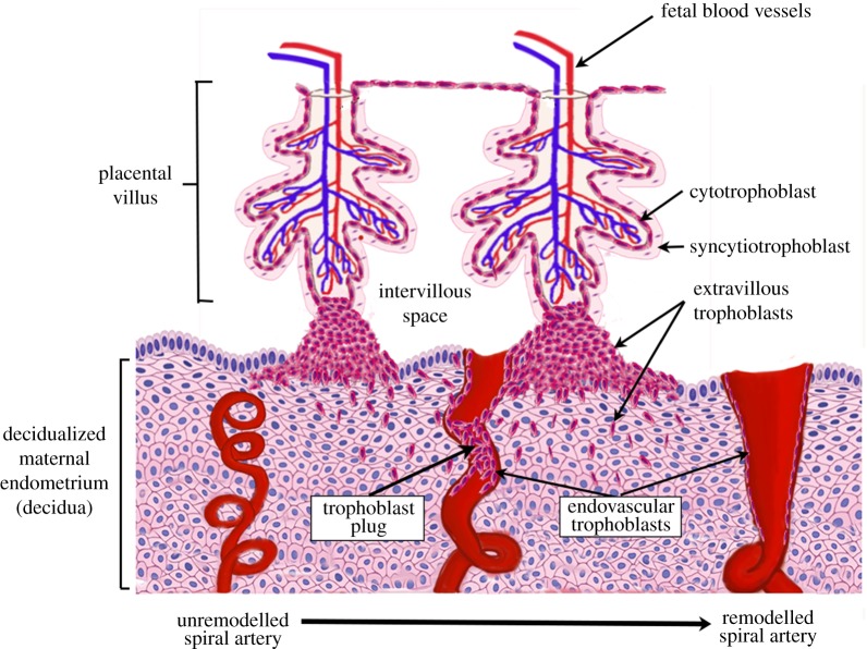
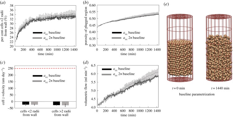
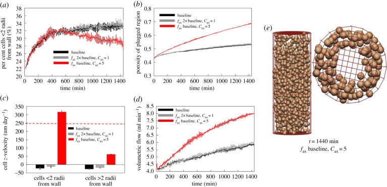
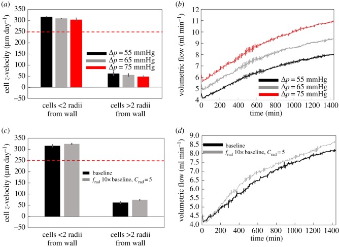
The ability of the baby to receive nutrients and oxygen in utero depends on the healthy development of the placenta. For maternal blood to adequately perfuse the placenta, it dramatically alters the arteries in the uterus that supply it with nutrient-rich blood right from the start of pregnancy. Placental cells (trophoblasts) invade both into the tissue of the uterus and into the maternal blood vessels nearest to the site of implantation (the spiral arteries (SAs)) and transform these allowing a relatively high and steady flow of nutrient-rich blood to perfuse the placenta. Trophoblasts also form plugs that occlude SAs, preventing maternal blood flow to the placenta until the late first trimester, at which point these plugs dislodge or disintegrate. Here we present an agent-based model of trophoblast migration within plugged SAs to tease apart the impact of chemical signals and mechanical factors on trophoblast behaviour. The model supports our previous in vitro hypothesis that plugging of the maternal arteries in early pregnancy can act to promote trophoblast invasion by providing a 'low flow' environment and extends our understanding by suggesting 'weak spots' in plug structure can lead to plug degeneration, allowing increased blood flow through the materno-fetal circulation.

Keywords: agent-based model; haemodynamics; placenta; spiral artery; trophoblast; utero-placental circulation.


Conflict of interest statement

We declare we have no competing interests.

Figures

Figure 1.
Figure 1.
A schematic of the process of spiral artery remodelling. Prior to trophoblast mediated remodelling the spiral arteries are small tortuous vessels. Invasive extravillous trophoblasts invade both into the maternal decidua and the lumen of the spiral arteries in the first trimester of pregnancy. The endovascular trophoblast initially plugs the lumen of the spiral arteries, preventing high-velocity blood flow to the placental surface in the first trimester. Ultimately, the spiral arteries are remodelled into large ‘funnel-like’ openings and can accommodate a high flow volume of maternal blood without excessive velocity. (Online version in colour.)
Figure 2.
Figure 2.
Schematics of how in vivo and in vitro scenarios are modelled, with an illustration of how they relate to the physical structures modelled and flow through them. In vitro, we simulate a micro-channel, of which a subsection is imaged using timelapse microscopy. The base of the micro-channel is the xz plane, and cells can break free of the base of the micro-channel in the y-direction and be carried with the flow. However, this does not happen commonly in the model or the in vitro data. In vivo, we simulate a portion of the plugged spiral artery as a cylindrical tube, with cells initially placed within the tube in a configuration that produces similar cell densities to those observed in anatomical imaging. (Online version in colour.)
Figure 3.
Figure 3.
(a) A schematic of the cell–cell and cell–wall interaction forces. When cells are far from one another, they are assumed to have a negligible impact on each other's motion; as they move closer they exert an attractive force on one another, until their centres are very close and this force becomes repulsive. (b) A schematic of environmental forces in the system. Blood flow acts along the negative z-axis in this model and is opposed by a chemoattractive force along the main axis of the vessel due to the production of chemoattractants upstream of the plug (e.g. in the myometrium). Chemoattractants may also be released by cells in or near to the vessel wall, and thus a radial force due to these (potentially different) chemoattractants is also modelled. (Online version in colour.)
Figure 4.
Figure 4.
Comparison of model trophoblast migration behaviours with experimentally determined behaviours. (ad) In experimental culture, trophoblast migrates preferentially in the direction of an induced blood flow, and the model fit to reproduce these behaviours across all culture conditions. Estimates of (e) diffusivity and (f) persistence time predicted by the model are consistent with experimental data.
Figure 5.
Figure 5.
With baseline parametrization and a range of cell–cell force strengths the model can be parametrized such that the plug stays intact over a long period. Grey black lines show model results with parameters as in table 1, and grey lines show predictions with acc at 2× baseline values. (a) The percentage of cells within two cell radii of the vessel wall is low and stable over time, except for an initial transient, (b) while porosity increases over time, changes are small and within the expected physiological range, (c) average z-velocity of cells is small and in the direction of flow (horizontal dashed line shows expected migration rate along the vessel wall), and (d) increases in predicted blood flow within the artery for a fixed pressure drop are small. (e) There are some changes in cell configuration from initial seeding; however, the plug remains relatively intact. (Online version in colour.)
Figure 6.
Figure 6.
When increasing the axial chemotaxis force alone (fax at 2× baseline, cax = 1), the plug generally stays intact, but moves as a whole either with or against the flow. By localizing the axial chemotaxis force at the wall (fax at 2× baseline, cax = 5), the model can predict physiological migration rates near the wall, and much lower migration rates for cells in the plug. However, plug break-up occurs more rapidly. (a) The percentage of cells within two cell radii of the vessel wall, (b) changes in plug porosity over time, (c) average z-velocity of cells (horizontal dashed line shows expected migration rate along the vessel wall) and (d) increases in predicted blood flow within the artery for a fixed pressure drop. (e) When axial chemotaxis is restricted to near the vessel wall, cells migrate primarily along the vessel wall. (Online version in colour.)
Figure 7.
Figure 7.
(a,b) Increasing the driving pressure across the system has a small impact on cell migration velocities (a), but increases volumetric flow through the spiral artery (b). (c,d) A strengthening radial chemotactic force draws cells away from the plugged region of the artery, resulting in a small increase in cell migration rates (c), increasing the porosity of the plugged region and so increasing blood flow through the spiral artery (d). (Online version in colour.)

Similar articles

Cited by

References

    1. Benirschke K, Kaufmann P, Baergen R. 2000. Pathology of the human placenta. New York, NY: Springer.
    1. Cunninham F. 2005. Implantation, embryogenesis, and placental development. In Williams obstetrics, 22nd edn, pp. 39–90. New York, NY: McGraw-Hill.
    1. Burton GJ. 2009. Oxygen, the Janus gas; its effects on human placental development and function. J. Anat. 215, 27–35. (10.1111/j.1469-7580.2008.00978.x) - DOI - PMC - PubMed
    1. Harris L. 2010. Trophoblast-vascular cell interactions in early pregnancy: how to remodel a vessel. Placenta 31, S93–S98. (10.1016/j.placenta.2009.12.012) - DOI - PubMed
    1. Burton G, Woods A, Jauniaux E, Kingdom J. 2009. Rheological and physiological consequences of conversion of the maternal spiral arteries for uteroplacental blood flow during human pregnancy. Placenta 30, 473–482. (10.1016/j.placenta.2009.02.009) - DOI - PMC - PubMed

LinkOut - more resources