Review
. 2019 Oct 6;9(5):20190026.
doi: 10.1098/rsfs.2019.0026. Epub 2019 Aug 16.

Mechanics of cervical remodelling: insights from rodent models of pregnancy

Affiliations
Review

Mechanics of cervical remodelling: insights from rodent models of pregnancy

Kyoko Yoshida et al. Interface Focus. .

Abstract

The uterine cervix undergoes a complex remodelling process during pregnancy, characterized by dramatic changes in both extracellular matrix (ECM) structure and mechanical properties. Understanding the cervical remodelling process in a term or preterm birth will aid efforts for the prevention of preterm births (PTBs), which currently affect 14.8 million babies annually worldwide. Animal models of pregnancy, particularly rodents, continue to provide valuable insights into the cervical remodelling process, through the study of changes in ECM structure and mechanical properties at defined gestation time points. Currently, there is a lack of a collective, quantitative framework to relate the complex, nonlinear mechanical behaviour of the rodent cervix to changes in ECM structure. This review aims to fill this gap in knowledge by outlining the current understanding of cervical remodelling during pregnancy in rodent models in the context of solid biomechanics. Here we highlight the collective contribution of multiple mechanical studies which give evidence that cervical softening coincides with known ECM changes throughout pregnancy. Taken together, mechanical tests on tissue from pregnant rodents reveal the cervix's remarkable ability to soften dramatically during gestation to allow for a compliant tissue that can withstand damage and can dissipate mechanical loads.

Keywords: cervix; extracellular matrix (ECM); growth and remodelling; review; soft tissue mechanics.


Conflict of interest statement

The authors have no competing interests to disclose.

Figures
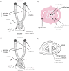
Figure 1.
Figure 1.
(a) Mouse reproductive tract and (b) its cervical cross-section, demonstrating the layers of the cervix. (c) Rat reproductive tract and (d) its cervical cross-section. Reproduced with permission from [38]. (Online version in colour.)
Figure 2.
Figure 2.
(a) Cervical tissue volume approximately doubles during pregnancy (in vivo volume assessed directly after dissection and swollen volume assessed after equilibrium in PBS). (b) Cervical dry mass doubles during pregnancy [48] while hydration slightly increases after d6. (c) Similar trends in dry weight and volume lead to a constant current apparent density of the solid component ρs during pregnancy. The referential apparent mass density, ρrs (solid tissue mass divided by referential (NP) tissue volume), decreases slightly from NP to d6 and increases steadily after d12, resulting in a significantly higher referential apparent mass density at d18. * represents p < 0.05 from NP, ** represents p < 0.05 from d6, *** represents p < 0.05 from d12, † represents p < 0.05 compared with all other groups. (Online version in colour.)
Figure 3.
Figure 3.
(a) Referential collagen apparent mass density, ρrcol, first decreases between NP and d6 and then increases between d12 and d18 of pregnancy [48]. (b) Mature intermolecular cross-link density (mature cross-links divided by collagen) steadily decreases throughout pregnancy, while immature cross-links increase between d6 and d18. These changes suggest that collagen fibres produced between d12 and d18 of pregnancy are less cross-linked than NP collagen fibres [48]. † represents p < 0.05 compared with all other groups and ** represents p < 0.05 from d6. (Online version in colour.)
Figure 4.
Figure 4.
Desmosine content normalized by cervical dry weight [28]. † represents p < 0.05 compared with all other groups and * represents p < 0.05 compared with day 12. (Online version in colour.)
Figure 5.
Figure 5.
(a) Referential apparent mass density of sGAGs (ρrsGAG) decreases slightly between NP and d6 followed by an increase between d6 and d18, resulting in a significantly higher ρrsGAG at d18 than at NP [66]. (b) Referential apparent density of HA (ρrHA) follows a similar trend to sGAGs but shows a dramatic increase between d15 and d18 [66]. ‡ represents p < 0.05 compared with all other groups except d15, † represents p < 0.05 compared with all other groups. (Online version in colour.)
Figure 6.
Figure 6.
Mechanical testing images of a mouse cervix at three gestation time points (day 6, 12 and 18) illustrate the changes in the mechanical response of the tissue to tensile loading and the dramatic extensibility of pregnant tissue. Images taken at time t = 0 and right before break for three different time points during mouse gestation. The scale box is 1 mm on each side.
Figure 7.
Figure 7.
ECM contributions to the mechanical behaviour of the cervix can be teased out with specific mechanical testing methodologies. (Online version in colour.)
Figure 8.
Figure 8.
The cervical geometry can be simplified as a cylinder. The following are two common mechanical test configurations for the cervix. (a) Ring test, where two sutures or pins are threaded into the canal and pulled apart to deform the cervix. An example experimental set-up from Yoshida et al. [17] is shown. (b) Strip uni-axial test, where the cervix is cut longitudinally and unfolded into a rectangle. Grips are attached to either end and pulled apart to deform the cervix. An example experimental set-up from Barnum et al. [75] is shown. Reproduced with permission from [17,75]. (Online version in colour.)
Figure 9.
Figure 9.
Comparing change in cervical softening for a normal mouse pregnancy across studies demonstrates significant differences in viscoelastic properties. (Online version in colour.)

References

    1. Centers for Disease Control and Prevention, Preterm birth, 2018. See https://www.cdc.gov/reproductivehealth/maternalinfanthealth/pretermbirth....
    1. Lee AC, Blencowe H, Lawn JE. 2018. Small babies, big numbers: global estimates of preterm birth. Lancet Glob. Health 7, e2–e3. (10.1016/S2214-109X(18)30484-4) - DOI - PubMed
    1. Chawanpaiboon S. et al. 2019. Global, regional, and national estimates of levels of preterm birth in 2014: a systematic review and modelling analysis. Lancet Glob. Health 7, e37–e46. (10.1016/S2214-109X(18)30451-0) - DOI - PMC - PubMed
    1. Vink J, Feltovich H. 2016. Cervical etiology of spontaneous preterm birth. Semin. Fetal Neonatal Med. 21, 106–112. (10.1016/j.siny.2015.12.009) - DOI - PMC - PubMed
    1. Mahendroo MS. 2012. Cervical remodeling in term and preterm birth: insights from an animal model. Reproduction 143, 429–438. (10.1530/REP-11-0466) - DOI - PubMed

LinkOut - more resources