

Computed tomographic analysis of maxillary sinus anatomy relevant to sinus lift procedures in edentulous ridges in Taiwanese patients
- PMID: 31485374
- PMCID: PMC6713807
- DOI: 10.5051/jpis.2019.49.4.237
Computed tomographic analysis of maxillary sinus anatomy relevant to sinus lift procedures in edentulous ridges in Taiwanese patients
Abstract
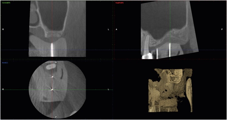
Purpose: To analyze the maxillary sinus anatomy over edentulous ridges in the bilateral posterior maxillary area in Taiwanese patients using cone-beam computed tomography (CBCT).
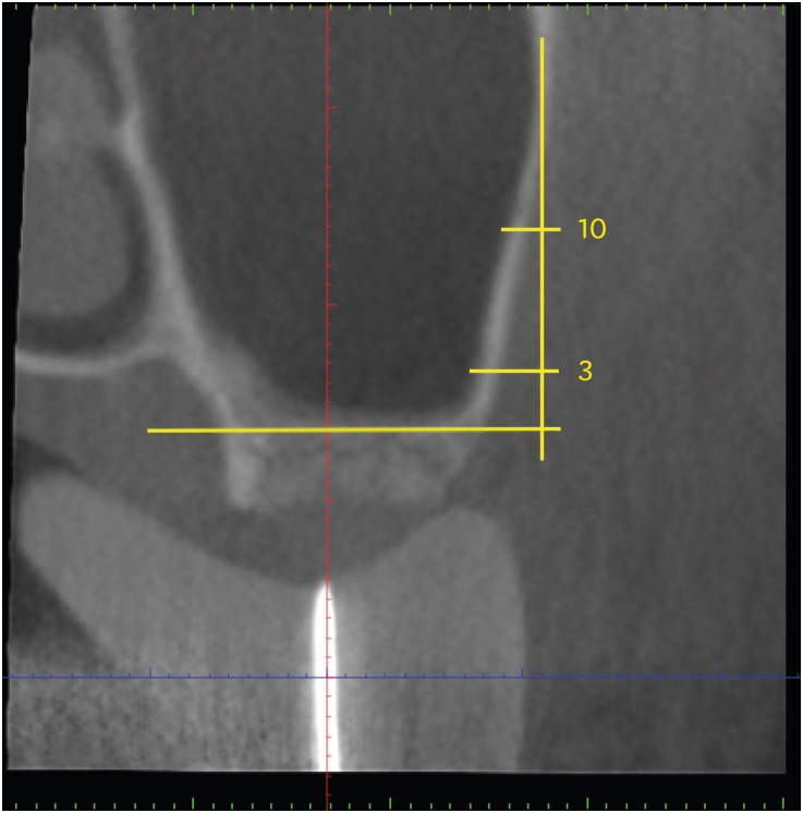
Methods: In total, 101 anatomical sites from 61 patients, including 32 premolar and 69 molar regions, were analyzed using CBCT. Measurements were made of the width and height of edentulous ridges, the thickness of the lateral wall of the maxillary sinus, and the presence of a sinus septum and the posterior superior alveolar artery (PSAA). A statistical analysis of the measurements was performed, and correlations among the measurements were assessed.
Results: The average ridge width was 10.26±3.16 mm, with a significantly greater ridge width in the second molar region than in the premolar region. The mean residual ridge height was 8.55±4.09 mm, and ridge height showed an opposite trend from ridge width for the premolar and molar regions. A sinus septum was present at 5.9% of the sites, and the PSAA was observed in 24.5%. The average thickness of the lateral wall of the maxillary sinus was 2.08±0.94 mm, with no significant difference between the tooth position and lateral wall thickness.
Conclusions: This study presents the anatomical features of the maxillary sinus, which should be considered in sinus lift procedures for implant placement, in the Taiwanese population. The use of CBCT is recommended to avoid intraoperative complications.
Keywords: Asians; Cone-beam computed tomography; Edentulous jaw; Maxillary sinus; Sinus floor augmentation.
Conflict of interest statement
Conflict of Interest: No potential conflict of interest relevant to this article was reported.
Figures


References
-
- Shanbhag S, Karnik P, Shirke P, Shanbhag V. Cone-beam computed tomographic analysis of sinus membrane thickness, ostium patency, and residual ridge heights in the posterior maxilla: implications for sinus floor elevation. Clin Oral Implants Res. 2014;25:755–760. - PubMed
-
- Kang SJ, Shin SI, Herr Y, Kwon YH, Kim GT, Chung JH. Anatomical structures in the maxillary sinus related to lateral sinus elevation: a cone beam computed tomographic analysis. Clin Oral Implants Res. 2013;24(Suppl A100):75–81. - PubMed
-
- Rosano G, Taschieri S, Gaudy JF, Weinstein T, Del Fabbro M. Maxillary sinus vascular anatomy and its relation to sinus lift surgery. Clin Oral Implants Res. 2011;22:711–715. - PubMed
-
- Neugebauer J, Ritter L, Mischkowski RA, Dreiseidler T, Scherer P, Ketterle M, et al. Evaluation of maxillary sinus anatomy by cone-beam CT prior to sinus floor elevation. Int J Oral Maxillofac Implants. 2010;25:258–265. - PubMed
-
- Acharya A, Hao J, Mattheos N, Chau A, Shirke P, Lang NP. Residual ridge dimensions at edentulous maxillary first molar sites and periodontal bone loss among two ethnic cohorts seeking tooth replacement. Clin Oral Implants Res. 2014;25:1386–1394. - PubMed
LinkOut - more resources
Full Text Sources
