. 2019 Jun;4(2):1950001.
doi: 10.1142/S2424905X19500016. Epub 2018 May 15.

System Integration and Preliminary Clinical Evaluation of a Robotic System for MRI-Guided Transperineal Prostate Biopsy

Affiliations

System Integration and Preliminary Clinical Evaluation of a Robotic System for MRI-Guided Transperineal Prostate Biopsy

Niravkumar A Patel et al. J Med Robot Res. 2019 Jun.

Abstract

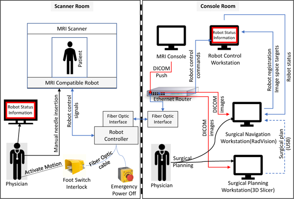
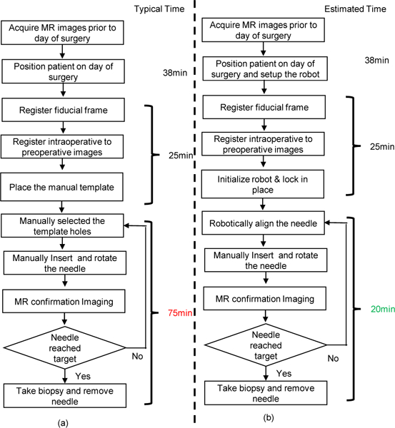
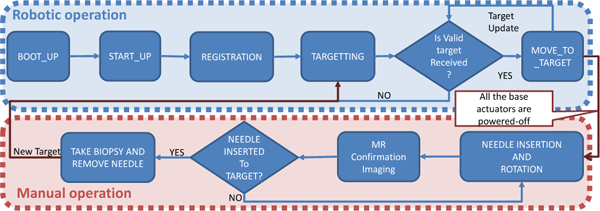
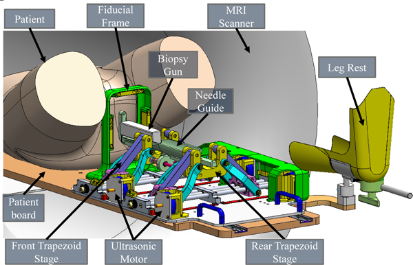
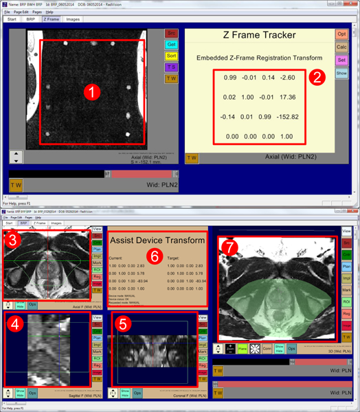
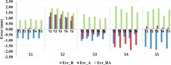
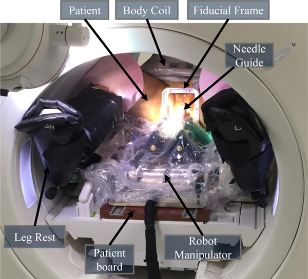
This paper presents the development, preclinical evaluation, and preliminary clinical study of a robotic system for targeted transperineal prostate biopsy under direct interventional magnetic resonance imaging (MRI) guidance. The clinically integrated robotic system is developed based on a modular design approach, comprised of surgical navigation application, robot control software, MRI robot controller hardware, and robotic needle placement manipulator. The system provides enabling technologies for MRI-guided procedures. It can be easily transported and setup for supporting the clinical workflow of interventional procedures, and the system is readily extensible and reconfigurable to other clinical applications. Preclinical evaluation of the system is performed with phantom studies in a 3 Tesla MRI scanner, rehearsing the proposed clinical workflow, and demonstrating an in-plane targeting error of 1.5mm. The robotic system has been approved by the institutional review board (IRB) for clinical trials. A preliminary clinical study is conducted with the patient consent, demonstrating the targeting errors at two biopsy target sites to be 4.0mm and 3.7mm, which is sufficient to target a clinically significant tumor foci. First-in-human trials to evaluate the system's effectiveness and accuracy for MR image-guide prostate biopsy are underway.

Keywords: MRI-compatible robotic system; MRI-guided prostate biopsy; image-guided surgery; piezoelectric actuation.


Figures

Fig. 1.
Fig. 1.
Clinical system configuration. In the control console room: (1) MRI scanner control, (2) surgical navigation user interface, and (3) robot control software. Inside the scanner room: (4)robot controller, (5) robotic manipulator inside the scanner bore covered with sterile drape, (6) patient lying inside the scanner bore in semi-lithotomy position, (7) fiberoptic foot-pedal interlock, and (8)display showing robot status to the clinician in the scanner room. Communication between the control room and scanner room is through (9) fiber optic cable.
Fig. 2.
Fig. 2.
System block diagram showing integration of all components and data flow between them. The robot status information display inside the scanner room is connected to the robot control workstation through the scanner console display system.
Fig. 3.
Fig. 3.
Workflow comparison of manual template-based approach and robot-assisted approach for MRI-guided prostate biopsy. (a) Workflow of a manual template-based prostate biopsy with measured average time per step. (b) Workflow of a robot-assisted prostate biopsy with estimated time per step.
Fig. 4.
Fig. 4.
Flowchart of the robot control workflow and robot operation modes, showing only valid transitions from one state to another.
Fig. 5.
Fig. 5.
Annotated CAD model of the parallel manipulator for transperineal prostate intervention inside the MRI scanner bore. The patient lies in the supine position, the robotic manipulator is placed between the legs, and a biopsy gun targets the prostate through the perineum. Note that the leg rest and motor covers are hidden on the left side to visualize the internal structure of the manipulator.
Fig. 6.
Fig. 6.
RadVision user interface showing (1) acquired MR images of fiducial frame, (2) calculated robot registration transform, (3) axial view, (4) sagittal view, (5) coronal view, (6) robot status, current robot pose, and desired target pose, and (7) 3D view with overlaid reachable robot workspace shown in light green. Also in all image views (3, 4, 5) light green boundary indicates reachable robot workspace.
Fig. 7.
Fig. 7.
Kinematic transformation chain for registering the robotic system to the MR scanner coordinate system (RAS coordinates) based on imaging of the fiducial frame (Z-Frame).
Fig. 8.
Fig. 8.
Phantom studies accuracy assessment: plot of measured needle placement accuracy in each of the five trials in each of the five sessions. Data is shown with errors in the lateral R-L direction (Err_R), vertical A-P direction (Err_A), and total in-plane error magnitude (Err_RA).
Fig. 9.
Fig. 9.
System configuration for the patient study. The patient lies in the supine position with legs supported by the leg rest on the patient board. The sterilized fiducial frame is fixed to the patient board between the patient’s legs. The robot manipulator is covered by the sterile plastic drape with sterile needle guide affixed, positioned on the patient board, and locked into place.
Fig. 10.
Fig. 10.
(a)Snapshot of 3D view with an MR image of prostate gland showing desired targets (green spheres) and actual needle trajectories segmented from the MRI volume images, (b- c)zoomed-in view of transverse image slice showing targets and intersection of the image slice with corresponding needle trajec- tories(the blue and red circles) from the confirmation images.

Similar articles

Cited by

References

    1. Kelloff GJ, Choyke P and Coffey DS, Challenges in clinical prostate cancer: role of imaging AJR., American journal of roentgenology 192(6) (2009) p. 1455. - PMC - PubMed
    1. Han M, Chang D, Kim C, Lee BJ, Zuo Y, Kim H-J, Petrisor D, Trock B, Partin AW, Rodriguez R et al., Geometric evaluation of systematic transrectal ultrasound guided prostate biopsy, The Journal of urology 188(6) (2012) 2404–2409. - PMC - PubMed
    1. Hricak H, Choyke PL, Eberhardt SC, Leibel SA and Scardino PT, Imaging prostate cancer: A multidisciplinary perspective 1, Radiology 243(1) (2007) 28–53. - PubMed
    1. Xu S, Kruecker J, Turkbey B, Glossop N, Singh AK, Choyke P, Pinto P and Wood BJ, Real-time mri-trus fusion for guidance of targeted prostate biopsies, Computer Aided Surgery 13(5) (2008) 255–264. - PMC - PubMed
    1. Kadoury S, Yan P, Xu S, Glossop N, Choyke P, Turkbey B, Pinto P, Wood BJ and Kruecker J, Realtime trus/mri fusion targeted-biopsy for prostate cancer: a clinical demonstration of increased positive biopsy rates, International Workshop on Prostate Cancer Imaging, Springer; (2010), pp. 52–62.

LinkOut - more resources