. 2019 Nov;42(5):1781-1792.
doi: 10.3892/or.2019.7293. Epub 2019 Aug 23.

Co‑culturing with hypoxia pre‑conditioned mesenchymal stem cells as a new strategy for the prevention of irradiation‑induced fibroblast‑to‑myofibroblast transition

Affiliations

Co‑culturing with hypoxia pre‑conditioned mesenchymal stem cells as a new strategy for the prevention of irradiation‑induced fibroblast‑to‑myofibroblast transition

Lei Zhuang et al. Oncol Rep. 2019 Nov.

Abstract

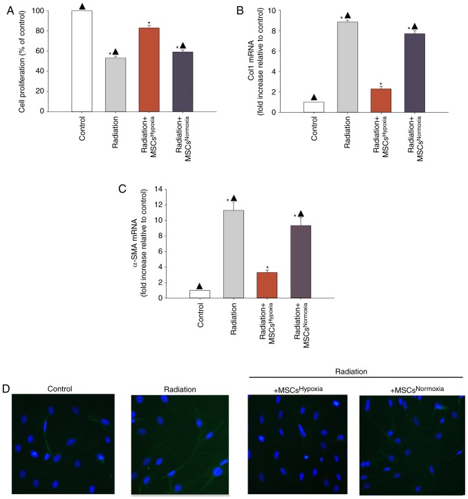
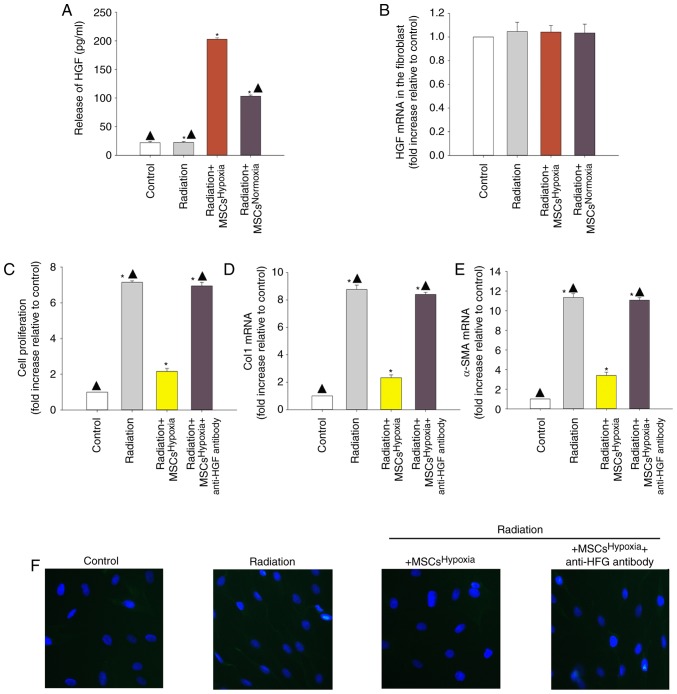
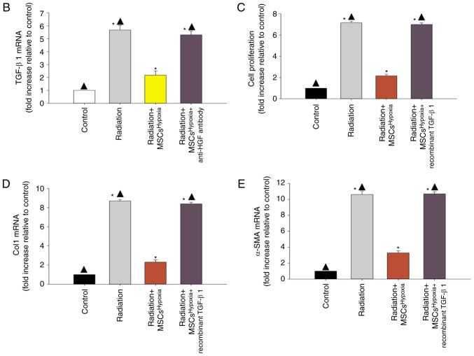
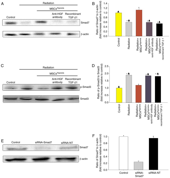
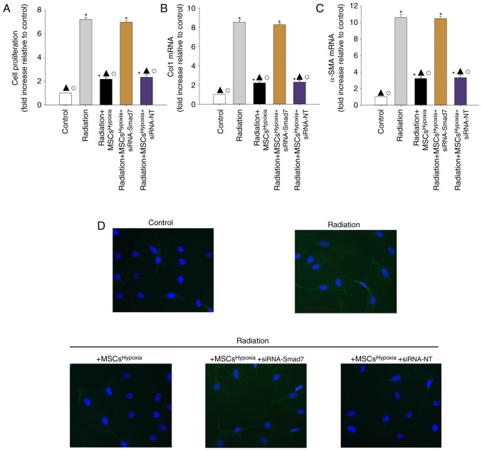
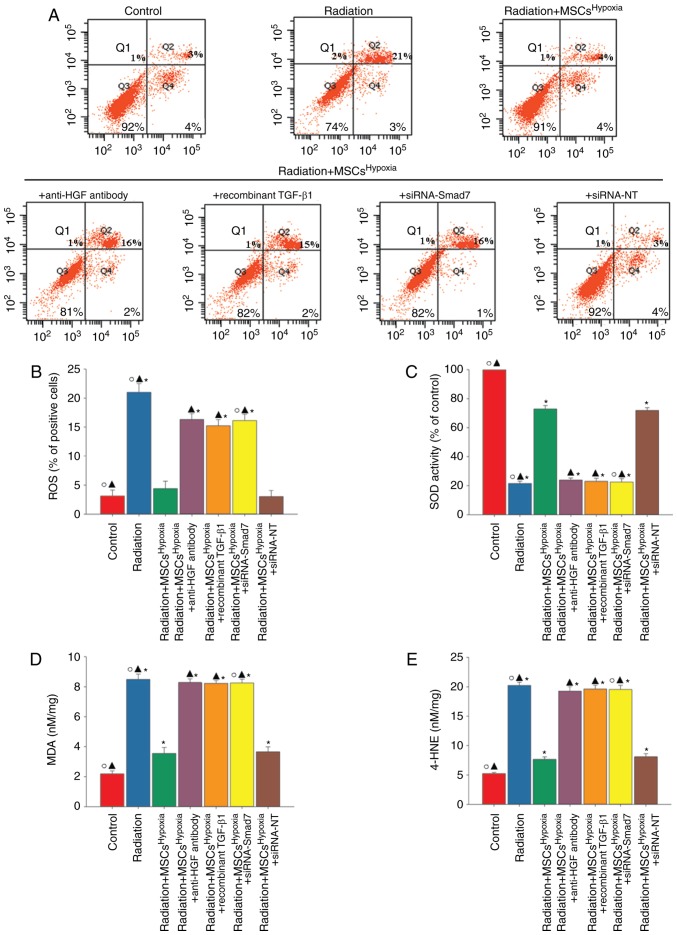
Cardiac fibrosis is a pathological consequence of radiation‑induced fibroblast proliferation and fibroblast‑to‑myofibroblast transition (FMT). Mesenchymal stem cell (MSC) transplantation has been revealed to be an effective treatment strategy to inhibit cardiac fibrosis. We identified a novel MSC‑driven mechanism that inhibited cardiac fibrosis, via the regulation of multiple fibrogenic pathways. Hypoxia pre‑conditioned MSCs (MSCsHypoxia) were co‑cultured with fibroblasts using a Transwell system. Radiation‑induced fibroblast proliferation was assessed using an MTT assay, and FMT was confirmed by assessing the mRNA levels of various markers of fibrosis, including type I collagen (Col1) and alpha smooth muscle actin (α‑SMA). α‑SMA expression was also confirmed via immunocytochemistry. The expression levels of Smad7 and Smad3 were detected by western blotting, and Smad7 was silenced using small interfering RNAs. The levels of oxidative stress following radiation were assessed by the detection of reactive oxygen species (ROS) and the activity of superoxide dismutase (SOD), malondialdehyde (MDA), and 4‑hydroxynonenal (HNE). It was revealed that co‑culturing with MSCsHypoxia could inhibit fibroblast proliferation and FMT. In addition, the present results indicated that MSCs are necessary and sufficient for the inhibition of fibroblast proliferation and FMT by functionally targeting TGF‑β1/Smad7/Smad3 signaling via the release of hepatocyte growth factor (HGF). Furthermore, it was observed that MSCs inhibited fibrosis by modulating oxidative stress. Co‑culturing with MSCsHypoxia alleviated fibroblast proliferation and FMT via the TGF‑β1/Smad7/Smad3 pathway. MSCs may represent a novel therapeutic approach for the treatment of radiation‑related cardiac fibrosis.


Figures

Figure 1.
Figure 1.
Effects of MSCsHypoxia on radiation-induced fibroblast proliferation and FMT. The following conditions were assessed: Fibroblasts alone, fibroblasts treated with radiation, and fibroblasts co-cultured with MSCsHypoxia or MSCsNormoxia in the presence of radiation. (A) Proliferation growth curves as determined by an MTT assay. (B and C) Col1 and α-SMA mRNA levels analyzed by RT-qPCR. (D) Expression of α-SMA was assessed by immunofluorescence staining. Each column represents the mean ± SD of three independent experiments; *P<0.05 vs. the Control; ▲P<0.05 vs. Radiation + MSCsHypoxia. MSCs, mesenchymal stem cells; FMT, fibroblast-to-myofibroblast transition; Col1, type I collagen; α-SMA, α-smooth muscle actin.
Figure 2.
Figure 2.
Involvement of HGF in MSCsHypoxia-driven inhibition of fibroblast proliferation and radiation-induced FMT. To explore the role of the anti-fibrotic effect of MSCsHypoxia on radiation-treated fibroblast cells, a Transwell co-culture system was used. (A) ELISA measuring the release of HGF into the condition media of fibroblasts, fibroblasts treated with radiation, and fibroblasts co-cultured with MSCsHypoxia or MSCsNormoxia in the presence of radiation. (B) HGF mRNA levels in fibroblasts, fibroblasts treated with radiation, and fibroblasts co-cultured with MSCsHypoxia or MSCsNormoxia in the presence of radiation were analyzed by RT-qPCR. Each column represents the mean ± SD from three independent experiments; *P<0.05 vs. the Control; ▲P<0.05 vs. Radiation + MSCsHypoxia. In fibroblasts, fibroblasts treated with radiation, and fibroblasts co-cultured with MSCsHypoxia or MSCsHypoxia + anti-HGF antibody in the presence of radiation, (C) proliferation growth curves were determined using an MTT assay. (D and E) Col1 and α-SMA mRNA levels as analyzed by RT-qPCR. (F) Expression of α-SMA was measured using immunofluorescence staining. Each column represents the mean ± SD from three independent experiments; *P<0.05 vs. the Control; ▲P<0.05 vs. Radiation + MSCsHypoxia. HGF, hepatocyte growth factor; MSCs, mesenchymal stem cells; FMT, fibroblast-to-myofibroblast transition; Col1, type I collagen; α-SMA, α-smooth muscle actin.
Figure 3.
Figure 3.
Identification of TGF-β1 as a modulator of radiation-induced fibroblast proliferation and FMT. (A) Heat map of RNAs differentially regulated by radiation in fibroblasts. ‘Red’ indicates upregulation, and ‘blue’ indicates downregulation. (B) RT-qPCR validation of differentially regulated RNAs in fibroblasts, fibroblasts treated with radiation, and fibroblasts co-cultured with MSCsHypoxia or MSCsHypoxia + anti-HGF antibody in the presence of radiation. *P<0.05 vs. the Control; P<0.05 vs. Radiation + MSCsHypoxia. In fibroblasts, fibroblasts treated with radiation, and fibroblasts co-cultured with MSCsHypoxia or MSCsHypoxia + recombinant TGF-β1 in the presence of radiation, (C) proliferation growth curves were determined using an MTT assay. (D and E) Col1 and α-SMA mRNA levels as analyzed by RT-qPCR. Each column represents the mean ± SD from three independent experiments; *P<0.05 vs. the Control; P<0.05 vs. Radiation + MSCsHypoxia. FMT, fibroblast-to-myofibroblast transition; MSCs, mesenchymal stem cells; HGF, hepatocyte growth factor; Col1, type I collagen; α-SMA, α-smooth muscle actin.
Figure 3.
Figure 3.
Identification of TGF-β1 as a modulator of radiation-induced fibroblast proliferation and FMT. (A) Heat map of RNAs differentially regulated by radiation in fibroblasts. ‘Red’ indicates upregulation, and ‘blue’ indicates downregulation. (B) RT-qPCR validation of differentially regulated RNAs in fibroblasts, fibroblasts treated with radiation, and fibroblasts co-cultured with MSCsHypoxia or MSCsHypoxia + anti-HGF antibody in the presence of radiation. *P<0.05 vs. the Control; P<0.05 vs. Radiation + MSCsHypoxia. In fibroblasts, fibroblasts treated with radiation, and fibroblasts co-cultured with MSCsHypoxia or MSCsHypoxia + recombinant TGF-β1 in the presence of radiation, (C) proliferation growth curves were determined using an MTT assay. (D and E) Col1 and α-SMA mRNA levels as analyzed by RT-qPCR. Each column represents the mean ± SD from three independent experiments; *P<0.05 vs. the Control; P<0.05 vs. Radiation + MSCsHypoxia. FMT, fibroblast-to-myofibroblast transition; MSCs, mesenchymal stem cells; HGF, hepatocyte growth factor; Col1, type I collagen; α-SMA, α-smooth muscle actin.
Figure 4.
Figure 4.
Co-culture with MSCsHypoxia induces modulation of the TGF-β1/Smad signaling pathway. Representative images of western blots of (A and B) Smad7 and β-actin expression and (C and D) p-Smad3 and Smad3 from fibroblasts, fibroblasts treated with radiation, and fibroblasts co-cultured with MSCsHypoxia or MSCsHypoxia + anti-HGF antibody or recombinant TGF-β1. Each column represents the mean ± SD from three independent experiments; *P<0.05 vs. the Control; P<0.05 vs. Radiation + MSCsHypoxia. (E and F) Fibroblasts were transfected with siRNA-Smad7, or with siRNA-NT as a control. siRNA-mediated transfection efficiency was determined by western blotting. Each column represents the mean ± SD from three independent experiments; *P<0.05 vs. siRNA-Smad7. MSCs, mesenchymal stem cells; HGF, hepatocyte growth factor.
Figure 5.
Figure 5.
Co-culturing with MSCsHypoxia functionally targets Smads downstream of TGF-β1 to reduce fibroblast proliferation and FMT. Fibroblasts were transfected with siRNA against Smad7, or with siRNA-NT as a control, followed by co-culture with MSCsHypoxia in the presence of radiation. In parallel experiments, fibroblasts were treated with radiation, or co-cultured with MSCsHypoxia in the presence of radiation. Fibroblasts under normal culture conditions were used as the control. (A) Proliferation growth curves as determined using an MTT assay. (B and C) Col1 and α-SMA mRNA levels as analyzed by RT-qPCR. (D) Expression of α-SMA as measured by immunofluorescence staining. Each column represents the mean ± SD from three independent experiments; *P<0.05 vs. the Control; P<0.05 vs. Radiation; P<0.05 vs. Radiation + MSCsHypoxia + siRNA-Smad7. MSCs, mesenchymal stem cells; FMT, fibroblast-to-myofibroblast transition; Col1, type I collagen; α-SMA, α-smooth muscle actin.
Figure 6.
Figure 6.
MSCHypoxia co-culturing attenuates radiation-induced oxidative stress in fibroblasts. Fibroblasts were transfected with siRNA against Smad7, or with siRNA-NT as a control, followed by co-culture with MSCsHypoxia in the presence of radiation. In parallel experiments, fibroblasts were treated with radiation, or co-cultured with MSCsHypoxia or MSCsHypoxia + anti-HGF antibody or MSCsHypoxia + recombinant TGF-β1 in the presence of radiation. Fibroblasts under normal culture conditions were used as the control. (A and B) Intracellular ROS production was assessed using a ROS detection kit, and was analyzed using flow cytometry. (C) SOD activity evaluated by a colorimetric assay. (D) Lipid peroxidation as evaluated by MDA formation. (E) Quantification of 4-HNE levels. *P<0.05 vs. the Control; P<0.05 vs. Radiation + MSCsHypoxia; P<0.05 vs. Radiation + MSCsHypoxia + siRNA-Smad7. MSCs, mesenchymal stem cells; HGF, hepatocyte growth factor; ROS, reactive oxygen species; SOD, superoxide dismutase; MDA, malondialdehyde; HNE, 4-hydroxynonenal.

References

    1. Swerdlow AJ, Higgins CD, Smith P, Cunningham D, Hancock BW, Horwich A, Hoskin PJ, Lister A, Radford JA, Rohatiner AZ, Linch DC. Myocardial infarction mortality risk after treatment for Hodgkin disease: A collaborative British cohort study. J Natl Cancer Inst. 2007;99:206–214. doi: 10.1093/jnci/djk029. - DOI - PubMed
    1. Darby SC, Ewertz M, McGale P, Bennet AM, Blom-Goldman U, Brønnum D, Correa C, Cutter D, Gagliardi G, Gigante B, et al. Risk of ischemic heart disease in women after radiotherapy for breast cancer. N Engl J Med. 2013;368:987–998. doi: 10.1056/NEJMoa1209825. - DOI - PubMed
    1. Darby S, McGale P, Peto R, Granath F, Hall P, Ekbom A. Mortality from cardiovascular disease more than 10 years after radiotherapy for breast cancer: Nationwide cohort study of 90000 Swedish women. BMJ. 2003;326:256–257. doi: 10.1136/bmj.326.7383.256. - DOI - PMC - PubMed
    1. Tukenova M, Guibout C, Oberlin O, Doyon F, Mousannif A, Haddy N, Guérin S, Pacquement H, Aouba A, Hawkins M, et al. Role of cancer treatment in long-term overall and cardiovascular mortality after childhood cancer. J Clin Oncol. 2010;28:1308–1315. doi: 10.1200/JCO.2008.20.2267. - DOI - PubMed
    1. Tapio S. Pathology and biology of radiation-induced cardiac disease. J Radiat Res. 2016;57:439–448. doi: 10.1093/jrr/rrw064. - DOI - PMC - PubMed

MeSH terms