. 2019 Oct;55(4):833-844.
doi: 10.3892/ijo.2019.4864. Epub 2019 Aug 30.

ROCK1 promotes migration and invasion of non‑small‑cell lung cancer cells through the PTEN/PI3K/FAK pathway

Affiliations

ROCK1 promotes migration and invasion of non‑small‑cell lung cancer cells through the PTEN/PI3K/FAK pathway

Changpeng Hu et al. Int J Oncol. 2019 Oct.

Abstract

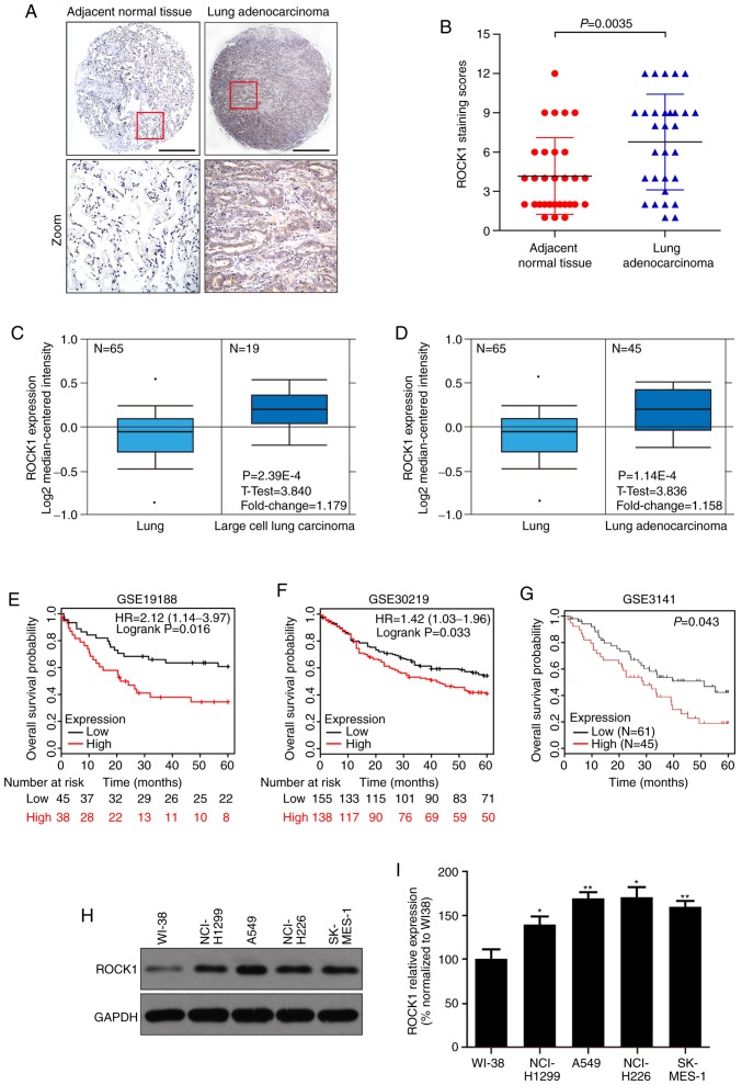
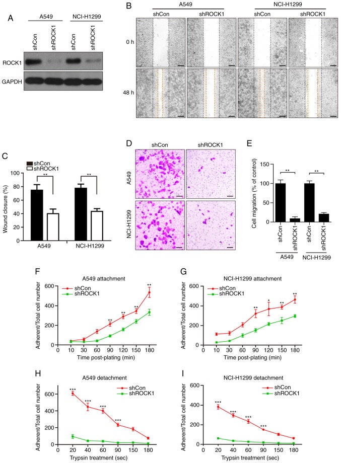
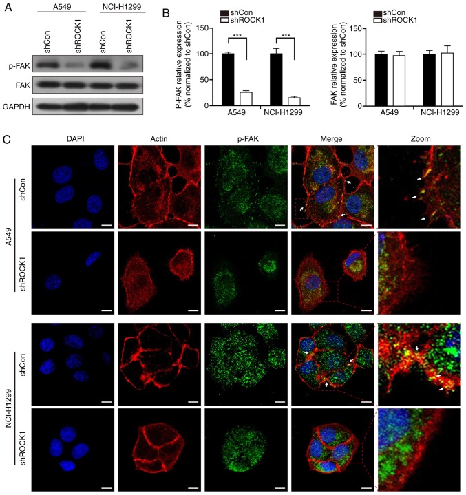
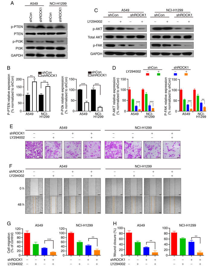
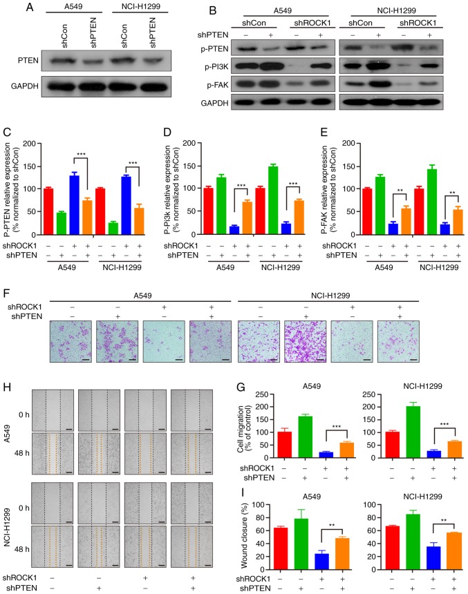
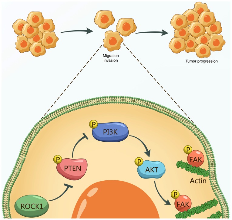
Rho‑associated protein kinase 1 (ROCK1), a member of the ROCK family, serves an important function in cell migration and invasion in neoplasms. ROCK1 has been found to be overexpressed in several types of cancers. However, the role of ROCK1 in non‑small‑cell lung cancer (NSCLC) is poorly understood. In the present study, ROCK1 was found to be overexpressed in NSCLC cells and tissues, and it was associated with poor survival of NSCLC patients. Subsequently, ROCK1 knockdown NSCLC cell lines were established using shRNA. ROCK1 knockdown significantly reduced the migration and invasion ability in the cell monolayer scratching and Transwell assays. ROCK1 knockdown was also found to markedly inhibit cell adhesion ability. Moreover, the phosphorylation of focal adhesion kinase (FAK) was inhibited by ROCK1 knockdown, reducing NSCLC cell migration and invasion ability. This mechanistic study revealed that ROCK1 significantly enhanced cell migration and invasion by inhibiting the phosphatase and tensin homolog (PTEN)/phosphoinositide 3‑kinase (PI3K)/FAK pathway. More importantly, the interruption of the PTEN/PI3K/FAK pathway markedly rescued the inhibition of cell migration and invasion mediated by ROCK1 knockdown. Taken together, these results suggest a novel role for ROCK1 in cell migration and invasion by inhibiting cell adhesion ability, and indicate that ROCK1 may be of value as a therapeutic target for the treatment of NSCLC.


Figures

Figure 1
Figure 1
ROCK1 is overexpressed in NSCLC, and higher expression of ROCK1 predicts worse survival. The expression of ROCK1 was measured in lung adenocarcinoma tissues and cells. (A) Representative immunohistochemical images of ROCK1 in lung adenocarcinoma and adjacent normal tissues. Scale bars: 100 µm. (B) Expression level of ROCK1 in lung adenocarcinoma and adjacent normal tissues (n=30, **P=0.0035). (C and D) The expression of ROCK1 is upregulated in NSCLC tissues compared with non-tumor tissues. All the data (fold change and P-values) were calculated from Oncomine (www.oncomine.org). (E-G) The survival analysis was performed by the Kaplan-Meier method in the GSE19188 (n=83, *P=0.016), GSE30219 (n=293, *P=0.033) and GSE3141 datasets (n=106, *P=0.043). (H-I) The expression level of ROCK1 was detected by western blotting in NSCLC cell lines (NCI-H1299, A549, NCI-H226 and SK-MES-1) and human embryonic lung fibroblasts WI38. The relative densities of ROCK1 were normalized to GAPDH by densitometric analysis using Quantity One software (*P<0.05, **P<0.01, vs. the WI-38 group). The results represent three independent experiments. ROCK1, Rho-associated protein kinase 1; NSCLC, non-small-cell lung cancer.
Figure 2
Figure 2
ROCK1 promotes the migration and invasion of NSCLC A549 and NCI-H1299 cells. (A) ROCK1 knockdown efficiency of the shRNA was measured by western blotting. (B and C) The role of ROCK1 in A549 and NCI-H1299 cell migration was evaluated by the wound healing assay. The wound closure distance was measured in 3 randomly selected fields. Scale bars: 200 µm. (D and E) The Transwell assay was employed to evaluate the effects of shROCK1 on cell invasion; the number of invading cells was counted from three independent experiments. Scale bars: 200 µm. (F) A549 and (G) NCI-H1299 cells were seeded in fibronectin-coated plates and cultured for 10-180 min; the adherent cells were trypsinized and counted at each time point. (H and I) Cells were cultured in 12-well plates (1×10³ cells per well) for 24 h, then trypsinized with a diluted trypsin solution (trypsin:PBS, 1:19) for different time intervals (20-180 sec). The remaining adherent cells were digested and counted. Each experiment was repeated three times. All data are presented as the mean ± standard deviation (*P<0.05, **P<0.01, ***P<0.001). ROCK1, Rho-associated protein kinase 1; NSCLC, non-small-cell lung cancer.
Figure 3
Figure 3
ROCK1 regulates the phosphorylation of FAK in NSCLC cells. (A and B) The expression levels of FAK and p-FAK were detected by western blotting after ROCK1 knockdown. The relative protein expression was analyzed by Quantity One software (***P<0.001). The results represent three independent experiments. (C) Cells were seeded on coverslips and immunostained with actin (red), p-FAK (green) and DAPI (blue). The arrows indicate the lamellipodia (colocalization of p-FAK with actin) on the cell membrane. Scale bars: 10 µm. ROCK1, Rho-associated protein kinase 1; NSCLC, non-small-cell lung cancer; FAK, focal adhesion kinase.
Figure 4
Figure 4
Effects of ROCK1 on the PI3K signaling pathway. (A) Following transfection with ROCK1 shRNA, the expression levels of p-PTEN/PTEN and p-PI3K/PI3K were analyzed by western blotting in A549 and NCI-H1299 cells. (B) The ratio of these proteins was analyzed by Quantity One software (**P<0.01, ***P<0.001). The results represent three independent experiments. (C) LY294002 was added to A549 and NCI-H1299 cells, and the expression of p-AKT/AKT and p-FAK was detected by western blotting. (D) The ratio of these protein expressions was analyzed by Quantity One software (**P<0.01, ***P<0.001). The results represent three independent experiments. (E and G) Invasion of A549 and NCI-H1299 cells was measured by the Transwell assay, and the invading cells were counted in three independent fields (**P<0.01). (F and H) Migration was measured by the cell monolayer scratching assay, and wound closure was measured in randomly selected fields (**P<0.01). Scale bars: 200 µm. ROCK1, Rho-associated protein kinase 1; FAK, focal adhesion kinase; PI3K, phosphoinositide 3 kinase; PTEN, phosphatase and tensin homolog.
Figure 5
Figure 5
PTEN is an important intermediate regulator in ROCK1-mediated PI3K/FAK pathway activation. (A) The PTEN knockdown efficiency of the shRNA was measured by western blotting. (B) Stable cell lines (shROCK1, shPTEN and shROCK1 + shPTEN) were prepared and analyzed for the expression of p-PTEN, p-PI3K and p-FAK by western blotting. The relative expression of (C) p-PTEN, (D) p-PI3K and (E) p-FAK was analyzed by Quantity One software (**P<0.01, ***P<0.001). The results represent three independent experiments. (F-G) Invasion of A549 and NCI-H1299 cells was measured by the Transwell assay, and the invading cells were analyzed in three independent experiments (***P<0.001). (H-I) Furthermore, migration was measured by the cell monolayer scratching assay, and the wound closure was measured at three randomly selected fields (**P<0.01). PTEN, phosphatase and tensin homolog; PI3K, phosphoinositide 3 kinase; ROCK1, Rho-associated protein kinase 1; FAK, focal adhesion kinase.
Figure 6
Figure 6
Mechanism of ROCK1 promoting tumor cell migration, invasion and progression in NSCLC. ROCK1 reduces the activation/phosphorylation of PTEN and then phosphorylates PI3K/AKT, resulting in FAK phosphorylation and leading to accelerated cell migration/invasion and promotion of NSCLC progression. ROCK1, Rho-associated protein kinase 1; NSCLC, non-small-cell lung cancer; PTEN, phosphatase and tensin homolog; PI3K, phosphoinositide 3 kinase; FAK, focal adhesion kinase.

References

    1. Chen W, Zheng R, Baade PD, Zhang S, Zeng H, Bray F, Jemal A, Yu XQ, He J. Cancer statistics in China, 2015. CA Cancer J Clin. 2016;66:115–132. doi: 10.3322/caac.21338. - DOI - PubMed
    1. Siegel RL, Miller KD, Jemal A. Cancer statistics, 2018. CA Cancer J Clin. 2018;68:7–30. doi: 10.3322/caac.21442. - DOI - PubMed
    1. She K, Yan H, Huang J, Zhou H, He J. miR-193b availability is antagonized by LncRNA-SNHG7 for FAIM2-induced tumour progression in non-small cell lung cancer. Cell Prolif: 2018;51 doi: 10.1111/cpr.12406. - DOI - PMC - PubMed
    1. Yang J, Zhao H, Xin Y, Fan L. MicroRNA-198 inhibits proliferation and induces apoptosis of lung cancer cells via targeting FGFR1. J Cell Biochem. 2014;115:987–995. doi: 10.1002/jcb.24742. - DOI - PubMed
    1. Kohler J. Second-line treatment of NSCLC-the Pan-ErbB inhibitor afatinib in times of shifting paradigms. Front Med (Lausanne) 2017;4:9. - PMC - PubMed

MeSH terms