. 2019 Oct;20(4):3487-3498.
doi: 10.3892/mmr.2019.10610. Epub 2019 Aug 23.

Rsf‑1 regulates malignant melanoma cell viability and chemoresistance via NF‑κB/Bcl‑2 signaling

Affiliations

Rsf‑1 regulates malignant melanoma cell viability and chemoresistance via NF‑κB/Bcl‑2 signaling

Jiani He et al. Mol Med Rep. 2019 Oct.

Abstract

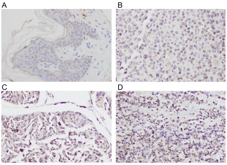
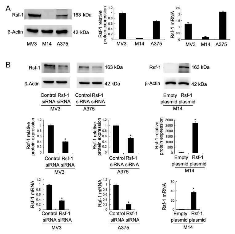
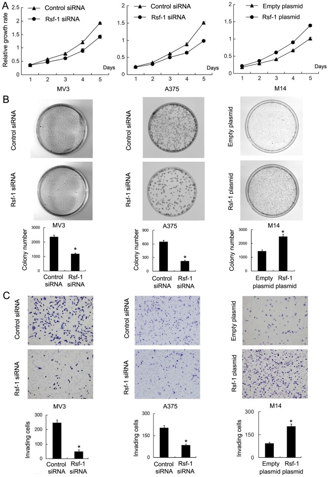
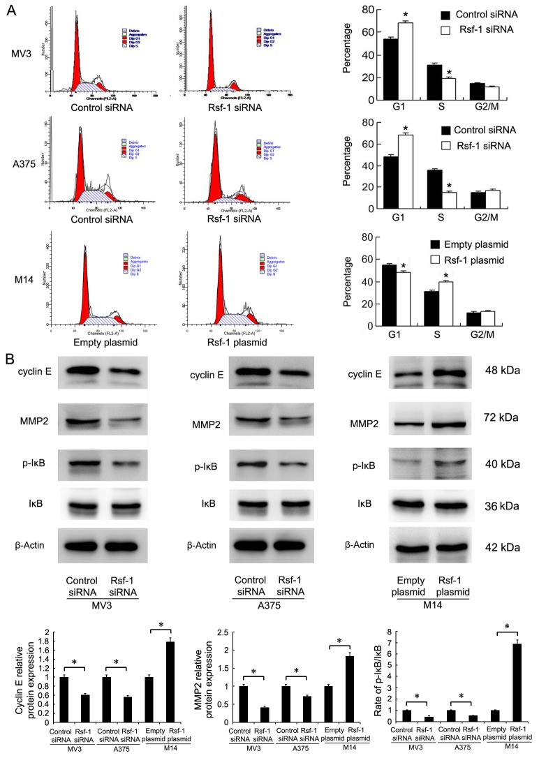
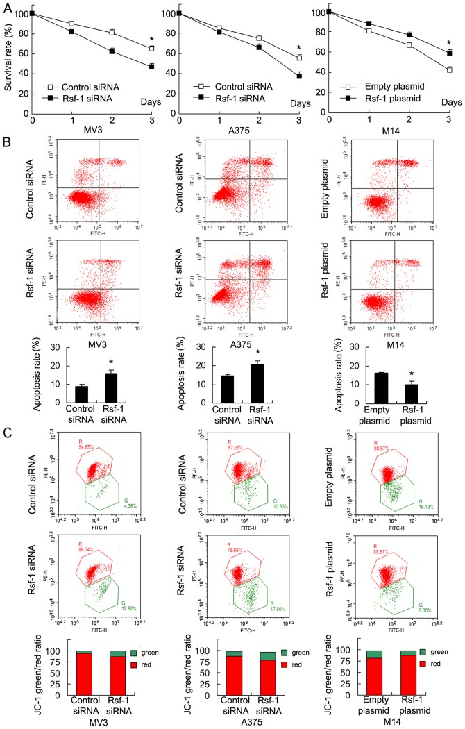
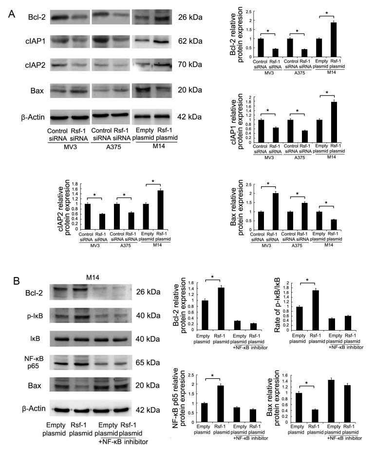
Remodeling and spacing factor 1 (Rsf‑1) has been reported as overexpressed in numerous cancers; however, its expression, biological functions and mechanisms in malignant melanoma remain unknown. In the present study, the expression of Rsf‑1 was investigated in 50 cases of malignant melanoma samples using immunohistochemistry. The results revealed that Rsf‑1 expression was elevated in 38% of specimens. MTT, colony formation, Transwell and flow cytometry assays were performed to investigate the functions of Rsf‑1. Knockdown of Rsf‑1 in the MV3 and A375 melanoma cell lines decreased the viability, invasion and cell cycle transition of cells. Conversely, overexpression of Rsf‑1 in M14 cells with low endogenous Rsf‑1 expression induced opposing effects. Further analysis revealed that Rsf‑1 knockdown decreased matrix metalloproteinase‑2, cyclin E and phosphorylated‑IκB expression. Additionally, Rsf‑1 depletion reduced cisplatin resistance and significantly increased the cisplatin‑associated apoptotic rate, whereas Rsf‑1 overexpression exhibited opposing effects. Rsf‑1 also maintained the mitochondrial membrane potential following cisplatin treatment. Analysis of apoptosis‑associated proteins revealed that Rsf‑1 positively regulated B‑cell lymphoma 2 (Bcl‑2), cellular inhibitor of apoptosis 1 (cIAP1) and cIAP2, and downregulated Bcl‑2‑associated X protein expression. Nuclear factor κ‑light‑chain‑enhancer of activated B‑cells (NF‑κB) inhibition reversed the effects of Rsf‑1 on Bcl‑2. In conclusion, Rsf‑1 was overexpressed in malignant melanoma and may contribute to the malignant behaviors of melanoma cells, possibly via the regulation of NF‑κB signaling. Therefore, Rsf‑1 may be a potential therapeutic target in the treatment of malignant melanoma.


Figures

Figure 1.
Figure 1.
Expression of Rsf-1 in melanoma tissue. (A) Negative Rsf-1 staining observed in normal skin tissue. Moderate Rsf-1 staining was detected in cases of melanoma tissue of stages (B) II and (C) III. (D) Strong Rsf-1 staining was detected in a case of melanoma tissue in stage IV. Magnification, ×400. Rsf-1, remodeling and spacing factor 1.
Figure 2.
Figure 2.
Rsf-1 expression in melanoma cell lines and Rsf-1 knockdown efficiency. (A) Western blotting and RT-qPCR analysis revealed the endogenous expression levels of Rsf-1 in three melanoma cell lines (MV3, M14 and A375). (B) Western blotting and RT-qPCR analysis demonstrated that Rsf-1 siRNA transfection significantly decreased Rsf-1 expression levels in MV3 and A375 cells, while Rsf-1 plasmid transfection upregulated the protein and mRNA expression of Rsf-1 in M14 cells. Data were presented as the mean ± standard deviation of at least three experiments. *P<0.05 vs. control. Rsf-1, remodeling and spacing factor 1; RT-qPCR, reverse transcription-quantitative polymerase chain reaction; siRNA, small interfering RNA.
Figure 3.
Figure 3.
Rsf-1 regulates melanoma cell viability and invasion. (A) An MTT assay (96-well plate) revealed that Rsf-1 depletion decreased the viability of MV3 and A375 cells; conversely, Rsf-1 overexpression increased M14 cell viability. (B) A colony formation assay (culture dish diameter, 6 cm) demonstrated that the colony number was reduced in MV3 and A375 cells transfected with Rsf-1 siRNA, while Rsf-1 overexpression promoted colony formation ability in M14 cells. (C) A Transwell invasion assay (24-well plate) revealed that the number of invading cells decreased following Rsf-1 depletion in MV3 and A375, and increased following Rsf-1 overexpression in M14 cells. Data were presented as the mean ± standard deviation of at least three experiments. *P<0.05 vs. control. Magnification, ×200. Rsf-1, remodeling and spacing factor 1; siRNA, small interfering RNA.
Figure 4.
Figure 4.
Rsf-1 regulates cell cycle progression of melanoma and expression of MMP2, cyclin E and p-IκB. (A) Cell cycle analysis revealed that Rsf-1 depletion increased the percentage of G1 phase cells and decreased that of S phase cells in MV3 and A375 cell groups; Rsf-1 overexpression in M14 cells exhibited opposing effects. (B) Western blotting demonstrated that Rsf-1 depletion decreased the levels of MMP2, cyclin E and p-IκB in MV3 and A375 cell lines. Rsf-1 overexpression upregulated expression of MMP2, cyclin E and p-IκB in M14 cells. Data were presented as the mean ± standard deviation of at least three experiments. *P<0.05 vs. control. MMP2, matrix metalloproteinase-2; p, phosphorylated; Rsf-1, remodeling and spacing factor 1; siRNA, small interfering RNA.
Figure 5.
Figure 5.
Rsf-1 regulates chemoresistance and the MMP of melanoma cells. (A) An MTT assay revealed that cell viability was decreased following Rsf-1 depletion in MV3 and A375 cells treated with cisplatin. Rsf-1 overexpression promoted cell viability in M14 cells treated with cisplatin. (B) Annexin V/propidium iodide analysis revealed that the percentage of apoptotic cells was significantly increased in Rsf-1-depleted MV3 and A375 cells compared with controls. Rsf-1 overexpression downregulated cisplatin-induced apoptosis in M14 cells. (C) Rsf-1 overexpression reduced MMP depolarization in M14 cells, while Rsf-1 depletion increased depolarization in MV3 and A375 cells treated with cisplatin. Data were presented as the mean ± standard deviation of at least three experiments. *P<0.05 vs. control. FITC, fluorescein isothiocyanate; MMP, mitochondrial membrane potential, Rsf-1, remodeling and spacing factor 1; siRNA, small interfering RNA.
Figure 6.
Figure 6.
Rsf-1 regulates Bcl-2 expression via NF-κB signaling. (A) Western blotting revealed that Bax expression levels increased, whereas cIAP1, cIAP2 and Bcl-2 expression decreased significantly following Rsf-1 depletion in MV3 and A375 cells. Rsf-1 overexpression in M14 cells exhibited opposing effects. (B) NF-κB inhibition significantly downregulated p-IκB and NF-κB p65 protein levels in M14 cells. NF-κB inhibition also eradicated the effects of Rsf-1 overexpression on Bcl-2 upregulation. Total IκB expression was markedly altered. Data were presented as the mean ± standard deviation of at least three experiments. *P<0.05 vs. control. Bax, Bcl-2-associated X protein; Bcl-2, B-cell lymphoma 2; cIAP1, cellular inhibitor of apoptosis protein 1; NF-κB, nuclear factor κ-light-chain-enhancer of activated B cells; p, phosphorylated; Rsf-1, remodeling and spacing factor 1; siRNA, small interfering RNA.

Similar articles

Cited by

References

    1. Merrill SJ, Subramanian M, Godar DE. Worldwide cutaneous malignant melanoma incidences analyzed by sex, age, and skin type over time (1955–2007): Is HPV infection of androgenic hair follicular melanocytes a risk factor for developing melanoma exclusively in people of European-ancestry? Dermatoendocrinol. 2016;8:e1215391. doi: 10.1080/19381980.2016.1215391. - DOI - PMC - PubMed
    1. Li H, Pedersen L, Nørgaard M, Ulrichsen SP, Thygesen SK, Nelson JJ. The occurrence of non-melanoma malignant skin lesions and non-cutaneous squamous-cell carcinoma among metastatic melanoma patients: An observational cohort study in Denmark. BMC Cancer. 2016;16:295. doi: 10.1186/s12885-016-2315-0. - DOI - PMC - PubMed
    1. Nahar VK, Allison Ford M, Brodell RT, Boyas JF, Jacks SK, Biviji-Sharma R, Haskins MA, Bass MA. Skin cancer prevention practices among malignant melanoma survivors: A systematic review. J Cancer Res Clin Oncol. 2016;142:1273–1283. doi: 10.1007/s00432-015-2086-z. - DOI - PMC - PubMed
    1. Peterson M, Albertini MR, Remington P. Remington, incidence, survival, and mortality of malignant cutaneous melanoma in wisconsin, 1995–2011. WMJ. 2015;114:196–201. - PubMed
    1. Johnson-Obaseki SE, Labajian V, Corsten MJ, McDonald JT. Incidence of cutaneous malignant melanoma by socioeconomic status in Canada: 1992–2006. J Otolaryngol Head Neck Surg. 2015;44:53. doi: 10.1186/s40463-015-0107-1. - DOI - PMC - PubMed