. 2019 Oct;20(4):3782-3792.
doi: 10.3892/mmr.2019.10636. Epub 2019 Sep 2.

lncRNA ROR promotes the progression of renal cell carcinoma through the miR‑206/VEGF axis

Affiliations

lncRNA ROR promotes the progression of renal cell carcinoma through the miR‑206/VEGF axis

Jianguo Shi et al. Mol Med Rep. 2019 Oct.

Retraction in

Abstract

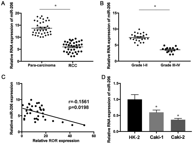
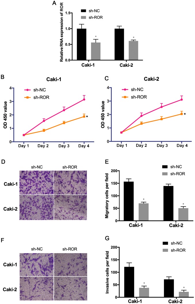
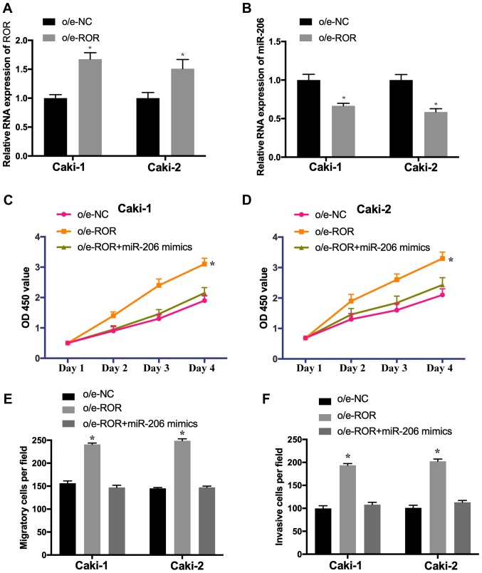
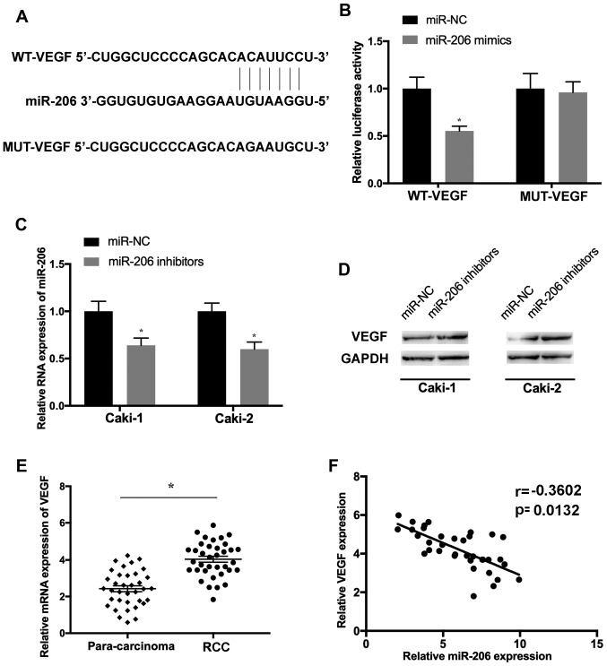
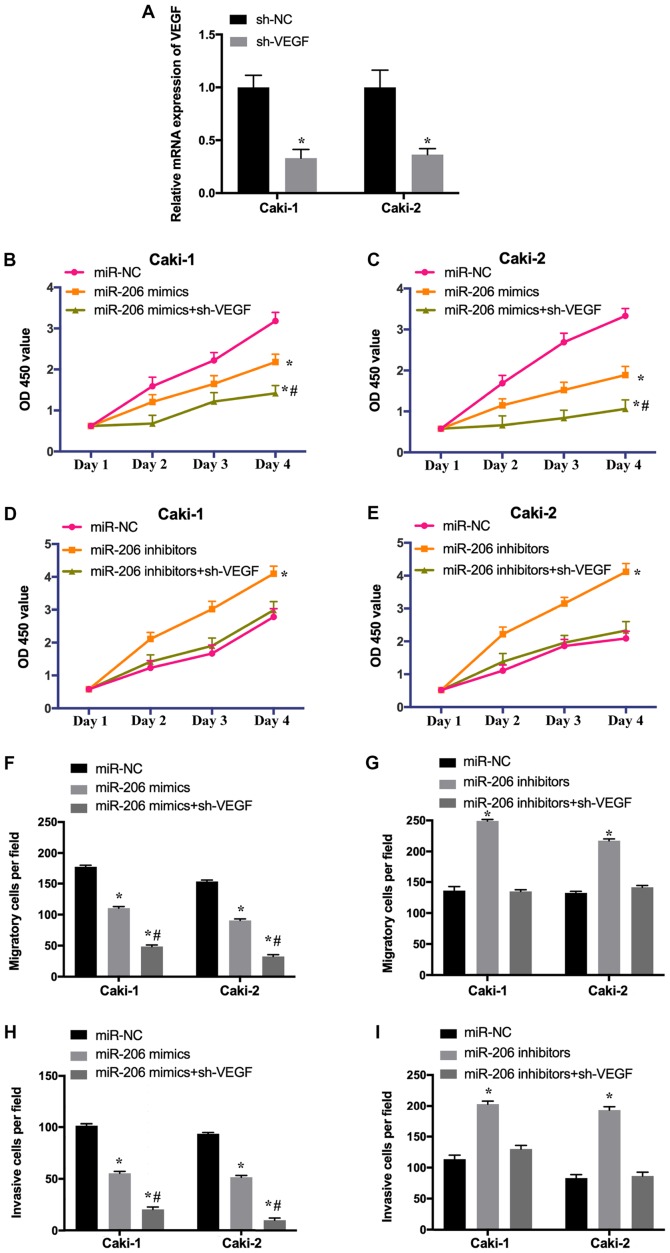
Renal cell carcinoma (RCC) is the most common kidney malignancy, responsible for ~80% of all cases in adults. The pathogenesis of RCC is complex, involving alterations at both the genetic and epigenetic levels. Numerous signaling pathways, such as PI3K/Akt/mTOR and Wnt‑β‑catenin have been demonstrated to be associated with the tumorigenesis and development of RCC. Long non‑coding RNAs (lncRNAs) are functional RNA molecules involved in the initiation and progression of cancer, and investigating the effects of lncRNA could facilitate the development of novel treatments. The lncRNA regulator of reprogramming (ROR) is aberrantly expressed in a variety of tumors. However, its underlying mechanisms remain largely unknown. In the present study, ROR was found to be upregulated and microRNA (miR)‑206 was found to be downregulated in RCC tissues and cells. Furthermore, the knockdown of ROR inhibited the proliferation, migration and invasion of RCC cells. It was found that ROR binds to miR‑206, and that ROR‑induced cell proliferation and metastasis were reversed by the overexpression of miR‑206. In addition, the levels of miR‑206 and ROR were negatively correlated in RCC tissues. Furthermore, the overexpression of miR‑206 notably suppressed the proliferation, migration and invasion of RCC cells, and these effects were enhanced by the knockdown of vascular endothelial growth factor (VEGF); cell growth and metastasis induced by miR‑206 inhibitors could be reversed by the knockdown of VEGF. In addition, the expression levels of miR‑206 and VEGF were inversely correlated in RCC samples. In summary, the results of the present study revealed that ROR was upregulated in RCC tissues, which promoted tumor progression by regulating the miR‑206/VEGF axis. The present findings provided a novel insight into the potential functions of ROR in RCC, and the ROR/miR‑206/VEGF pathway may be a promising therapeutic target for the treatment of patients with RCC.


Figures

Figure 1.
Figure 1.
miR-206 is downregulated in RCC tissues and cells. (A) The expression level of miR-206 determined in 36 RCC and matched non-tumor samples using reverse transcription-quantitative PCR. (B) miR-206 expression was determined in patients with different stages of RCC. (C) The RNA levels of ROR and miR-206 were inversely correlated in RCC tissues (r=−0.1561; P=0.0198). (D) The expression levels of miR-206 were determined in normal human kidney cells (HK-2) and RCC cell lines (Caki-1 and Caki-2). *P<0.05 vs. HK-2 cell line. miR, microRNA; RCC, renal cell carcinoma; ROR, long non-coding RNA regulator of reprogramming.
Figure 2.
Figure 2.
Downregulation of ROR suppresses the proliferation, migration and invasion of RCC cells. (A) The transfection efficiency of sh-ROR was accessed using reverse transcription quantitative PCR. The proliferation rate of (B) Caki-1 and (C) Caki-2 cells transfected with sh-ROR or sh-NC was determined using the Cell Counting Kit-8 assay. (D) The migration rate of transfected Caki-1 and Caki-2 cells was evaluated (magnification, ×200) and (E) quantified using a Transwell assay. (F) The invasive abilities of Caki-1 and Caki-2 cells transfected with sh-ROR or sh-NC were determined (magnification, ×200) and (G) quantified. *P<0.05 vs. sh-NC. NC, negative control; sh-, short hairpin RNA; ROR, long non-coding RNA regulator of reprogramming.
Figure 3.
Figure 3.
miR-206 is a target of ROR in RCC. (A) The putative binding sites of miR-206 on the ROR transcript. (B) Overexpression of miR-206 significantly reduced the luciferase activity of WT-ROR but not of MUT-ROR. The expression levels of miR-206 in Caki-1 and Caki-2 cells transfected with (C) sh-ROR and (D) miR-206 mimics were determined using RT-qPCR. *P<0.05 vs. respective control. miR, microRNA; NC, negative control; MUT, mutant; WT, wild-type; sh-, short hairpin RNA; ROR, long non-coding RNA regulator of reprogramming; RCC, renal cell carcinoma.
Figure 4.
Figure 4.
o/e-ROR promotes the proliferation, migration and invasion of RCC cells through miR-206. The expression levels of (A) ROR and (B) miR-206 in Caki-1 and Caki-2 cells transfected with o/e-NC or o/e-ROR were evaluated using reverse-transcription-quantitative PCR. The proliferation of (C) Caki-1 and (D) Caki-2 cells transfected with o/e-NC, o/e-ROR or co-transfected with o/e-ROR and miR-206 mimics was evaluated using the Cell Counting Kit-8 assay. The (E) migration and (F) invasion of transfected Caki-1 and Caki-2 cells were evaluated using a Transwell assay. *P<0.05 vs. o/e-NC. miR, microRNA; NC, negative control; sh-, short hairpin RNA; ROR, long non-coding RNA regulator of reprogramming; RCC, renal cell carcinoma; o/e, overexpression.
Figure 5.
Figure 5.
VEGF is a target gene of miR-206 in RCC cells. (A) The putative binding sites of VEGF in the miR-206 transcript. (B) The overexpression of miR-206 resulted in a significant decrease in the luciferase activity of WT-VEGF, whereas no change was observed in MUT-VEGF. (C) The expression levels of miR-206 in Caki-1 and Caki-2 cells transfected with miR-NC or miR-206 inhibitors were evaluated using reverse transcription-quantitative PCR. (D) The protein level of VEGF in transfected Caki-1 and Caki-2 cells was determined using western blot analysis. (E) The expression of VEGF was assessed in RCC and matched para-carcinoma tissues. (F) The RNA levels of miR-206 and VEGF were inversely correlated in RCC samples (r=−0.3602; P=0.0132). *P<0.05 vs. respective control. RCC, renal cell carcinoma; miR, microRNA; NC, negative control; VEGF, vascular endothelial growth factor; MUT, mutant; WT, wild-type.
Figure 6.
Figure 6.
Downregulation of VEGF enhances the effects of the overexpression of miR-206 and reverses the effects of the miR-206 inhibitor in RCC cells. (A) The transfection efficiency of sh-VEGF was determined by reverse transcription-quantitative PCR. The proliferation of (B) Caki-1 and (C) Caki-2 cells transfected with miR-NC, miR-206 mimic or co-transfected with miR-206 mimic and sh-VEGF was evaluated using the Cell Counting Kit-8 assay. The proliferation of (D) Caki-1 and (E) Caki-2 cells transfected with miR-NC, miR-206 inhibitors or co-transfected with miR-206 inhibitors and sh-VEGF was assessed. The migration of Caki-1 and Caki-2 cells transfected with (F) miR-206 mimic or (G) miR-206 inhibitor and co-transfected with sh-VEGF were determined using a Transwell assay. The invasion of Caki-1 and Caki-2 cells transfected with (H) miR-206 mimic or (I) miR-206 inhibitor and co-transfected with sh-VEGF were was evaluated. *P<0.05 vs. miR-NC; #P<0.05 vs. miR-206 mimic. RCC, renal cell carcinoma; miR, microRNA; NC, negative control; VEGF, vascular endothelial growth factor; sh-, short hairpin.

References

    1. Dabestani S, Beisland C, Stewart GD, Bensalah K, Gudmundsson E, Lam TB, Gietzmann W, Zakikhani P, Marconi L, Fernandéz-Pello S, et al. Intensive imaging-based follow-up of surgically treated localised renal cell carcinoma does not improve post-recurrence survival: Results from a European multicentre database (RECUR) Eur Urol. 2019;75:261–264. doi: 10.1016/j.eururo.2018.10.007. - DOI - PubMed
    1. Sheth S, Scatarige JC, Horton KM, Corl FM, Fishman EK. Current concepts in the diagnosis and management of renal cell carcinoma: Role of multidetector ct and three-dimensional CT. Radiographics. 2001;21:S237–S254. doi: 10.1148/radiographics.21.suppl_1.g01oc18s237. - DOI - PubMed
    1. Chen W, Zheng R, Baade PD, Zhang S, Zeng H, Bray F, Jemal A, Yu XQ, He J. Cancer statistics in China, 2015. CA Cancer J Clin. 2016;66:115–132. doi: 10.3322/caac.21338. - DOI - PubMed
    1. Vera-Badillo FE, Conde E, Duran I. Chromophobe renal cell carcinoma: A review of an uncommon entity. Int J Urol. 2012;19:894–900. doi: 10.1111/j.1442-2042.2012.03079.x. - DOI - PubMed
    1. Di Cristofano C, Minervini A, Menicagli M, Salinitri G, Bertacca G, Pefanis G, Masieri L, Lessi F, Collecchi P, Minervini R, et al. Nuclear expression of hypoxia-inducible factor-1alpha in clear cell renal cell carcinoma is involved in tumor progression. Am J Surg Pathol. 2007;31:1875–1881. doi: 10.1097/PAS.0b013e318094fed8. - DOI - PubMed

Publication types