

Phosphorylated abacavir analogue (ABC-1) has ameliorative action against Newcastle disease virus induced pathogenesis in chicken
- PMID: 31486095
- PMCID: PMC7161887
- DOI: 10.1002/bab.1814
Phosphorylated abacavir analogue (ABC-1) has ameliorative action against Newcastle disease virus induced pathogenesis in chicken
Abstract
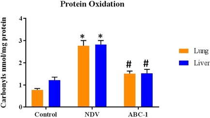
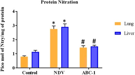
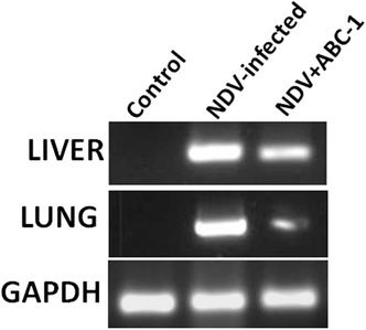
Newcastle disease virus (NDV) causes huge economic loss to the poultry industry due to high mortality and morbidity. The present study aimed to assess the protective role of novel phosphorylated analogue ABC-1 in vivo in NDV-infected chickens through the inhibition of fusion protein. Both NDV-induced oxidative damage and protective role of novel phosphorylated ABC-1 were evaluated in vital organs such as the liver and lung of chickens. Enzyme linked immunosorbent assay (ELISA) results showed that protein oxidation and nitration levels were significantly raised in NDV-infected tissues compared to healthy controls, whereas these levels were reduced significantly (P < 0.05) in birds treated with phosphorylated compounds compared to the NDV-infected group alone. Additional investigation with double immunofluorescence showed that the large amount of immuno colocalization and Western blot analysis also confirmed this observation through its band pattern in NDV-infected birds compared to healthy birds, whereas these alterations were reduced in treatment with novel phosphorylated ABC-1. The expression of fusion glycoprotein was studied by immuno colocalization, PCR, and flow cytometry, and results demonstrated that the novel phosphorylated analogues reduced the expression of fusion glycoprotein. These results put forth that novel phosphorylated ABC-1 protects chickens from NDV-induced pathogenesis, protein oxidation/nitration, and exerts potent antiviral activity.
Keywords: Newcastle disease virus; fusion protein; oxidative stress; phosphorylated compound.
© 2019 International Union of Biochemistry and Molecular Biology, Inc.
Conflict of interest statement
The authors declare no conflict of interest.
Figures
 ) = 50 µm.
 ) = 50 µm.
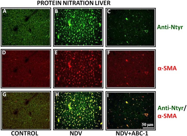 ) = 50 µm.
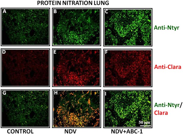 ) = 50 µm.
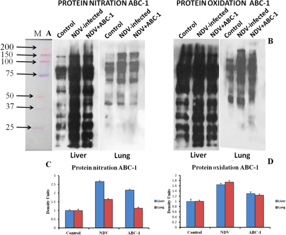
 ) = 50 µm.
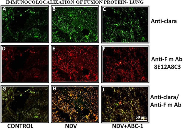 ) = 50 µm.
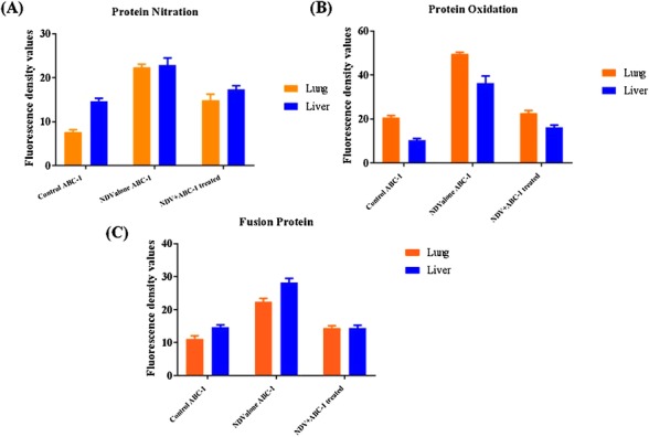
Similar articles
-
Design, Synthesis and Biological Evaluation of Novel Phosphorylated Abacavir Derivatives as Antiviral Agents Against Newcastle Disease Virus Infection in Chicken.Appl Biochem Biotechnol. 2016 Sep;180(2):361-81. doi: 10.1007/s12010-016-2104-x. Epub 2016 May 3. Appl Biochem Biotechnol. 2016. PMID: 27142273
-
Newcastle disease virus (NDV) induces protein oxidation and nitration in brain and liver of chicken: Ameliorative effect of vitamin E.Int J Biochem Cell Biol. 2015 Jul;64:97-106. doi: 10.1016/j.biocel.2015.03.019. Epub 2015 Apr 4. Int J Biochem Cell Biol. 2015. PMID: 25849457
-
Synthesis and Antiviral Activity of Novel Phosphorylated Derivatives of Didanosine Against Newcastle Disease Virus in Chicken.Arch Pharm (Weinheim). 2016 Jun;349(6):442-55. doi: 10.1002/ardp.201600038. Epub 2016 Apr 29. Arch Pharm (Weinheim). 2016. PMID: 27128998
-
Vitamin E Supplementation Ameliorates Newcastle Disease Virus-Induced Oxidative Stress and Alleviates Tissue Damage in the Brains of Chickens.Viruses. 2018 Apr 3;10(4):173. doi: 10.3390/v10040173. Viruses. 2018. PMID: 29614025 Free PMC article.
-
Immune responses of poultry to Newcastle disease virus.Dev Comp Immunol. 2013 Nov;41(3):447-53. doi: 10.1016/j.dci.2013.04.012. Epub 2013 Apr 25. Dev Comp Immunol. 2013. PMID: 23623955 Review.
Cited by
-
Antiviral Chemotherapy in Avian Medicine-A Review.Viruses. 2024 Apr 12;16(4):593. doi: 10.3390/v16040593. Viruses. 2024. PMID: 38675934 Free PMC article. Review.
References
-
- Zhu, W. , Dong, J. , Xie, Z. , Liu, Q. , and Khan, M. I. (2010) Virus Genes 40, 231–235. - PubMed
-
- Wentao, F. , Yang, W. , Shenghua, W. , Ziqiang, C. , Huijun, G. , Xiaona, Z. , and Jianzhu, L. (2017) Res. Vet. Sci. 111, 49–54.
-
- Kuiken, T. , Heckert, R. A. R. J. , Leighton, F. A. , and Wobeser, G. (1998) Avian Pathol. 27, 541–546. - PubMed
-
- Alexander, D. J. (2000) Avian Pathol. 29, 95–100. - PubMed
-
- King A. M. Q., Adams M. J., Carstens E. B., and Lefkowitz E. J. (eds.). (2011) In Virus Taxonomy—Ninth Report of the International Committee on Taxonomy of Viruses. Pp. 1273–1277, Elsevier/Academic Press, London.
MeSH terms
Substances
Grants and funding
LinkOut - more resources
Full Text Sources
Medical
