. 2019 Dec;33(12):3228-3241.
doi: 10.1002/ptr.6497. Epub 2019 Sep 4.

Citrus unshiu peel suppress the metastatic potential of murine melanoma B16F10 cells in vitro and in vivo

Affiliations

Citrus unshiu peel suppress the metastatic potential of murine melanoma B16F10 cells in vitro and in vivo

Eun Ok Choi et al. Phytother Res. 2019 Dec.

Abstract

The peel of Citrus unshiu Marcow. fruits (CU) has long been used as a traditional medicine that has therapeutic effects against pathogenic diseases, including asthma, vomiting, dyspepsia, blood circulation disorders, and various types of cancer. In this study, we investigated the effect of CU peel on metastatic melanoma, a highly aggressive skin cancer, in B16F10 melanoma cells, and in B16F10 cells inoculated-C57BL/6 mice. Our results show that ethanol extracts of CU (EECU) inhibited cell growth and increased the apoptotic cells in B16F10 cells. EECU also stimulated the induction of mitochondria-mediated intrinsic pathway, with reduced mitochondrial membrane potential and increased generation of intracellular reactive oxygen species. Furthermore, EECU suppressed the migration, invasion, and colony formation of B16F10 cells. In addition, the oral administration of EECU reduced serum lactate dehydrogenase activity without weight loss, hepatotoxicity, nor nephrotoxicity in B16F10 cell-inoculated mice. Moreover, EECU markedly suppressed lung hypertrophy, the number and expression of metastatic tumor nodules, and the expression of inflammatory tumor necrosis factor-alpha in lung tissue. In conclusion, our findings suggest that the inhibitory effect of EECU on the metastasis of melanoma indicates that it may be regarded as a potential therapeutic herbal drug for melanoma.

Keywords: B16F10 cells; Citrus unshiu (CU); apoptosis; melanoma; metastasis.


Conflict of interest statement

The authors declare that there are no conflicts of interest.

Figures

Figure 1
Figure 1
Ethanol extracts of Citrus unshiu Marcow. fruits (EECU) induced apoptotic cell death in B16F10 melanoma cells. (a) Cells were incubated with EECU of 20 to 100 μg/mL for 24 hr. The cell viability was assessed by MTT assay. Data are expressed as the mean ± SD (n = 3). The statistical analyses were conducted using analysis of variance (ANOVA‐Tukey's post hoc test) between groups. ** p < .01 and *** p < .001 when compared with control. (b; top) The morphological change of B16F10 cells treated with EECU for 24 hr was observed under a microscope at 40× magnification. (Bottom) The nuclear morphological change was observed using DAPI staining, and was photographed under a fluorescence microscope at 400× magnification. (c) Apoptosis of B16F10 cells treated with EECU was measured by flow cytometric analysis using annexin V‐fluorescein isothiocyanate (V‐FITC) and propidium iodide (PI). The percentage of annexin V+/PI+ cells in the top and annexin V+/PI cells in the bottom right quadrant are indicated. Each point represents the mean of three independent experiments. (d) The percentage of apoptotic cells are shown in the bar diagram as the mean ± SD (n = 3). The statistical analyses were conducted using analysis of variance (ANOVA‐Tukey's post hoc test) between groups. ** p < .01 and *** p < .001 when compared with control [Colour figure can be viewed at http://wileyonlinelibrary.com]
Figure 2
Figure 2
The effect of ethanol extracts of Citrus unshiu Marcow. fruits (EECU) on the mitochondrial dysfunction in B16F10 melanoma cells. (a) mitochondrial membrane potential (Δψ m) was assessed 24 hr after cells were treated with EECU of 20–100 μg/mL. The cells were stained with JC‐1 dye and then analyzed at 488/575 nm using a flow cytometer. Representative FL1/FL2 profiles with green/red fluorescence are shown. A plot of red fluorescence (FL2‐H) from living cells with intact mitochondrial membrane potential and green fluorescence (FLl‐H) from cells with loss of mitochondrial membrane potential was recorded. (b and c) Intracellular reactive oxygen species generation was measured by flow cytometry using DCF‐DA dye. The cells were treated with EECU for 30 min and were incubated with 10 μM DCF‐DA in the last 20 min of treatment. B16F10 cells were gated on the basis of forward‐scatter characteristics (FSC) and side‐scatter characteristic (SSC). (d and e) The quantitative data are expressed in the bar diagram as the mean ± SD (n = 3). The statistical analyses were conducted using analysis of variance (ANOVA‐Tukey's post hoc test) between groups. ** p < .01 and *** p < .001 when compared with control. (f) The expression of apoptosis‐related proteins in B16F10 melanoma cells treated with EECU. After the cells were incubated with EECU of 20–100 μg/mL for 24 hr, the expression of Bax, Bcl‐2, caspase‐3, and PARP was evaluated by Western blot analysis with whole cell lysates
Figure 3
Figure 3
Ethanol extracts of Citrus unshiu Marcow. fruits (EECU) suppress the motility of B16F10 melanoma cells via the regulation of matrix metalloproteinases (MMPs) activity and expression. (a; top) Cell migration of B16F10 cells was assessed by wound healing assay at 24 hr after EECU treatment. Representative photographs are shown from three independent cell migration experiments. The dotted line indicates the baseline immediately at wound scratch, whereas the solid line indicates the migration at 24 hr after EECU treatment. (Middle) Cell invasion assay was evaluated using the trans‐well chamber system. B16F10 cells were placed in the upper chamber of trans‐well insert, and complement medium supplemented with EECU of 0, 20, and 40 μg/mL was added in the lower chamber, and then cells were incubated for 24 hr. (Bottom) B16F10 cells were exposed to EECU of 0, 20, and 40 μg/mL for 15 days, followed by colony formation assay. Cells were stained with 0.1% crystal violet solution, and visualized colonies were observed under microscope. (b) The mobility of B16F10 cells was calculated for EECU‐treated cells, as compared with the nontreated control cells for each experiment in different field. (c) The numbers of invading cells in EECU‐treated cells, as compared with the nontreated control cells, for each experiment. (d) Rates of colony formation were detected by microplate reader at 650 nm. The quantitative data are expressed in the bar diagram as the mean ± SD (n = 3). The statistical analyses were conducted using analysis of variance (ANOVA‐Tukey's post hoc test) between groups. *** p < .0001 when compared to control. (e) Determination of MMP‐2 and MMP‐9 enzymatic activity by ELISA assay. Relative activities of MMP‐2 and MMP‐9 in EECU‐treated cells are indicated as ‐fold of control. The data are expressed as the mean ± SD (n = 3), and the statistical analyses were conducted using analysis of variance (ANOVA‐Tukey's post hoc test) between groups. *** p < .0001 when compared with control. (f) mRNA expression of extracellular matrix remodeling‐related enzymes by EECU in B16F10 cells. At 24 hr after EECU treatment, total RNA was isolated from cells, and quantitative reverse transcription polymerase chain reaction analysis performed of MMP‐2, MMP‐9, TIMP‐1, and TIMP‐2 mRNA expression, using the indicated primers. GAPDH was used as the internal control. (g) Protein expression of ECM remodeling‐related enzymes by EECU in B16F10 cells. After the cells were incubated with EECU of 20–100 μg/mL for 24 hr, the cellular protein expression of MMP‐2, MMP‐9, TIMP‐1, and TIMP‐2 was measured by Western blot analysis with whole cell lysates [Colour figure can be viewed at http://wileyonlinelibrary.com]
Figure 4
Figure 4
The effects of oral administration of ethanol extracts of Citrus unshiu Marcow. fruits (EECU) in B16F10 cells‐inoculated C57BL/6 mice. (a) Experimental design of in vivo study. For 28 mice, the mouse model of metastatic lung cancer was experimentally induced in 8 weeks old C57BL/6 mice by intravenous injections into the tail vein with 3 × 10⁵ B16F10 cells/100 μL phosphate‐buffered saline (PBS). At the same time, 16 mice were injected in the same area with PBS. After 1 day of tumor inoculation, B16F10 cell‐injected mice were randomly divided into three groups: the B16 + control group (n = 10, 100 μL of distilled water), the B16 + EECU 100 group (n = 8, 100 μL of EECU 100 mg/kg/day), and the B16 + EECU 200 group (n = 8, 100 μL of EECU 200 mg/kg/day). PBS‐injected 18 mice were also randomly divided into two groups: the normal group (n = 8, 100 μL of distilled water) and the EECU 200 group (n = 8, 200 μL of EECU 200 mg/kg/day). All treatments were administrated orally once per day in the morning for 21 days. (b) Kaplan‐Meier graph representing the cumulative survival of mice in the indicated treatment groups. The data shows the survival rate of the experimental period and were analyzed using Kaplan‐Meier survival analysis (n = 8–10 per group). (c) Body weight change of mice with the oral administration of EECU in B16F10‐induced metastatic lung cancer. Body weight was measured every 7 days. There were no differences in body weight between the groups. (d) Mice were sacrificed at Day 21 after B16F10 melanoma cells injection, and liver, kidney, spleen, lung, and thymus were immediately surgically excised and then measured the weight. The statistical analyses were conducted using analysis of variance (ANOVA‐Tukey's post hoc test) between groups. *** p < .0001 when compared with normal group on weight of lung. (e) plasma lactate dehydrogenase (LDH), (f) alanine aminotransferase (ALT), and (g) aspartate aminotransferase (AST) activities and (h) blood urea nitrogen (BUN) levels after 21 days EECU treatment in B16F10‐induced metastatic lung cancer. The data are expressed as the mean ± SD (n = 7–8). The statistical analyses were conducted using analysis of variance (ANOVA‐Tukey's post hoc test) between groups. * p < .05 and *** p < .001 when compared with normal group. ### p < .001 when compared with B16F10 cell‐injected control group [Colour figure can be viewed at http://wileyonlinelibrary.com]
Figure 5
Figure 5
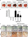
Orally administration of ethanol extracts of Citrus unshiu Marcow. fruits (EECU) suppressed B16F10 cells‐induced metastatic lung cancer. (a) Photographs of lung tissue at Day 21 after treatment. (b) The metastatic nodules were counted, and data presented as the mean ± SD (n = 7–8). The statistical analyses were conducted using analysis of variance (ANOVA‐Tukey's post hoc test) between groups. ** p < .01 and *** p < .001 when compared with normal group. ### p < .001 when compared with B16F10 cell‐injected control group. (c; top) Immunopathological damage assessed by hematoxylin and eosin (H&E) staining of the lung sections. The images of the sections were photographed by microscope (Carl Zeiss). Black arrow indicates lung metastatic foci. Original magnification: 200×. (Bottom) The expression of tumor necrosis factor alpha (TNF‐α) of lung sections. Tumor tissues of lung metastasis were immunohistochemistry stained with TNF‐α. Black arrow indicates the TNF‐α‐expressed area shown in brown color, and marks the metastasis nodule. Original magnification: 200 × [Colour figure can be viewed at http://wileyonlinelibrary.com]
Figure 6
Figure 6
The phytochemical compound chromatogram from high‐performance liquid (HPL) analysis of ethanol extracts of Citrus unshiu Marcow. fruits (EECU). (a) Three reference components of naringin, hesperidin, and neohesperidin were analyzed using high‐performance liquid chromatography (HPLC). (b) The chromatograms from the HPLC analysis of EECU

References

    1. Ahn, K. I. , Choi, E. O. , Kwon, D. H. , HwangBo, H. , Kim, M. Y. , Kim, H. J. , … Choi, Y. H. (2017). Induction of apoptosis by ethanol extract of Citrus unshiu Markovich peel in human bladder cancer T24 cells through ROS‐mediated inactivation of the PI3K/Akt pathway. Bioscience Trends, 11(5), 565–573. 10.5582/bst.2017.01218 - DOI - PubMed
    1. Aroui, S. , Aouey, B. , Chtourou, Y. , Meunier, A. C. , Fetoui, H. , & Kenani, A. (2015). Naringin suppresses cell metastasis and the expression of matrix metalloproteinases (MMP‐2 and MMP‐9) via the inhibition of ERK‐P38‐JNK signaling pathway in human glioblastoma. Chemico‐Biological Interactions, 244, 195–203. 10.1016/j.cbi.2015.12.011 - DOI - PubMed
    1. Aroui, S. , Najiaoui, F. , Chtourou, Y. , Meunier, A. C. , Laaijimi, A. , Kenani, A. , … Fetoui, H. (2016). Naringin inhibits the invasion and migration of human glioblastoma cell via downregulation of MMP‐2 and MMP‐9 expression and inactivation of p38 signaling pathway. Tumour Biology, 37(3), 3831–3839. 10.1007/s13277-015-4230-4 - DOI - PubMed
    1. Bhatia, S. , Tykodi, S. S. , & Thompson, J. A. (2009). Treatment of metastatic melanoma: An overview. Oncology (Williston Park), 23(6), 488–496. - PMC - PubMed
    1. Bold, R. J. , Termuhlen, P. M. , & McConkey, D. J. (1997). Apoptosis, cancer and cancer therapy. Surgical Oncology, 6(3), 133–142. 10.1016/S0960-7404(97)00015-7 - DOI - PubMed