. 2019 Sep;22(9):e25384.
doi: 10.1002/jia2.25384.

Predominance of the heterozygous CCR5 delta-24 deletion in African individuals resistant to HIV infection might be related to a defect in CCR5 addressing at the cell surface

Affiliations

Predominance of the heterozygous CCR5 delta-24 deletion in African individuals resistant to HIV infection might be related to a defect in CCR5 addressing at the cell surface

Vic Arendt et al. J Int AIDS Soc. 2019 Sep.

Abstract

Introduction: The chemokine receptor CCR5 is the main co-receptor for R5-tropic HIV-1 variants. We have previously described a novel 24-base pair deletion in the coding region of CCR5 among individuals from Rwanda. Here, we investigated the prevalence of hCCR5Δ24 in different cohorts and its impact on CCR5 expression and HIV-1 infection in vitro.

Methods: We screened hCCR5Δ24 in a total of 3232 individuals which were either HIV-1 uninfected, high-risk HIV-1 seronegative and seropositive partners from serodiscordant couples, Long-Term Survivors, or HIV-1 infected volunteers from Africa (Rwanda, Kenya, Guinea-Conakry) and Luxembourg, using a real-time PCR assay. The role of the 24-base pair deletion on CCR5 expression and HIV infection was assessed in cell lines and PBMC using mRNA quantification, confocal analysis, flow and imaging cytometry.

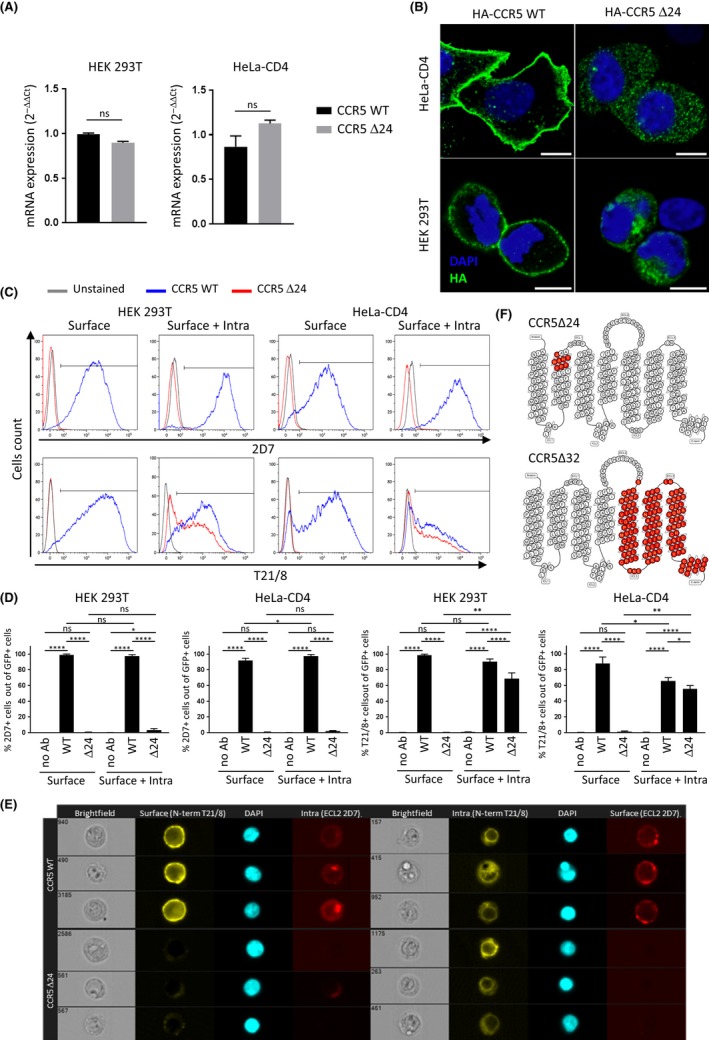
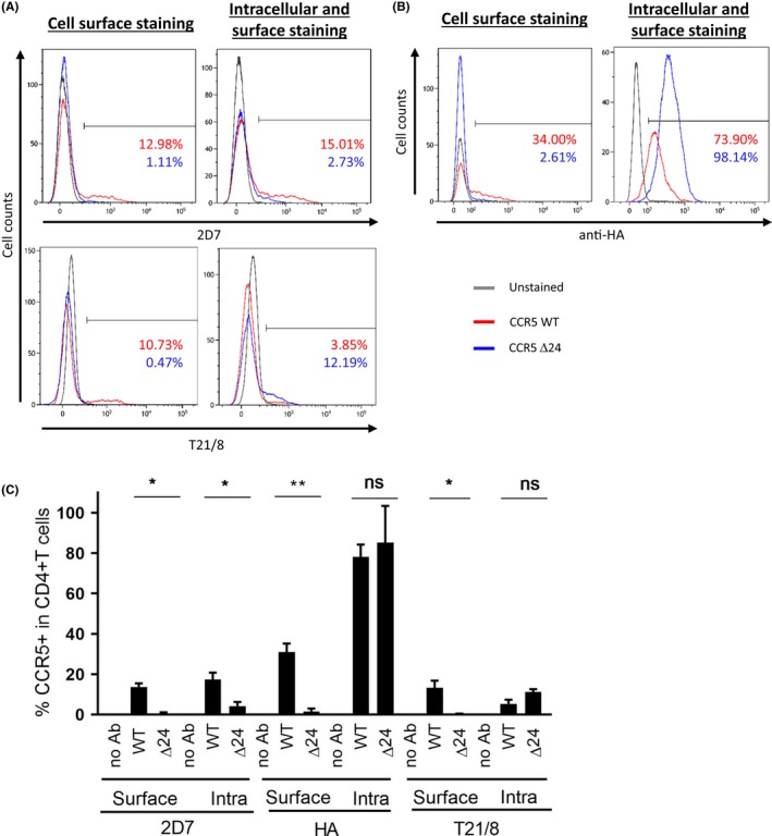
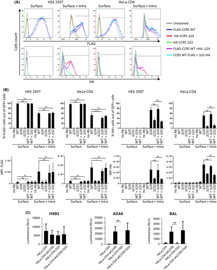
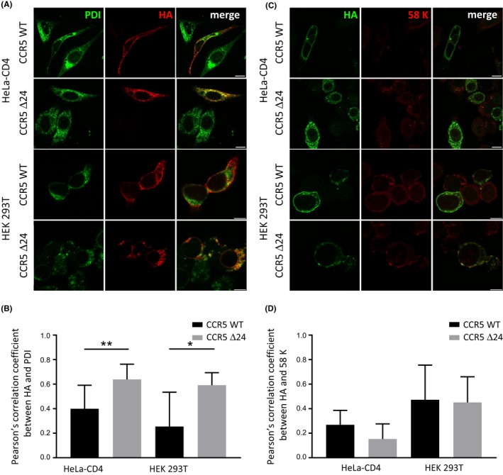
Results and discussion: Among the 1661 patients from Rwanda, 12 individuals were heterozygous for hCCR5Δ24 but none were homozygous. Although heterozygosity for this allele may not confer complete resistance to HIV-1 infection, the prevalence of the mutation was 2.41% (95%CI: 0.43; 8.37) in 83 Long-Term Survivors (LTS) and 0.99% (95%CI: 0.45; 2.14) in 613 HIV-1 exposed seronegative members as compared with 0.35% (95% Cl: 0.06; 1.25) in 579 HIV-1 seropositive members. The prevalence of hCCR5Δ24 was 0.55% (95%CI: 0.15; 1.69) in 547 infants from Kenya but the mutation was not detected in 224 infants from Guinea-Conakry nor in 800 Caucasian individuals from Luxembourg. Expression of hCCR5Δ24 in cell lines and PBMC showed that the hCCR5Δ24 protein is stably expressed but is not transported to the plasma membrane due to a conformational change. Instead, the mutant receptor was retained intracellularly, colocalized with an endoplasmic reticulum marker and did not mediate HIV-1 infection. Co-transfection of hCCR5Δ24 and wtCCR5 did not indicate a transdominant negative effect of CCR5Δ24 on wtCCR5.

Conclusions: Our findings indicate that hCCR5Δ24 is not expressed at the cell surface. This could explain the higher prevalence of the heterozygous hCCR5Δ24 in LTS and HIV-1 exposed seronegative members from serodiscordant couples. Our data suggest an East-African localization of this deletion, which needs to be confirmed in larger cohorts from African and non-African countries.

Keywords: AIDS; Africa; CCR5; HIV-1; mutation; receptor expression.


Figures

Figure 1
Figure 1
hCCR5Δ24 mutant is not expressed at the cell surface in cell lines. (A) The hCCR5Δ24 deletion does not affect the relative mRNA expression levels of the CCR5 receptor in HEK 293T and HeLa‐CD4 cells transiently transfected with pCMV5/HA‐wtCCR5 and pCMV5/HAhCCR5Δ24 and measured by qRTPCR. GAPDH was used as a reference gene. (B) Confocal immunofluorescence analysis reveals hCCR5Δ24 mutant accumulates in the intracellular compartment but not at the cell surface. CCR5 was stained using anti‐HA mAb and the nucleus was stained using DAPI. 10 μm scale. (C and D) wtCCR5 and hCCR5Δ24 surface or surface +intracellular expression measured by flow cytometry using Ab targeting the NH2‐terminal region or ECL2. (D) A GFP reporter vector was added to each transfection in order to analyse CCR5 expression in transfected populations. (E) Intracellular hCCR5Δ24 is detectable in transfected HEK 293T cells by imaging cytometry using an Ab targeting the NH2‐terminal region but not ECL2. CCR5 surface or surface + intracellular expression were measured using anti‐CCR5 2D7 (ECL 2) and anti‐CCR5 T21/8 (N‐term) mAbs (C, D and E). Statistical significance was considered when  0.05 (**** 0.0001, *** 0.001, ** 0.01, * 0.05; N = 3 independent experiments). Error bars denote mean ± SD. (F) Snake diagram depicting CCR5 topology and identifying the positions occupied by missing residues (red) in the delta 24 (upper panel) and delta 32 (lower level) receptors respectively.
Figure 2
Figure 2
hCCR5Δ24 is not expressed at the cell surface of CD4+ T cells in PBMCs.(A and B). CCR5 surface and intracellular expression was measured by flow cytometry in human CD4+ T cells from PBMCs transiently transfected with pCMV5/HA‐wtCCR5 and pCMV5/HAhCCR5Δ24 using anti‐CCR5 2D7 (ECL 2) and anti‐CCR5 T21/8 (N‐term) mAbs (A) and anti‐HA mAbs (C). Statistical analyses of the flow cytometry experiments from (A). Statistical significance was considered when  0.05 (** 0.01, * 0.05; N = 3 independent experiments). Error bars denote mean ± SD.
Figure 3
Figure 3
hCCR5Δ24 mutant has no transdominant negative effect on wtCCR5.(A) HEK‐293T and HeLa‐CD4 cells were transfected with FLAG‐wtCCR5, HAhCCRΔ24 and HAhCCR5Δ32 alone or cotransfected with FLAG‐wtCCR5 and HAhCCRΔ24 or HAhCCR5Δ32 in equimolar ratio. (A) GFP reporter vector was added to each transfection in order to analyse CCR5 expression in transfected populations. CCR5 surface or surface + intracellular expression was analysed by flow cytometry using anti‐FLAG and anti‐HA mAbs. (B) Quantification of the flow cytometry experiment from A. (C) HeLa‐CD4 cells were transfected with FLAG‐wtCCR5, HAhCCRΔ24 alone or cotransfected with FLAG‐wtCCR5 and HAhCCRΔ24 in equimolar ratios. Transfected cells were infected with HXB2, ADA8 or BaL pseudovirus expressing a Luciferase reporter gene. HIV‐1 infection was quantified by measuring Luciferase‐dependent luminescence. Statistical significance was considered when  0.05 (**** 0.0001, *** 0.001, ** 0.01, * 0.05; N = 3 independent experiments). Error bars denote mean±SD.
Figure 4
Figure 4
The hCCR5Δ24 mutation increases CCR5 colocalization with Endoplasmic Reticulum but not Golgi markers. HEK 293T and HeLa‐CD4 cells were transiently transfected with pCMV5/HA‐wtCCR5 and pCMV5/HAhCCR5Δ24. Representative confocal micrographs of CCR5, ER (A) and Golgi (C) staining using anti‐HA, anti‐PDI and anti‐58K mAbs respectively. Pearson's colocalization correlation coefficients between anti‐HA and anti‐PDI (B) or anti‐58k (D) staining were calculated for regions of interest (ROI) corresponding to anti‐HA positive cells using Colocalization Threshold (Image J). Statistical significance was considered when  0.05, ** 0.01, * 0.05; N = 3 independent experiments). Error bars denote mean ± SD. 10 um scale.

References

    1. Berger EA, Murphy PM, Farber JM. Chemokine receptors as HIV‐1 coreceptors: roles in viral entry, tropism, and disease. Annu Rev Immunol. 1999;17:657–700. - PubMed
    1. Shaik MM, Peng H, Lu J, Rits‐Volloch S, Xu C, Liao M, et al. Structural basis of coreceptor recognition by HIV‐1 envelope spike. Nature. 2019;565:318–23. - PMC - PubMed
    1. Keele BF, Giorgi EE, Salazar‐Gonzalez JF, Decker JM, Pham KT, Salazar MG, et al. Identification and characterization of transmitted and early founder virus envelopes in primary HIV‐1 infection. Proc Natl Acad Sci USA. 2008;105(21):7552–7. - PMC - PubMed
    1. van't Wout AB, Kootstra NA, Mulder‐Kampinga GA, Albrecht‐van Lent N, Scherpbier HJ, Veenstra J, et al. Macrophage‐tropic variants initiate human immunodeficiency virus type 1 infection after sexual, parenteral, and vertical transmission. J Clin Investig. 1994;94(5):2060–7. - PMC - PubMed
    1. Verhofstede C, Nijhuis M, Vandekerckhove L. Correlation of coreceptor usage and disease progression. Curr Opin HIV AIDS. 2012;7(5):432–9. - PubMed

Publication types

MeSH terms