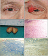
Palpebral Tarsal Solitary Neurofibroma
- PMID: 31486611
- PMCID: PMC6761380
- DOI: 10.4274/tjo.galenos.2019.47124
Palpebral Tarsal Solitary Neurofibroma
Abstract
Solitary neurofibroma is a rare, benign tumor of the peripheral nerve sheath, and is often associated with neurofibromatosis type 1. Herein, a case of palpebral tarsal solitary neurofibroma in a patient without neurofibromatosis is presented, with a review of the literature. A 68-year-old man presented with a subcutaneous mass in the right upper eyelid of 6 months’ duration. Eversion of the eyelid revealed a round, reddish mass on the lateral part of the tarsal plate which measured 12x8 mm in size. The lesion was excised with its tarsal base, diagnosed histologically, and did not recur during a follow-up of 34 months. Isolated, solitary neurofibroma of the eyelid has been reported in a total of 7 cases, including the case presented herein. The tumors arose from the eyelid margin in 4 cases, from the tarsal plate in 2 cases, and from the supratarsal conjunctiva in 1 case. The tumor did not recur after surgical excision in 5 cases for which follow-up data were available.
Keywords: Eyelid; neurofibroma; tarsus; tumor.
Conflict of interest statement
Figures

Similar articles
-
Solitary eyelid neurofibroma presenting as tarsal cyst: Report of a case and review of literature.Am J Ophthalmol Case Rep. 2018 Feb 6;10:71-73. doi: 10.1016/j.ajoc.2018.02.002. eCollection 2018 Jun. Am J Ophthalmol Case Rep. 2018. PMID: 29780919 Free PMC article.
-
Isolated neurofibroma of the eyelid mimicking recurrent chalazion.Indian J Ophthalmol. 2018 Mar;66(3):451-453. doi: 10.4103/ijo.IJO_852_17. Indian J Ophthalmol. 2018. PMID: 29480265 Free PMC article.
-
Solitary neurofibroma of eyelid masquerading as chalazion.Int Med Case Rep J. 2017 May 23;10:177-179. doi: 10.2147/IMCRJ.S136255. eCollection 2017. Int Med Case Rep J. 2017. PMID: 28579839 Free PMC article.
-
Solitary Neurofibroma with Malignant Transformation: Case Report and Review Of Literature.Conn Med. 2015 Apr;79(4):217-9. Conn Med. 2015. PMID: 26259300 Review.
-
Solitary neurofibroma confined to inferior rectus muscle tendon: a case report.Orbit. 2025 Jun;44(3):317-320. doi: 10.1080/01676830.2024.2377248. Epub 2024 Jul 17. Orbit. 2025. PMID: 39018161 Review.
References
-
- Henderson JW, Campbell RJ, Farrow GM, Garrity JA. Orbital Tumors (3rd ed) New York; NY: Raven Press; 1994:221–237.
-
- Shibata N, Kitagawa K, Noda M, Sasaki H. Solitary neurofibroma without neurofibromatosis in the superior tarsal plate simulating a chalazion. Graefes Arch Clin Exp Ophthalmol. 2012;250:309–310. - PubMed
-
- Kehoe NJ, Reid RP, Semple JC. Solitary benign peripheral-nerve tumours: review of 32 years’ experience. J Bone Joint Surg. 1995;77:497–500. - PubMed
-
- Rose GE, Wright JE. Isolated peripheral nerve sheath tumours of the orbit. Eye (Lond). 1991;5:668–673. - PubMed
-
- Fenton S, Mourits MP. Isolated conjunctival neurofibromas at the puncta, an unusual cause of epiphora. Eye (Lond). 2003;17:665–666. - PubMed
Publication types
MeSH terms
LinkOut - more resources
Full Text Sources
Research Materials
