. 2019 Nov;68(11):981-992.
doi: 10.1007/s00011-019-01280-6. Epub 2019 Sep 5.

TIPE2 ameliorates lipopolysaccharide-induced apoptosis and inflammation in acute lung injury

Affiliations

TIPE2 ameliorates lipopolysaccharide-induced apoptosis and inflammation in acute lung injury

Xiaojing Wu et al. Inflamm Res. 2019 Nov.

Abstract

Objective: Tumour necrosis factor-α-induced protein 8-like 2 (TIPE2) has strong anti-inflammatory properties. However, it is unknown whether increased TIPE2 is protective against lipopolysaccharide (LPS)-induced ALI. In the current study, we aimed to investigate whether increased TIPE2 can exert protective effects in a mouse model of ALI induced by LPS.

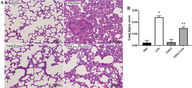
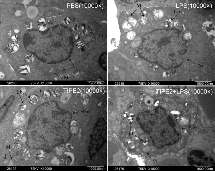
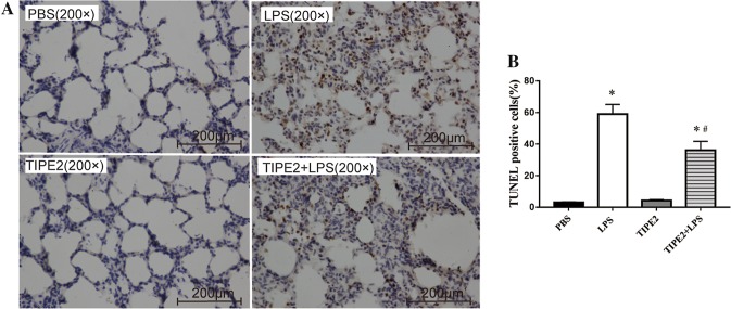
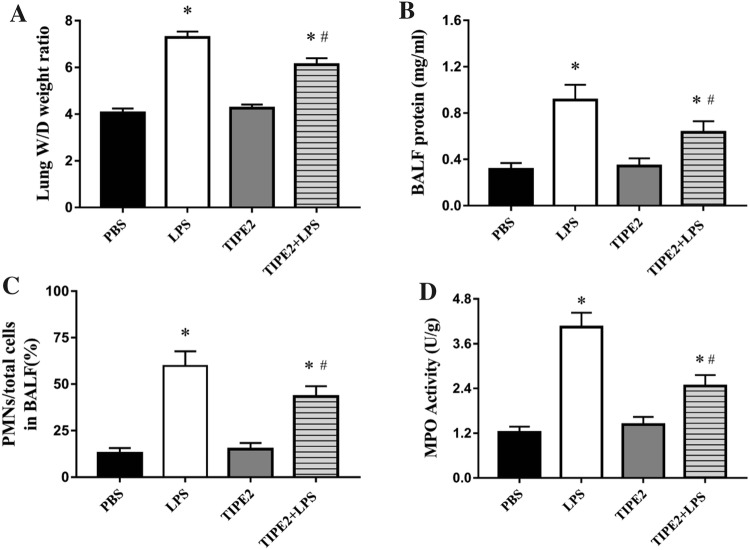
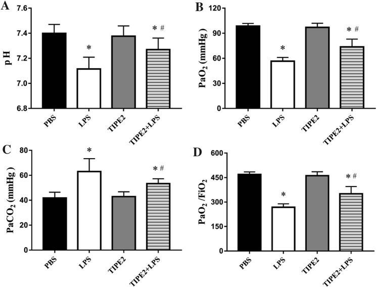
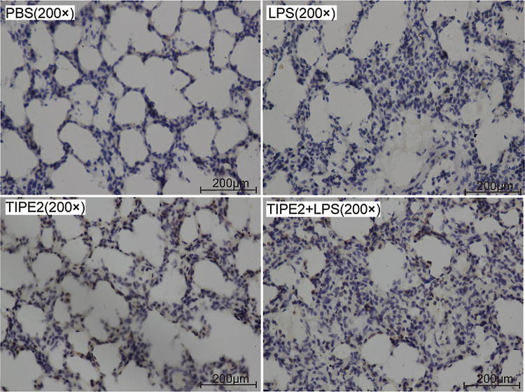
Methods: We administered TIPE2 adeno-associated virus (AAV-TIPE2) intratracheally into the lungs of mice. Three weeks later, ALI was induced by intratracheal injection of LPS into BALB/c mice. Twenty-four hours later, lung bronchoalveolar lavage fluid (BALF) was acquired to analyse cells and protein, arterial blood was collected for arterial blood gas analysis and the determination of pro-inflammatory factor levels, and lung issues were collected for histologic examination, transmission electron microscopy (TEM), TUNEL staining, wet/dry (W/D) weight ratio analysis, myeloperoxidase (MPO) activity analysis and blot analysis of protein expression.

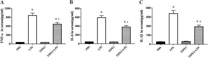
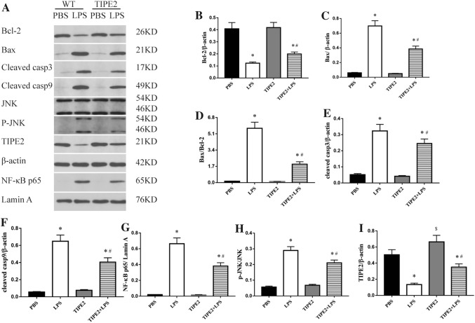
Results: We found that TIPE2 overexpression markedly mitigated LPS-induced lung injury, which was evaluated by the deterioration of histopathology, histologic scores, the W/D weight ratio, and total protein expression in the BALF. Moreover, TIPE2 overexpression markedly attenuated lung inflammation, as evidenced by the downregulation of polymorphonuclear neutrophils (PMNs) in the BALF, lung MPO activity, and pro-inflammatory cytokine levels in the serum. Moreover, TIPE2 overexpression not only dramatically prevented LPS-induced pulmonary cell apoptosis in mice but also blocked LPS-activated JNK phosphorylation and NF-κB p65 nuclear translocation.

Conclusions: Our study shows that the increased expression of AAV-mediated TIPE2 in the lungs of mice inhibits acute inflammation and apoptosis and suppresses the activation of NF-κB and JNK in a murine model of ALI.

Keywords: Acute lung injury; Apoptosis; Cytokines; Inflammation; JNK; NF-κB; TIPE2.


Conflict of interest statement

The authors declare no conflict of interest.

Figures

Fig. 1
Fig. 1
TIPE2 overexpression attenuates the lung histopathological changes in LPS-challenged mice. Acute lung injury was induced by i.t. administration of LPS at a dose of 5 mg/kg body weight. Control mice were intratracheally administered 50 μl of sterile PBS. 24 h after the LPS/PBS treatment, the mice were sacrificed by an i.p. injection of pentobarbital, the lung tissues without lavage were collected for measuring the histopathology, histologic injury scores and ultrastructural changes of the lung. a Representative histology sections of lung tissues under light microscope (H&E staining, magnification, × 200); b Lung histologic injury score. The data are presented as mean ± SEM. n = 10/group, *P < 0.05 versus PBS group; #P < 0.05 versus LPS group
Fig. 2
Fig. 2
TIPE2 overexpression attenuates the lung ultrastructural changes in LPS-challenged mice. Ultrastructural changes of lung tissues under transmission electron microscope (× 10,000). Lb lamellar body, Nu nucleus, M mitochondria
Fig. 3
Fig. 3
TIPE2 overexpression prevented the lung cell apoptosis in LPS-challenged mice. The mice were treated as described in Fig. 1. The lung samples were collected for measuring TUNEL staining at 24 h after PBS/LPS administration. a Representative lung TUNEL staining (× 400); b Percentage of TUNEL-positive cells. The data are presented as mean ± SEM. n = 10/group, *P < 0.05 versus PBS group; #P < 0.05 versus LPS group
Fig. 4
Fig. 4
TIPE2 overexpression reduced Lung W/D ratio, BALF protein concentration, PMNs/total cells in BALF as well as lung MPO activity in LPS-challenged mice. The mice were treated as described in Fig. 1. BALF and lung tissues were collected at 24 h after LPS or PBS administration. a Lung W/D ratio; b BALF protein concentration; c PMNs/total cells in BALF; d lung MPO activity. The data are presented as mean ± SEM. n = 10/group, *P < 0.05 versus PBS group; #P < 0.05 versus LPS group
Fig. 5
Fig. 5
TIPE2 overexpression improved pulmonary dysfunction during ALI induced by LPS. The mice were treated as described in Fig. 1. Arterial blood was collected for blood gas analysis at 24 h after LPS or PBS administration. a pH; b PaO2; c PaO2/FiO2; d PaCO2. The data are presented as mean ± SEM. n = 10/group, *P < 0.05 versus PBS group; #P < 0.05 versus LPS group
Fig. 6
Fig. 6
TIPE2 overexpression reduced LPS-induced proinflammatory cytokine levels in mice. The mice were treated as described in Fig. 1. Blood sample was collected for measuring the pro-inflammatory cytokines levels at 24 h after LPS or PBS administration. a TNF-α; b IL-6; c IL-1β. The data are presented as mean ± SEM. n = 10/group, *P < 0.05 versus PBS group; #P < 0.05 versus LPS group
Fig. 7
Fig. 7
TIPE2 overexpression suppressed the activation of JNK and NF-κB as well as the protein expression of genes involved in apoptosis and lung injury induced by LPS. The mice were treated as described in Fig. 1. Lung tissues were collected for western blotting analysis at 24 h after LPS or PBS administration. a Western blotting analysis of protein expression of Bax, cleaved caspase-3, cleaved caspase-9, Bcl-2, JNK, p-JNK, nuclear NF-κB p65, and TIPE2; bh The relative ratio of Bax, cleaved caspase-3, cleaved caspase-9, Bcl-2, p-JNK, nuclear NF-κB p65, and TIPE2 protein expression. The data are presented as mean ± SEM. n = 10/group, *P < 0.05 versus PBS group; #P < 0.05 versus LPS group
Fig. 8
Fig. 8
Immunohistochemical staining of TIPE2 protein expression in the lung. The mice were treated as described in Fig. 1. Lung tissues were collected for immunohistochemical staining at 24 h after LPS or PBS administration. Representative images of lung tissues under light microscope (Immunohistochemical staining, magnification, × 200)

References

    1. Ware LB, Matthay MA. The acute respiratory distress syndrome. N Engl J Med. 2000;342(18):1334–1349. doi: 10.1056/NEJM200005043421806. - DOI - PubMed
    1. Li C, Yang D, Cao X, Wang F, Jiang H, Guo H, Du L, Guo Q, Yin X. LFG-500, a newly synthesized flavonoid, attenuates lipopolysaccharide-induced acute lung injury and inflammation in mice. Biochem Pharmacol. 2016;113:57–69. doi: 10.1016/j.bcp.2016.05.007. - DOI - PubMed
    1. Zhao Z, Tang X, Zhao X, Zhang M, Zhang W, Hou S, et al. Tylvalosin exhibits anti-inflammatory property and attenuates acute lung injury in different models possibly through suppression of NF-kappaB activation. Biochem Pharmacol. 2014;90(1):73–87. doi: 10.1016/j.bcp.2014.04.015. - DOI - PMC - PubMed
    1. Georgene S, George G, Tony TC, Nagamani S. Incidence and outcome of acute lung injury and acute respiratory distress syndrome in the surgical intensive care unit. Indian J Crit Care Med. 2014;18(10):659–665. doi: 10.4103/0972-5229.142175. - DOI - PMC - PubMed
    1. Zgraggen BR, Tornic J, Müller-Edenborn B, Reyes L, Booy C, Beck-Schimmer B. Acute lung injury: apoptosis in effector and target cells of the upper and lower airway compartment. Clin Exp Immunol. 2010;161(2):324–331. - PMC - PubMed

MeSH terms