Review
. 2019:1169:95-117.
doi: 10.1007/978-3-030-24108-7_6.

Heterogeneity of Pulmonary Stem Cells

Affiliations
Review

Heterogeneity of Pulmonary Stem Cells

Thomas J Lynch et al. Adv Exp Med Biol. 2019.

Abstract

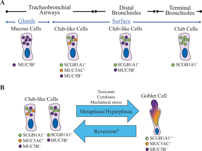
Epithelial stem cells reside within multiple regions of the lung where they renew various region-specific cells. In addition, there are multiple routes of regeneration after injury through built-in heterogeneity within stem cell populations and through a capacity for cellular plasticity among differentiated cells. These processes are important facets of respiratory tissue resiliency and organism survival. However, this regenerative capacity is not limitless, and repetitive or chronic injuries, environmental stresses, or underlying factors of disease may ultimately lead to or contribute to tissue remodeling and end-stage lung disease. This chapter will review stem cell heterogeneity among pulmonary epithelia in the lower respiratory system, discuss recent findings that may challenge long-held scientific paradigms, and identify several clinically relevant research opportunities for regenerative medicine.

Keywords: Alveolar type II cell; Basal stem cell; Chronic rejection; Club cell; Goblet cell; Ionocyte; Myoepithelial cell; Obliterative bronchiolitis; Stem cell; Submucosal gland.


Figures

Fig. 6.1
Fig. 6.1
Lineage relationships of the surface airway epithelium in the trachea and bronchi. (a) Basal cells self-renew and give rise to all of the cell types present in the surface airway epithelium. Basal cells differentiate into luminal lineages through an intermediate suprabasal cell state [10]. Basal cells can give rise to self-renewing neuroendocrine cells in addition to brush (or tuft) cells and ionocytes [1, 2]. In addition, a subset of basal cells that display intracellular Notch2 activation (N2ICD) gives rise to secretory cells, whereas basal cells that express low levels of c-myb are able to lineage commit toward multiciliated cells [11]. Recent, single-cell RNA sequencing experiments have suggested that basal cells give rise to multiciliated cells through a differentiation of an intermediate population of pre-ciliated cells [12]. If basal cells are ablated, club-like cells can renew basal cells through a process of dedifferentiation (dashed line) [13]. (b) Secretory cells in large airways consist of club-like cells and goblet cells. Club-like cells are similar to bronchiolar club cells in that they express proteins commonly used to identify bronchiolar club cells, such as SCGB1A1 (a.k.a. club cell secretory protein); however, unlike bronchiolar club cells, large airway club-like cells also express mucins such as MUC5B and MUC5AC [14]. Secretory goblet cells have a distinct goblet or cup-like morphology but express many of the same phenotypic markers that club-like cells express; thus, it is unclear if club-like cells and goblet cells are distinct secretory cell fates or if a single population of secretory cells fluctuates between displaying a club-like or goblet cell morphology depending on environmental factors. Multiciliated cell renewal is primarily accomplished by club-like progenitors at steady state [10] and following injury [1, 12]. In addition, it has recently been argued that goblet cells may act as a differentiation intermediate between club-like secretory cells and multiciliated cells [12]
Fig. 6.2
Fig. 6.2
Proliferative stress requirements of stem cells at steady state and following moderate and severe injury. (a) At steady state, the majority of mitotic stem cells undergo asymmetric cell division (green cells), which is capable of compensating for cell loss during a low rate of turnover without depleting the stem cell population. (b) Following injury, basal cells may undergo symmetric differentiation (yellow cells) in order to more rapidly compensate for the loss of many cells during a higher rate of turnover. Stem cells that undergo symmetric differentiation are removed from the stem cell pool, but other stem cells may compensate for this loss through symmetric self-renewal (blue cells). (c) If the proliferative stress is sustained long enough or the injury is severe enough, the capacity of the stem cell population to self-renew is insufficient to compensate for the loss of stem cells through differentiation. In this case, reserve stem cells from neighboring regenerative stem cell pools may attempt to compensate for stem cell loss. However, in the case of chronic injury, stem cells are ultimately depleted leading to fibrosis and disease
Fig. 6.3
Fig. 6.3
Submucosal gland myoepithelial cell lineages at steady state and following injury. The tracheobronchial airways possess epithelial submucosal glands that secrete mucous and serous fluids that help regulate mucociliary clearance on the airway surface. (a) Myoepithelial cells within the glands are self-renewing stem cells. At steady state, myoepithelial cells can differentiate into glandular serous and mucous cells [–46]. (b) Following injury to the airway surface epithelium by naphthalene or SO2 (red arrows), myoepithelial cells activate a Lef1 transcriptional program that promotes their migration to the airway surface where they are capable of establishing long-lived basal cell progenitors [45, 46]. In addition, ectopic overexpression of LEF1 (blue arrows) is sufficient to initiate this process without injury. Initially, following injury, myoepithelial-derived basal-like cells are less likely to renew into SCGB1A1+ secretory club-like cells and have a lineage bias toward multiciliated and secretory goblet cells (mucus-positive cells). However, with increasing time after injury and distance away from the submucosal glands, myoepithelial-derived basal-like cells become increasingly able to differentiate into SCGB1A1+ secretory club-like cells (dashed red line). In addition, ectopic overexpression of LEF1 accelerates this process [45]
Fig. 6.4
Fig. 6.4
Small airway bronchiolar and alveolar lineages. (a) Basal-like progenitors are rare in small airways, yet they play an important role in restoring the structural integrity of alveolar epithelium after catastrophic viral injury (green arrow) by generating KRT5+ alveolar pods [–60]. Recent evidence also suggests that small airway basal-like cells give rise to bronchiolar club cells [7]. (b) A rare subtype of club cells expressing Uroplakin3a (U-Club cells) is capable of renewing the larger club cell population at steady state. Following naphthalene injury (red arrow), U-club cells can also give rise to multiciliated cells, and following bleomycin injury (yellow arrow), U-Club cells renew alveolar type 2 (AT2) cells [61]. (c) The alveolar epithelium consists of AT2 cells and alveolar type 1 (AT1) cells, and AT2 cells are self-renewing and can lineage commit to AT1 cells [62]. Recently, a population of highly clonogenic stem cells called alveolar epithelial progenitors (AEPs) has been identified within the AT2 cell population [63]. In addition, AT1 cells are able to dedifferentiate following severe injury (dashed red arrow) into AT2 cells [64, 65]. (d) In addition, small airways harbor neuroepithelial bodies, which consist of clusters of self-renewing neuroendocrine cells. Forced induction of notch signaling in the context of injury induces neuroendocrine to club-like cell transdifferentiation [66]. During development (blue arrow), solitary neuroendocrine cells residing in proximal airways migrate to small airway branch points where they organize into neuroepithelial bodies [67]
Fig. 6.5
Fig. 6.5
Nuances among pulmonary secretory cells. Recent studies have provided compelling data for anatomical specificity of secretory cell types in the human lung. (a) Based on the expression of MUC5B, MUC5AC, and SCGB1A1 (CCSP), there are at least four distinct secretory cell types. Submucosal glands possess MUC5B-expressing secretory mucous cells. In proximal large airways, secretory club-like cells express MUC5B, MUC5AC, and SCGB1A1. In small bronchial airways, secretory club-like cells express MUC5B and SCGB1A1 but not MUC5AC, and in terminal bronchioles, secretory club-like cells express only SCGB1A1 [14]. (b) Given that club-like cells express many of the phenotypic characteristic of goblet cells, it may be necessary to experimentally challenge the paradigm that goblet cells are indeed a divergent cell type rather than simply being a hypersecretory state of club-like cells. For example, if goblet cell metaplasia/hyperplasia is readily reversible back to a club-like cell phenotype, this may suggest that goblet cells and club-like cells are the same population of secretory cells

References

    1. Montoro DT et al. (2018) A revised airway epithelial hierarchy includes CFTR-expressing ionocytes. Nature 560:319–324. 10.1038/s41586-018-0393-7 - DOI - PMC - PubMed
    1. Plasschaert LW et al. (2018) A single-cell atlas of the airway epithelium reveals the CFTR-rich pulmonary ionocyte. Nature 560:377–381. 10.1038/s41586-018-0394-6 - DOI - PMC - PubMed
    1. Nabhan AN, Brownfield DG, Harbury PB, Krasnow MA, Desai TJ (2018) Single-cell Wnt signaling niches maintain stemness of alveolar type 2 cells. Science 359:1118–1123. 10.1126/science.aam6603 - DOI - PMC - PubMed
    1. Yu M et al. (2019) Highly efficient transgenesis in ferrets using CRISPR/Cas9-mediated homology-independent insertion at the ROSA26 locus. Sci Rep 9(1):1971. - PMC - PubMed
    1. Treutlein B et al. (2014) Reconstructing lineage hierarchies of the distal lung epithelium using single-cell RNA-seq. Nature 509:371–375. 10.1038/nature13173 - DOI - PMC - PubMed