. 2019 Dec;1866(12):118552.
doi: 10.1016/j.bbamcr.2019.118552. Epub 2019 Sep 2.

LDL receptor related protein 1 requires the I3 domain of discs-large homolog 1/DLG1 for interaction with the kinesin motor protein KIF13B

Affiliations

LDL receptor related protein 1 requires the I3 domain of discs-large homolog 1/DLG1 for interaction with the kinesin motor protein KIF13B

Joslyn Mills et al. Biochim Biophys Acta Mol Cell Res. 2019 Dec.

Abstract

KIF13B, a kinesin-3 family motor, was originally identified as GAKIN due to its biochemical interaction with human homolog of Drosophila discs-large tumor suppressor (hDLG1). Unlike its homolog KIF13A, KIF13B contains a carboxyl-terminal CAP-Gly domain. To investigate the function of the CAP-Gly domain, we developed a mouse model that expresses a truncated form of KIF13B protein lacking its CAP-Gly domain (KIF13BΔCG), whereas a second mouse model lacks the full-length KIF13A. Here we show that the KIF13BΔCG mice exhibit relatively higher serum cholesterol consistent with the reduced uptake of [3H]CO-LDL in KIF13BΔCG mouse embryo fibroblasts. The plasma level of factor VIII was not significantly elevated in the KIF13BΔCG mice, suggesting that the CAP-Gly domain region of KIF13B selectively regulates LRP1-mediated lipoprotein endocytosis. No elevation of either serum cholesterol or plasma factor VIII was observed in the full length KIF13A null mouse model. The deletion of the CAP-Gly domain region caused subcellular mislocalization of truncated KIF13B concomitant with the mislocalization of LRP1. Mechanistically, the cytoplasmic domain of LRP1 interacts specifically with the alternatively spliced I3 domain of DLG1, which complexes with KIF13B via their GUK-MBS domains, respectively. Importantly, double mutant mice generated by crossing KIF13A null and KIF13BΔCG mice suffer from perinatal lethality showing potential craniofacial defects. Together, this study provides first evidence that the carboxyl-terminal region of KIF13B containing the CAP-Gly domain is important for the LRP1-DLG1-KIF13B complex formation with implications in the regulation of metabolism, cell polarity, and development.

Keywords: Cholesterol; DLG1; Endocytosis; GAKIN; KIF13A; KIF13B; Kinesin; LRP1; Molecular motor; Protein-protein interaction.


Conflict of interest statement

Conflict-of-interest disclosure

The authors declare no competing financial interests.

Figures
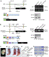
Fig. 1.
Fig. 1.
Generation of KIF13A knockout and KIF13B mutant mouse models. (A & B) Cartoons of KIF13A (A) and KIF13B (B) mouse models generated by β-geo cassette insertion and their verification by PCR (top right panel) and Western blotting (bottom right panel). (C) The LacZ staining showing expression of truncated KIF13B protein in KIF13BΔCG embryo E12.5. (D) A representative image of newborn WT and DKO (KIF13A and KIF13BΔCG) pups found dead shortly after birth. Arrowheads indicate face deformity (short snout). Black arrow indicates milk in the stomach whereas white arrow shows stomach full of air. (E) Co-IP of GFP-KIF13A and FLAG-KIF13B co-transfected into FIEK293T cells. (F) Microtubule binding assay using the C-terminus of human KIF13B or human KIF13A tagged with TRX.
Fig. 2.
Fig. 2.
Characterization of lipid and factor VIII levels. (A) Lipid panel including total cholesterol, direct measurement of LDL, HDL, and VLDL of WT, KIF13A FLKO, and KIF13BΔCG serum from mice after 6 hours of fasting, n = 10 WT, 11 KIF13BΔCG, 6 KIF13A. Significant or nearly significant p values between WT and KIF13BΔCG: Total cholesterol, p = 0.033; LDL, p = 0.0042, HDL, p = 0.067. (B) Timed clot assay to measure factor VIII activity in WT and KIF13BΔCG mouse plasma, n = 5 for each genotype. (C) Time course of [3H]CO-LDL uptake by MEFs from WT and KIF13BΔCG mice. MEFs were stimulated with 50 ug/ml of [3H]CO-LDL for 3, 4.5, 6, and 7.5 hours, and the amount of [3H]CO taken into the cells was measured. Measurements are from triplicate wells.
Fig. 3.
Fig. 3.
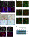
Subcellular localization of KIF13B and LRP1. (A, B, C) Localization of KIF13B in brain tissue from WT or KIF13BΔCG mice. (A & B) Paraffin-embedded sections of brain were stained with a polyclonal Ab that recognizes the N-terminus of KIF13B (red), which is present in both WT and KIF13BΔCG proteins, and nuclear counterstain with Hoechst or DAPI (blue) (A) and double stained with organelle specific markers (green) LAMP1 (B, top), PDI (B, middle), or TGN38 (B, bottom). Images were taken at 400X (A) or 600X (B), and insets are magnifications of up to two cells. Each image is a representativetion of truncated KIF13B in confluent culture of MEFs (left) and after the scratch wound in MEFs to initiate cell migration (right). KIF13BΔCG can migrate to the leading edge of the fibroblasts (red arrows). Top panels were co-stained with β-tubulin and bottom panels show counter staining with Nuclear Fast Red. Of note, we performed γ-tubulin staining in fibroblasts to detect the position of MTOC. However, the double staining of γ-tubulin and LacZ activity was not feasible because the blue signal from LacZ stain completely masks the faint signal from the γ-tubulin (data reviewed but not shown). The blue dots appear to be reminiscent of the MTOC. (D) LRP1 subcellular localization in MEFs from WT or KIF13BΔCG mice. MEFs were stained with LRP1 (red) and nuclear counterstain with Hoechst (blue). Images were taken at 400X and a representative frame from three independent observations is shown. (E) LRP1 steady state protein expression in MEFs detected by Western blotting. Lysate from HUVECs served as a negative control for the LRP1 expression, and β-actin was used as a loading control.
Fig. 4.
Fig. 4.
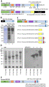
Localization of 4A5 monoclonal antibody epitope in KIF13B. (A) Schematic representation of human KIF13B and mouse mutant KIF13BΔCG. Mouse mutant KIF13B truncates at the position corresponding to 1520 AA in the human sequence. (B) Western blot analysis of TRX-CT11. Purified recombinant TRX-CT11 protein was detected by Coomassie Brilliant Blue (CBB) stain (lane 1), and Western blotting with anti-TRX mAb (lane 2) and anti-KIF13B mAb 4A5 (lane 3). (C) Recombinant protein constructs of KIF13B used to map the epitope of 4A5 mAb by Western blotting. (D) Total cell lysates of bacterially expressed proteins were analyzed by CBB stain (left panel) and Western blotting with 4A5 mAb (right panel). (E) Position of the 4A5 mAb epitope relative to the CAP-Gly domain within human KIF13B sequence. The 4A5 mAb epitope is deleted in KIF13BΔCG.
Fig. 5.
Fig. 5.
Identification of I3 domain of hDLG1 as the binding site for LRP1. (A) Schematics of multiple GST-hDLG1 constructs used in the pull-down assays. Ponceau staining (below) shows expression of each GST fusion protein marked by an*. GST= tag only, FLI2= full length hDLG1 with insert 2, PDZ = PDZ domains 1,2, and 3, SI3G=SH3-insert 3-GUK domains, SI2G=SH3-insert 2-GUK domains, GUK=GUK domain, GUK-FL=GUK domain and C-terminus, I3=insert 3 only, I3-pY=I3 insert with pY segment, I3-GUK= Insert 3-GUK domains. All constructs contained the N-terminal GST tag. (B) Cartoon of LRP1 focusing on the LRPI-β segment to identify the boundary of LRP1-Herz construct and location of NPxY motifs. The LRP1-Y4507 is mutated to LRP1-Y4507F. PM = plasma membrane. (C) Immunoblotting of LRP1-Herz in pull down assays from transfected FIEK293T cell lysates. LRP1 antibody detected exogenous LRPI-β (65 kDa) in the input lane, and in lanes corresponding to the beads that contained the I3 domain of hDLG1. (D) Immunoblotting of LRP1-Herz or LRP1-Y4507F in the pull-down assays from transfected HEK293T cell lysates. LRP1 mAb detected exogenous LRPI-β (65 kDa) in the input lane, and in lanes corresponding to the beads that contained the I3 domain of DLG1. Ponceau staining verified even loading and expression of GST constructs marked by an*. (E) Co-IP of LRP1 using an anti-hDLG1 mAb, anti-KIF13B mAb, and mouse IgG as negative control in the membrane fraction of HepG2 cells (top). Co-IP of DLG1 using an anti-LRP1 mAb, anti-KIF13B mAb, and pre-immune rabbit serum as negative control. The input lane is from the same membrane, but a shorter exposure is presented.
Fig. 6.
Fig. 6.
A proposed model for the LRP1-DLG1-KIF13B complex formation. (A) DLG1 serves as an adaptor by linking LRP1 with KIF13B. The LRP1-CT binds to the I3 domain of DLG1, which in turn utilizes its GUK domain to bind to the MBS domain of KIF13B. This model predicts that phosphorylation of Y4507 of LRP1-CT may induce a conformational change in its cytoplasmic domain that regulates its binding to DLG1. (B) A cartoon of truncated KIF13B expressed in KIF13BΔCG mice. The truncated KIF13B is mislocalized or destabilized at the plasma membrane preventing its interaction with DLG1 and suppressing LRP1 endocytosis.

Similar articles

Cited by

References

    1. Hanada T, Lin L, Tibaldi EV, Reinherz EL, Chishti AH, GAKIN, a novel kinesin-like protein associates with the human homologue of the Drosophila discs large tumor suppressor in T lymphocytes, J. Biol. Chem, 275 (2000) 28774–28784. - PubMed
    1. Miki H, Setou M, Kaneshiro K, Hirokawa N, All kinesin superfamily protein, KIF, genes in mouse and human, Proc. Natl. Acad. Sci. U. S. A, 98 (2001) 7004–7011. - PMC - PubMed
    1. Horiguchi K, Hanada T, Fukui Y, Chishti AH, Transport of PIP3 by GAKIN, a kinesin-3 family protein, regulates neuronal cell polarity, J. Cell Biol, 174 (2006) 425–436. - PMC - PubMed
    1. Venkateswarlu K, Hanada T, Chishti AH, Centaurin-alpha1 interacts directly with kinesin motor protein KIF13B, J. Cell Sci, 118 (2005) 2471–2484. - PubMed
    1. Asaba N, Hanada T, Takeuchi A, Chishti AH, Direct interaction with a kinesin-related motor mediates transport of mammalian discs large tumor suppressor homologue in epithelial cells, J. Biol. Chem, 278 (2003) 8395–8400. - PubMed

Publication types