Review
. 2019 Sep 4;11(9):2101.
doi: 10.3390/nu11092101.

Selenium, Selenoproteins and Viral Infection

Affiliations
Review

Selenium, Selenoproteins and Viral Infection

Olivia M Guillin et al. Nutrients. .

Abstract

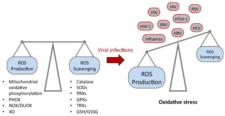
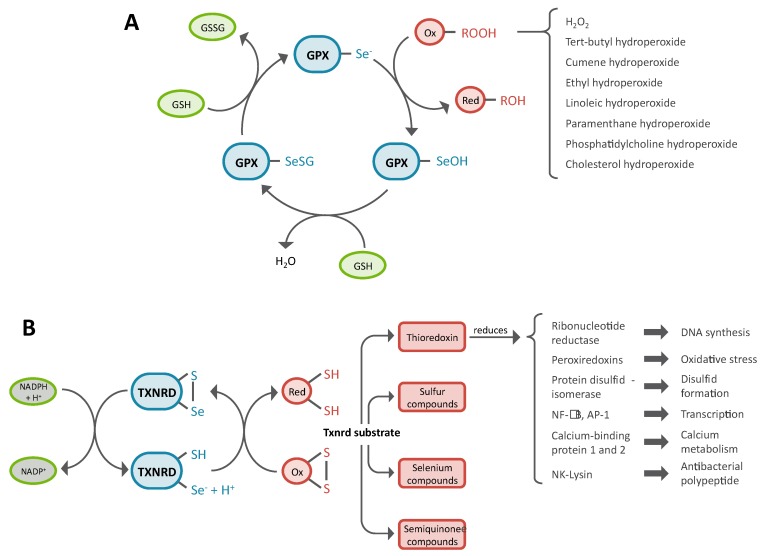
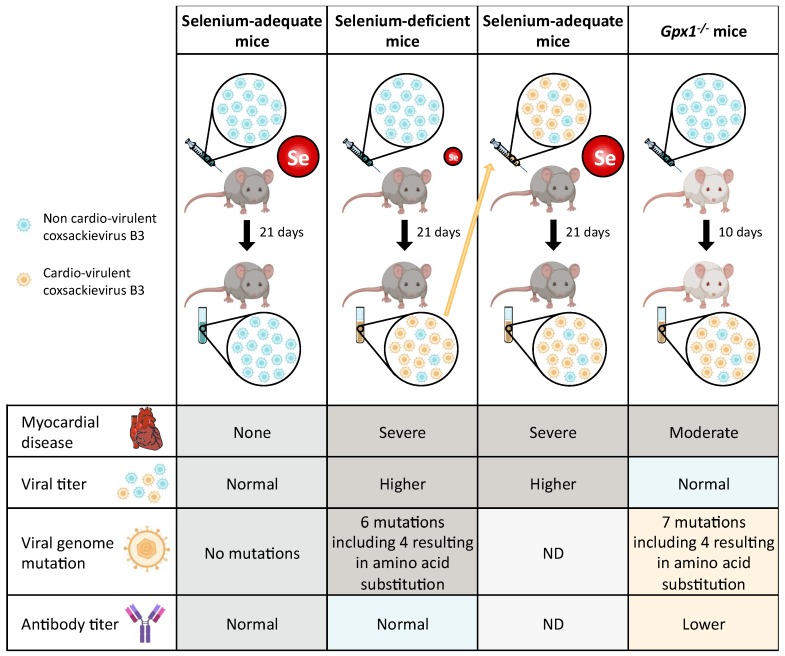
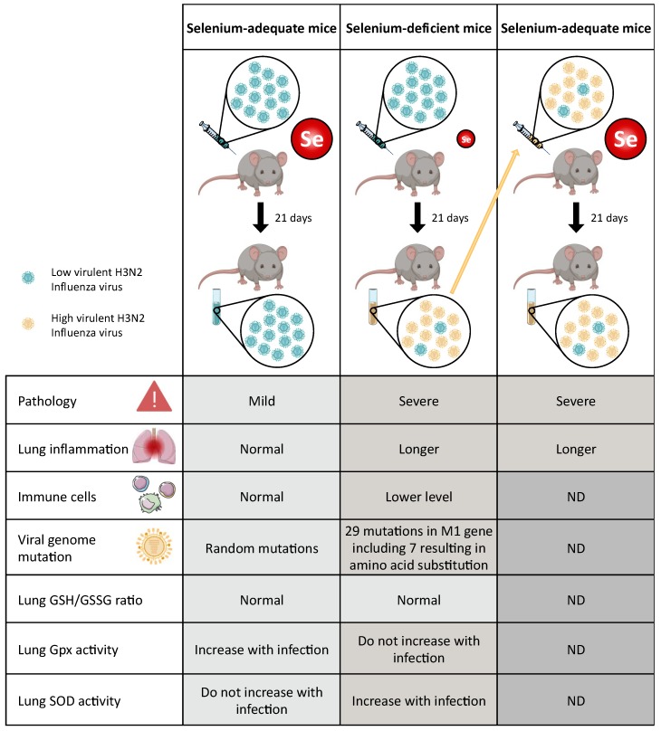
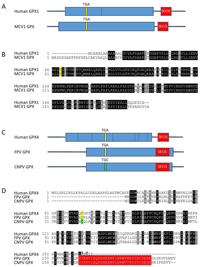
Reactive oxygen species (ROS) are frequently produced during viral infections. Generation of these ROS can be both beneficial and detrimental for many cellular functions. When overwhelming the antioxidant defense system, the excess of ROS induces oxidative stress. Viral infections lead to diseases characterized by a broad spectrum of clinical symptoms, with oxidative stress being one of their hallmarks. In many cases, ROS can, in turn, enhance viral replication leading to an amplification loop. Another important parameter for viral replication and pathogenicity is the nutritional status of the host. Viral infection simultaneously increases the demand for micronutrients and causes their loss, which leads to a deficiency that can be compensated by micronutrient supplementation. Among the nutrients implicated in viral infection, selenium (Se) has an important role in antioxidant defense, redox signaling and redox homeostasis. Most of biological activities of selenium is performed through its incorporation as a rare amino acid selenocysteine in the essential family of selenoproteins. Selenium deficiency, which is the main regulator of selenoprotein expression, has been associated with the pathogenicity of several viruses. In addition, several selenoprotein members, including glutathione peroxidases (GPX), thioredoxin reductases (TXNRD) seemed important in different models of viral replication. Finally, the formal identification of viral selenoproteins in the genome of molluscum contagiosum and fowlpox viruses demonstrated the importance of selenoproteins in viral cycle.

Keywords: coxsackie virus; glutathione peroxidases; hepatitis C virus; human immunodeficiency virus; immunity; influenza virus; molluscum contagiosum virus; reactive oxygen species; thioredoxin reductases; viral selenoproteins.


Conflict of interest statement

The authors declare no conflict of interest.

Figures

Figure 1
Figure 1
Balance between the generation of reactive oxygen species (ROS) and their scavenging systems in human. This equilibrium can be unbalanced during viral infections, resulting in oxidative stress. The main ROS producing systems include the mitochondrial oxidative phosphorylation, the phagocytic cell NAPDH oxidases (PHOX), the NADPH oxidases/dual oxidases (NOX/DUOX) and the xanthine oxidase (XO). The main ROS scavenging systems include the catalase, the superoxide dismutases (SODs), the peroxiredoxins (PRXs), the glutathione peroxidases (GPXs), the thioredoxins (TRXs) and the balance between reduced and oxidized glutathione (GSH/GSSG). The viruses for which an oxidative stress has been reported are herpes simplex virus type 1 (HSV-1), influenza viruses, vesicular stomatitis virus (VSV), Epstein-Barr virus (EBV), human immunodeficiency virus (HIV), human T cell leukaemia virus type 1 (HTLV-1), hepatitis B virus (HBV), respiratory syncytial virus (RSV) and hepatitis C virus (HCV) [34].
Figure 2
Figure 2
Enzymatic activities of the two most important families of selenoproteins involved in antioxidant defense in mammalian cells: the glutathione peroxidase (GPX) and the thioredoxin reductase (TXNRD). (A) GPXs use two molecules of reduced glutathione (GSH) to reduce hydrogen peroxides and organic hydroperoxides (ROOH) in their respective alcohols (ROH). The various peroxide substrates of mammalian GPXs are listed next to the bracket; (B) TXNRDs use NADPH to catalyze the reduction of thioredoxins and therefore participate in many cellular functions but can also reduce other sulfur or selenium containing compounds. Ox, oxidized molecule; Red, reduced molecule.
Figure 3
Figure 3
Evolution of the pathogenicity of Coxsackie virus as a function of selenium intake or selenoprotein knockout [53,93,94,96,97,98,99,107,108,109,110,111]. Coxsackie virus B3 (CVB3) infection of mice can cause myocarditis, similarly to that found in human disease. A non-virulent stain of CVB3 (referred to as CVB3/0, and shown in blue) does not lead to myocarditis in this animal model, although replicating in the mice heart fed with adequate selenium diet (left column). In case of selenium deficient mice, a group of animals was fed with a selenium-deficient diet for four weeks before infection with the benign strain CVB3/0 (second column from the left). A control group of animals was fed with an adequate-selenium diet and infected in parallel [98]. In case of selenium-deficient mice, they developed severe myocarditis. The sequencing of the CVB3 viral genome isolated from the heart of selenium-deficient mice showed mutations at nucleotide positions known to co-vary with cardio-virulence of CVB3 strains (shown in yellow). In comparison, the sequence of CVB3 isolated from selenium adequate mice showed no genetic variation (first column). To determine the consequences of the genetic alterations of the virus, CVB3 isolated from selenium deficient mice was inoculated in animals fed with a selenium-adequate diet (third column from the left) [98]. This experiment confirmed that the mutations of the viral genome increased the cardio-virulence of the virus, which can now induce severe myocarditis even in selenium adequate mice. To investigate whether the most abundant selenoprotein, GPX1, which expression correlates with selenium intake, is involved in the virulence of CVB3, a similar study was performed with Gpx1-/- mice (right column) [108]. These mice, infected with the benign strain CVB3/0, developed myocarditis and nucleotide mutations of the viral genome isolated from their heart, similarly to selenium deficient mice.
Figure 4
Figure 4
Evolution of the pathogenicity of influenza virus as a function of dietary selenium intake in mice. Influenza A/Bangkok/1/79 (H3N2) virus was inoculated in mice that were previously fed with selenium adequate or deficient diet for four weeks. This virus induces mild pneumonitis in selenium-adequate mice but a severe lung pathology in selenium deficient mice [95,121,122,123,124]. Various parameters, including the time of lung inflammation, the number of immune cells, the nucleotide mutations of the isolated influenza viruses, the oxidation status of glutathione (reduced/oxidized), the GPX and SOD enzymatic activities in the lung, were evaluated and compared between selenium adequate (left column) and deficient (middle column) mice. The low and high virulent H3N2 viruses are represented in blue and yellow respectively. The virus recovered from selenium-deficient mice was inoculated in selenium-adequate mice to evaluate its pathogenicity. Consistent with the observations made with coxsackie virus, the mutations of the influenza viral genome increased the pathogenicity of the virus, which can now induce severe lung pathology even in selenium adequate mice [95,121,122,123,124].
Figure 5
Figure 5
Gene structures and amino acid sequences of the selenoproteins present in the viral genomes of molluscum contagiosum virus subtype 1 (MCV1) (A,B) and of fowlpox virus (FPV) (C,D) in comparison with their respective human orthologs, GPX1 and GPX4 [175,176]. (A) Location of the typical features of a selenoprotein gene (coding sequence, TGA codon and SECIS element) in human GPX1 gene in comparison with those of MCV1 GPX gene. For clarity reasons, the introns of human gene have been removed, but the position of splice sites is indicated by a dashed bar. (B) Amino acid sequence alignment of human GPX1 (P07203) with MCV1 GPX (Q98234). Identical and similar amino acids in both sequences are highlighted in black and grey, respectively. The selenocysteine amino acid (U, in one-letter code) is highlighted in yellow. (C) Comparison of the location of selenoprotein gene features in human GPX4 gene with those of FPV GPX and Canarypox virus (CPV) GPX genes. The replacement of the TGA (selenocysteine) codon by a TGC (Cysteine) one in CPV GPX gene is indicated in green. (D) Amino acid sequence alignment of human GPX4 (P36969) with FPV GPX (Q70H87) and CPV GPX (Q6VZR0). In-frame SECIS elements in the C-terminal region of FPV GPX are highlighted in red.

Similar articles

Cited by

References

    1. Rayman M.P. Selenium and human health. Lancet. 2012;379:1256–1268. doi: 10.1016/S0140-6736(11)61452-9. - DOI - PubMed
    1. Rayman M.P. The importance of selenium to human health. Lancet. 2000;356:233–241. doi: 10.1016/S0140-6736(00)02490-9. - DOI - PubMed
    1. Hatfield D.L., Tsuji P.A., Carlson B.A., Gladyshev V.N. Selenium and selenocysteine: Roles in cancer, health, and development. Trends Biochem. Sci. 2014;39:112–120. doi: 10.1016/j.tibs.2013.12.007. - DOI - PMC - PubMed
    1. Hatfield D.L., Carlson B.A., Xu X.M., Mix H., Gladyshev V.N. Selenocysteine incorporation machinery and the role of selenoproteins in development and health. Prog. Nucleic Acid Res. Mol. Biol. 2006;81:97–142. - PubMed
    1. Meplan C., Hesketh J. Selenium and cancer: A story that should not be forgotten-insights from genomics. Cancer Treat. Res. 2014;159:145–166. doi: 10.1007/978-3-642-38007-5_9. - DOI - PubMed