. 2019 Sep 4;11(9):819.
doi: 10.3390/v11090819.

Role of Herpes Simplex Envelope Glycoprotein B and Toll-Like Receptor 2 in Ocular Inflammation: An ex vivo Organotypic Rabbit Corneal Model

Affiliations

Role of Herpes Simplex Envelope Glycoprotein B and Toll-Like Receptor 2 in Ocular Inflammation: An ex vivo Organotypic Rabbit Corneal Model

Andreana Marino et al. Viruses. .

Abstract

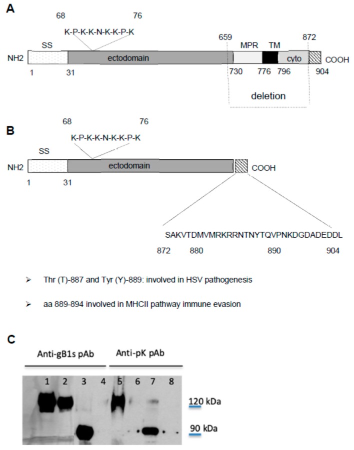
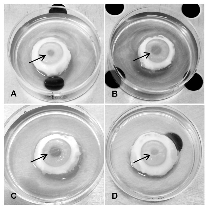
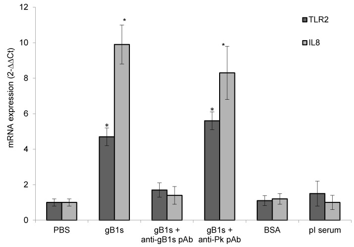
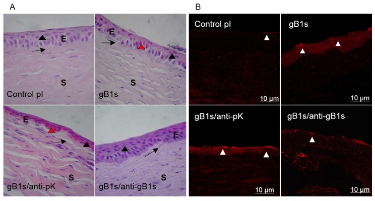
It has been recently reported, using in vitro studies, that the herpes simplex virus 1 (HSV-1) encoded envelope glycoprotein B (gB1) interacts with cell surface toll-like receptor 2 (TLR2) and induces the secretion of interleukin-8 (IL8), a representative marker of inflammatory cytokine activation. The purpose of this study is to investigate the role of gB1 in activating host inflammatory responses by using a secreted form of gB1 (gB1s) and an ex vivo organotypic rabbit corneal model. Abraded corneas exposed to gB1s alone or to the recombinant protein mixed with anti gB polyclonal antibody were cultured in an air-liquid interface. The corneas exposed to gB1s show the appearance of mydriasis and high levels of TLR2 and IL-8 mRNAs transcripts were detected in the superficial layer of corneal epithelial cells. Histological stain and immunohistochemical analyses revealed morphological changes in the epithelium of the treated corneas and variations in expression and localization of TLR2. Collectively these findings provide new insight into the pathogenesis of HSV-1 ocular infection by demonstrating the leading role of gB in activating an inflammatory response and in the appearance of mydriasis, a sign of HSV-1 anterior uveitis.

Keywords: HSV-1 gB; TLR2; ex vivo corneal model; mydriasis.


Conflict of interest statement

The authors declare no conflict of interest. The funder has no role in study design, data collection, and analysis, decision to publish, or preparation of the manuscript.

Figures

Figure 1
Figure 1
Schematic representation of KOS gB1 (A) and gB1s (B) and western blot analysis using anti-gB1s and anti-pK pAbs (C). SS, signal sequence; MPR, membrane-proximal region; TM, trans-membrane region; ectodomain and (cyto) cytodomain regions. Schematic representation of (A) full-length gB1 and (B) gB1s secreted protein obtained from the gB1 gene encoding the MPR and the TM regions, after having deleted 639 nucleotides and reconstructed the gene with the ectodomain and COOH cytodomain. The sequence from 872 to 904 represents the terminal sequence of the cytodomain present in gB1s. Sequence 68 to 76 is the lysine-rich region (pK) that binds HSPG. (C) WB analysis of gB1s recombinant protein (≈90 kD) using the anti-gB1s and anti-pK pAb (lane 3 and 7) and, as controls, full length KOS wt gB1 (120 kDa) (lane 1 and 5) or gBpK mutant (lane 2 and 6) using the anti-gB1s and anti-pK pAbs respectively and mock uninfected cells tested with anti-gB1s and anti-pK pAbs (lanes 4 and 8). The anti-pK pAb is not able to recognize the KOS gBpK mutant.
Figure 2
Figure 2
Clinical signs in treated corneas. The sclero-corneal rings were randomly divided into groups of three rings each treated as follows: (A), pI (pre-immune) rabbit serum as control; (B), gB1s recombinant protein; (C), gB1s recombinant protein preincubated with the anti-pK rabbit polyclonal specific antibody; (D), gB1s recombinant protein preincubated with the anti-gB1s rabbit polyclonal specific antibody. Arrows are indicating the pupil. For each group n = 3.
Figure 3
Figure 3
TLR2 and IL8 mRNA expression. Corneal epithelial cells were treated with 125 ng of gB1s alone or gB1s pre-treated with anti-pK pAb or gB1s pre-treated with anti-gB1s pAb or PBS, BSA, and pre-immune (pI) rabbit serum as control samples. For each group n = 3. Gene expression was expressed as 2ΔΔCt normalized to PBS control. Results were expressed as means ± SD from three different experiments and statistically analyzed by a one-way ANOVA test, followed by Tukey’s HSD. Differences in groups and treatments were considered significant for p <0.05. * p < 0.05 vs PBS control.
Figure 4
Figure 4
Morphological observations (A) and expression of TLR2 on the cell surface (B) in sections of rabbit corneal epithelium samples. (A) Hematoxylin-Eosin (H/E) stain. Epithelium (E), Stroma (S). The suprabasal layer (black arrowhead) and the basal layer (arrow) are indicated. Magnification 100×. control; pre-immune (pI) rabbit serum as control sample; epithelial cells are columnar in basal layer while wing and surface cells are flattened. gB1s; in cornea treated with recombinant protein, epithelial cells are flattened, disarranged in all layers, some cells present Cowdry bodies (red arrowhead). gB1s/anti-pK; in cornea treated with recombinant protein preincubated with anti-pK gB1s pAb, epithelial cells are flattened and strongly eosin stained. gB1s/anti-gB1s; with recombinant protein preincubated with anti-gB1s pAb, cells of the basal layer became columnar and in the suprabasal layer are flattened (as in the control in pI). (B) Immunofluorescence staining with anti-TLR2 Ab on abraded cornea exposed to: control pI; pre-immune rabbit serum as control sample; gB1s recombinant protein; gB1s/anti-pK, gB1s preincubated with anti-pK gB1s pAb; gB1s/anti-gB1s, gB1s preincubated with anti-gB1s pAb.

Similar articles

Cited by

References

    1. Farooq A.V., Shukla D. Herpes simplex epithelial and stromal keratitis: An epidemiologic update. Surv. Ophthalmol. 2012;57:448–462. doi: 10.1016/j.survophthal.2012.01.005. - DOI - PMC - PubMed
    1. Looker K.J., Magaret A.S., May M.T., Turner K.M., Vickerman P., Gottlieb S.L., Newman L.M. Global and Regional Estimates of Prevalent and Incident Herpes Simplex Virus Type 1 Infections in 2012. PLoS ONE. 2015;10:e0140765. doi: 10.1371/journal.pone.0140765. - DOI - PMC - PubMed
    1. Rowe A.M., St Leger A.J., Jeon S., Dhaliwal D.K., Knickelbein J.E., Hendricks R.L. Herpes keratitis. Prog. Retin. Eye Res. 2013;32:88–101. doi: 10.1016/j.preteyeres.2012.08.002. - DOI - PMC - PubMed
    1. Tsatsos M., MacGregor C., Athanasiadis I., Moschos M.M., Hossain P., Anderson D. Herpes simplex virus keratitis: An update of the pathogenesis and current treatment with oral and topical antiviral agents-response. Clin. Exp. Ophthalmol. 2017;45:317. doi: 10.1111/ceo.12835. - DOI - PubMed
    1. Hlinomazova Z., Loukotova V., Horackova M., Sery O. The treatment of HSV1 ocular infections using quantitative real-time PCR results. Acta Ophthalmol. 2012;90:456–460. doi: 10.1111/j.1755-3768.2010.01933.x. - DOI - PubMed

Publication types

Substances