Review
. 2019 Sep 4;8(9):1385.
doi: 10.3390/jcm8091385.

Relationship Between Oxidative Stress, ER Stress, and Inflammation in Type 2 Diabetes: The Battle Continues

Affiliations
Review

Relationship Between Oxidative Stress, ER Stress, and Inflammation in Type 2 Diabetes: The Battle Continues

Estefania Burgos-Morón et al. J Clin Med. .

Abstract

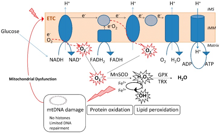
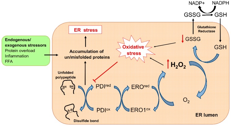
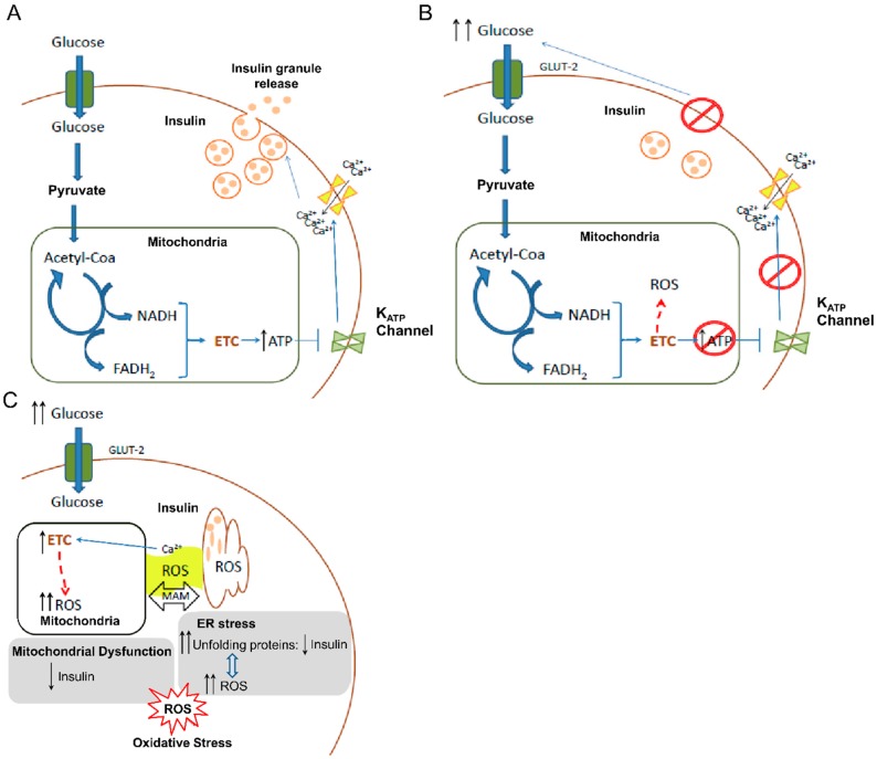
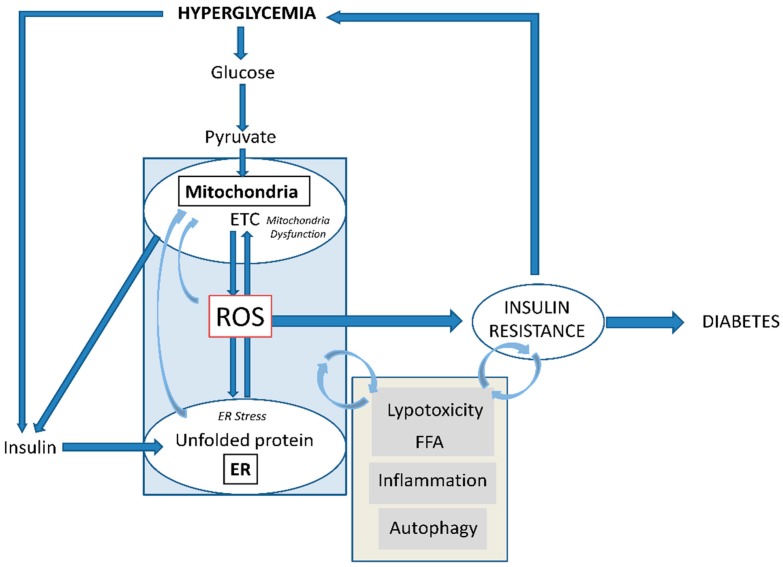
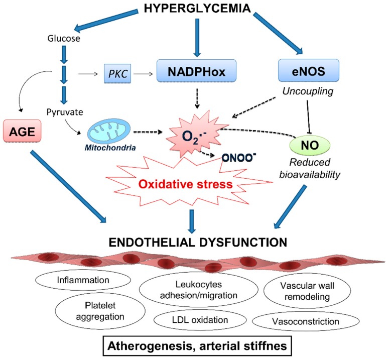
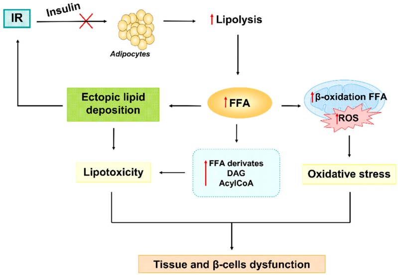
Type 2 diabetes (T2D) is a metabolic disorder characterized by hyperglycemia and insulin resistance in which oxidative stress is thought to be a primary cause. Considering that mitochondria are the main source of ROS, we have set out to provide a general overview on how oxidative stress is generated and related to T2D. Enhanced generation of reactive oxygen species (ROS) and oxidative stress occurs in mitochondria as a consequence of an overload of glucose and oxidative phosphorylation. Endoplasmic reticulum (ER) stress plays an important role in oxidative stress, as it is also a source of ROS. The tight interconnection between both organelles through mitochondrial-associated membranes (MAMs) means that the ROS generated in mitochondria promote ER stress. Therefore, a state of stress and mitochondrial dysfunction are consequences of this vicious cycle. The implication of mitochondria in insulin release and the exposure of pancreatic β-cells to hyperglycemia make them especially susceptible to oxidative stress and mitochondrial dysfunction. In fact, crosstalk between both mechanisms is related with alterations in glucose homeostasis and can lead to the diabetes-associated insulin-resistance status. In the present review, we discuss the current knowledge of the relationship between oxidative stress, mitochondria, ER stress, inflammation, and lipotoxicity in T2D.

Keywords: ER stress; ROS; antioxidants; insulin resistance; mitochondria; oxidative stress; type 2 diabetes.


Conflict of interest statement

References

Figures

Figure 1
Figure 1
Mitochondrial superoxide (O2•−) generation by the electron transport chain (ETC) and the implication of the enzyme manganese superoxide dismutase (MnSOD), the only superoxide dismutase enzyme located in the mitochondrial matrix, in its detoxification. The elevated levels of O2•− induce damage to macromolecules, including lipids, proteins, and nucleic acids, and promote mitochondrial dysfunction. Absence of histones in mitochondrial DNA (mtDNA) and limited DNA repair mechanisms make mitochondria highly susceptible to DNA damage induced by O2•−. ADP: Adenoxine diphosphate; ATP: Adenosine triphosphate; GPX: Glutathione peroxidase; H2O2: Hydrogen peroxide; IMM: Inner mitochondrial membrane; IMS: Intermembrane space; O2•−: Superoxide anion; OH: Hydroxyl radical; TRX: Thioredoxin reductase.
Figure 2
Figure 2
Oxidative protein machinery and ER stress. During disulfide bond formation, two electrons are transferred to the pair of cysteines in the polypeptide by the PDI active site. Thereafter, reduced PDI receive electrons from O2 through ERO1-mediated redox reaction, resulting in H2O2 formation. The GSH/GSSG system then recovers the redox status by scavenging H2O2. Several stimuli including increased protein synthesis demand overwhelm ER-folding capacity and disturb redox balance, leading to the accumulation of misfolded proteins and triggering ER stress. ER: Endoplasmic reticulum; ERO1: ER oxidoreductin 1; FFA: Free fatty acids; GSH: Glutathione; GSSG: Glutathione disulphide; NADPH: Nicotinamide adenine dinucleotide phosphate; PDI: Protein disulfide isomerase.
Figure 3
Figure 3
Cellular mechanisms in pancreatic β-cells involving mitochondrial ROS generation and their implication in insulin release and diabetes onset. (A) Mechanism of insulin release in β-cells in normal conditions. (B) Impaired insulin release by β-cells under hyperglycemic conditions. High glucose concentration in blood implies high ROS generation by mitochondria leading to alterations in insulin release. (C) Scheme of how hyperglycemia promotes ROS generation by ETC (electron transport chain) in mitochondria inducing oxidative stress. Oxidative stress is boosted by the ER stress as consequence of the accumulation of unfolded insulin peptides due to the enhanced demand of this hormone. In addition, the implication of Ca2+ ions from ER as a factor that increases ROS in mitochondria and the interconnection of both organelles through MAM (mitochondria-associated ER membranes) are depicted. ATP: Adenosine triphosphate; ER: Endoplasmic reticulum; GLUT2: Glucose transporter-2; ROS: Reactive oxygen species.
Figure 4
Figure 4
Development of insulin resistance and the relevant role of mitochondrial ROS generation. Frequent hyperglycemia condition promotes ROS production by mitochondria through the ETC. ER function and folding capacity is affected by mitochondrial ROS production. This process is especially important in pancreatic β-cells, in charge of insulin production and secretion. Initially, excess nutrient overload increases insulin synthesis demand. Perpetuated hyperglycemia and hyperinsulinemia progress to insulin resistance in peripheral tissues. This fact forces β-cells to produce more insulin promoting ER stress, in parallel with increased oxidative stress and mitochondrial dysfunction. Oxidative stress in mitochondria and ER stress feedback each other directly through ROS and also indirectly (curve arrow), aggravating the oxidative stress and promoting further insulin resistance. This situation can progress to β-cell failure and the impairment of insulin release, thus provoking the inability to control glucose levels in blood characteristic of T2D patients. Insulin resistance is also associated with processes such to inflammation, lipotoxicity, and autophagy impairment that make oxidative stress and insulin resistance characteristic of T2D worse. ETC: Electron transport chain; ER: Endoplasmic reticulum; FFA: Free fatty acids; ROS: Radical oxygen species.
Figure 5
Figure 5
Mechanisms of ROS-induced endothelial dysfunction in response to hyperglycemia. Vascular damage caused by elevated glucose levels is mainly derived by an imbalance between ROS production and NO bioavailability in the endothelium and by the direct damaged caused by the accumulation of AGE. Resulting endothelial dysfunction is characterized by the activation of several deleterious mechanisms, including proinflammatory response, recruitment of leukocytes, accumulation of oxidized LDL particles and impaired vasodilatation, in the onset of cardiovascular complications. AGE: Advanced glycation end-products; eNOS: Endothelial nitric oxide synthase; LDL: Low density lipoprotein particles; NADPHox: Nicotinamide adenine dinucleotide phosphate oxidase; NO: Nitric oxide; O2•−: Superoxide anion; ONOO: Peroxynitrite; PKC: Protein kinase C; RNS: Radical nitrogen species.
Figure 6
Figure 6
Under insulin resistance condition, adipocytes are not able to respond to insulin stimuli and downregulate lipolysis. In this way, an uncontrolled release of FFA causes ectopic lipid deposition in body tissues and an augment of lipid intermediates derivate from metabolism (DAG and AcylCoA), which together lead to lipotoxicity and further insulin resistance. On the other hand, FFA are not fully processed by mitochondria triggering incomplete β-oxidation of them, further ROS production and oxidative stress. Finally, lipotoxicity and oxidative stress they both concur to alterations in cells homeostasis and β-cells failure. AcylCoA: Acetyl coenzyme A; DAG: Dyacylglicerol; FFA: Free fatty acids; IR: Insulin resistance; ROS: Radical oxygen species.

References

    1. Cho N.H., Shaw J.E., Karuranga S., Huang Y., da Rocha Fernandes J.D., Ohlrogge A.W., Malanda B. IDF Diabetes Atlas: Global estimates of diabetes prevalence for 2017 and projections for 2045. Diabetes Res. Clin. Pract. 2018;138:271–281. doi: 10.1016/j.diabres.2018.02.023. - DOI - PubMed
    1. Zheng Y., Ley S.H., Hu F.B. Global aetiology and epidemiology of type 2 diabetes mellitus and its complications. Nat. Rev. Endocrinol. 2018;14:88–98. doi: 10.1038/nrendo.2017.151. - DOI - PubMed
    1. Einarson T.R., Acs A., Ludwig C., Panton U.H. Prevalence of cardiovascular disease in type 2 diabetes: A systematic literature review of scientific evidence from across the world in 2007–2017. Cardiovasc. Diabetol. 2018;17:83. doi: 10.1186/s12933-018-0728-6. - DOI - PMC - PubMed
    1. McBride H.M., Neuspiel M., Wasiak S. Mitochondria: More than just a powerhouse. Curr. Biol. 2006;16:R551–R560. doi: 10.1016/j.cub.2006.06.054. - DOI - PubMed
    1. Brehm A., Krssak M., Schmid A.I., Nowotny P., Waldhäusl W., Roden M. Increased lipid availability impairs insulin-stimulated ATP synthesis in human skeletal muscle. Diabetes. 2006;55:136–140. doi: 10.2337/diabetes.55.01.06.db05-1286. - DOI - PubMed

LinkOut - more resources